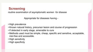
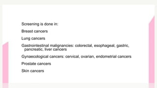
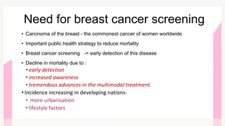
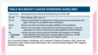
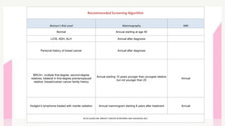
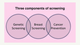
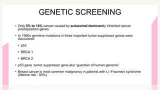
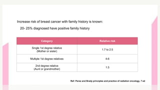
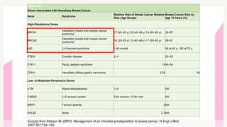
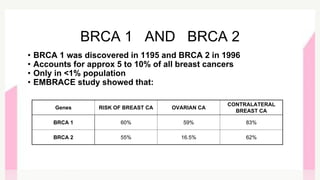
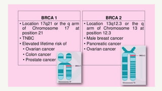
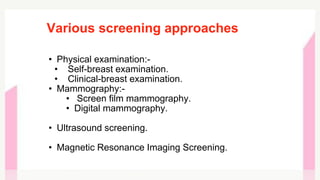
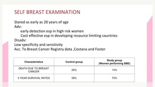
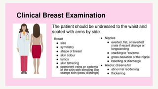
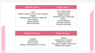
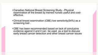
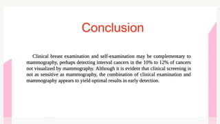
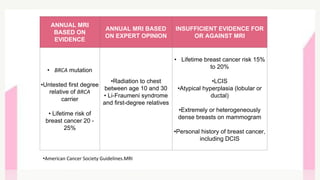
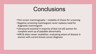
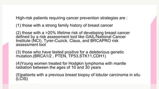
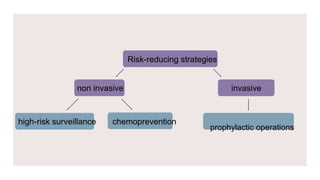
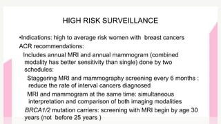
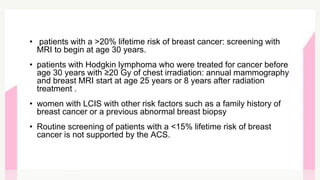
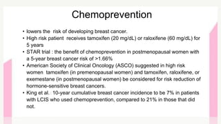
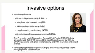
-DR. PALLAVI JAIN
MODERATOR- DR. ISHA JAISWAL
BREAST CANCER SCREENING
AND PREVENTION
• SCREENING PRINCIPLES
• BREAST CANCER SCREENING
• SCREENING GUIDELINES
• SCREENING TECHNIQUES
• CANCER PREVENTION
CONTENTS
Screening
routine examination of asymptomatic women for disease
Appropriate for diseases having -
• High prevalence
• Known natural history, precursor lesion and course of progression
• If detected in early stage, amenable to cure
• Methods used must be simple, cheap, specific and sensitive, acceptable,
risk-free and accessible.
• High sensitivity
• High specificity
Screening is done in:
Breast cancers
Lung cancers
Gastrointestinal malignancies: colorectal, esophageal, gastric,
pancreatic, liver cancers
Gynaecological cancers: cervical, ovarian, endometrial cancers
Prostate cancers
Skin cancers
• Carcinoma of the breast - the commonest cancer of women worldwide
• Important public health strategy to reduce mortality
• Breast cancer screening -> early detection of this disease
• Decline in mortality due to :
• early detection
• increased awareness
• tremendous advances in the multimodal treatment.
• Incidence increasing in developing nations-
• more urbanisation
• lifestyle factors
Need for breast cancer screening
Recommended Screening Algorithm
Woman's Risk Level Mammography MRI
Normal Annual starting at age 40
LCIS, ADH, ALH Annual after diagnosis
Personal history of breast cancer Annual after diagnosis
BRCA+; multiple first-degree, second-degree
relatives; bilateral in first-degree premenopausal
relative; breast/ovarian cancer family history
Annual starting 10 years younger than youngest relative
but not younger than 25
Annual
Hodgkin's lymphoma treated with mantle radiation Annual mammogram starting 8 years after treatment Annual
NCCN GUIDELINE :BREAST CANCER SCREENING AND DIAGNOSIS 2021
GENETIC SCREENING
• Only 5% to 10% cancer caused by autosomal dominantly inherited cancer
predisposition genes
• In 1990s germline mutations in three important tumor suppressor genes were
discovered
• p53
• BRCA 1
• BRCA 2
• p53 gene :tumor suppressor gene aka “guardian of human genome”
• Breast cancer is most common malignancy in patients with Li -Fraumeni syndrome
(lifetime risk - 90%)
Ref: Perez and Brady principles and practice of radiation oncology, 7 ed
Increase risk of breast cancer with family history is known:
20- 25% diagnosed have positive family history
Category Relative risk
Single 1st degree relative
(Mother or sister)
1.7 to 2.5
Multiple 1st degree relatives 4-6
2nd degree relative
(Aunt or grandmother)
1.5
TABLE 34.1
Genes Associated with Hereditary Breast Cancer
Gene Syndrome
Relative Risk of Breast Cancer Relative
Risk (Age Range)
Breast Cancer Risk by
Age 70 Years (%)
High-Penetrance Genes
BRCA1
Hereditary breast and ovarian cancer
syndrome
17 (20–29 y) 32 (40–49 y) 14 (60–69 y) 39–87
BRCA2
Hereditary breast and ovarian cancer
syndrome
19 (20–29 y) 10 (40–49 y) 11 (60–69 y) 26–91
p53 Li-Fraumeni syndrome 1.46 overall 56 at 45 y; >90 at 70 y
PTEN Cowden disease 2–4 25–50
STK11 Peutz-Jeghers syndrome 1545–54
CDH1 Hereditary diffuse gastric carcinoma 3.25 39
Low- to Moderate-Penetrance Genes
ATM Ataxia-telangiectasia 3–4 NA
CHEK2 Li-Fraumeni variant 2 for women; 10 for men NA
BRIP1 Fanconi anemia 2NA
PALB2 None 2.3NA
Excerpt from Robson M, Offit K. Management of an inherited predisposition to breast cancer. N Engl J Med
2007;357:154–162.
• BRCA 1 was discovered in 1195 and BRCA 2 in 1996
• Accounts for approx 5 to 10% of all breast cancers
• Only in <1% population
• EMBRACE study showed that:
BRCA 1 AND BRCA 2
Genes RISK OF BREAST CA OVARIAN CA
CONTRALATERAL
BREAST CA
BRCA 1 60% 59% 83%
BRCA 2 55% 16.5% 62%
• About 20-35% of breast tumors are due to p53 mutant
• Point missense mutation leading to synthesis of malfunctioned protein
• Associated with aggressive type of breast cancer- triple negative
breast cancer
• Germline mutation of p53 -Li Fraumani syndrome :
• Rare autosomal dominant
• Involves breast ,colon ,osteosarcomas, soft tissue sarcoma, brain
tumors ,leukaemia and adrenocortical cancers
• 50% LFS pt will develop invasive cancer by 30 years and 90% by 70
years
• Mc cancer in LFS- BREAST CANCER
• lifetime risk of developing breast cancer in LFS - 90%
p53
Indications Of Genetic Testing
•Family history of known BRCA1/BRCA2 mutations
•Personal h/o breast cancer plus (one of following)
1. age 45 years or younger
2. age <= 50 years with :
•One or more close relatives with breast or prostate or pancreatic cancer
3. age <= 60 years with :
•triple negative (TN) BREAST CANCER
4. any age with :
• 2 or more close blood relative with breast pancreatic, or prostate ca
•>=1 close blood relative with breast cancer >= 50 years
•>= 1 close relative with ovarian cancer
•Close male blood relative with breast cancer
•Ethnicity ass. with huge mutation frequency (eg. Ashkenazi Jewish)
•Personal h/o epithelial ovarian/fallopian tube/ primary peritoneal cancer
•Personal h/o male breast cancer
REF:NCCN GUIDELINES FOR GENETIC SCREENING
BREAST CANCER
SCREENING
Various screening approaches
• Physical examination:-
• Self-breast examination.
• Clinical-breast examination.
• Mammography:-
• Screen film mammography.
• Digital mammography.
• Ultrasound screening.
• Magnetic Resonance Imaging Screening.
SELF BREAST EXAMINATION
Stared as early as 20 years of age
Adv:
early detection esp in high risk women
Cost effective esp in developing resource limiting countries
Disadv:
Low specificity and sensitivity
Acc. To Breast Cancer Registry data ,Costana and Foster
Characteristics Control group
Study group
(Woman performing SBE)
DEATH DUE TO BREAST
CANCER
26% 14%
5 YEAR SURVIVAL RATES 59% 75%
Malignant masses Benign masses
Hard
Painless: painful in only 10-15% of patients.
Irregular
Possibly fixed to the skin or chest wall
Skin dimpling
Nipple retraction
Bloody discharge
Firm, rubbery mass
Frequently painful
Regular margins
Not fixed to skin or chest wall,
mobile
No skin dimpling
No nipple retraction
No bloody discharge
Malignant discharge Benign discharge
Unilateral
Spontaneous
One duct orifice
Bloody, serosanguineous, or serous
Bilateral
Spontaneous or induced
Multiple duct orifices
Thick green or yellow, induced and
bilateral (duct ectasia)
• Canadian National Breast Screening Study - Physical
examination of the breast by trained nurses useful and cost-
effective.
• Clinical breast examination (CBE) low sensitivity(54%) as a
screening tool .
• CBE has been recommended based on lack of conclusive
evidence against it and it can be used as a tool to discuss
early breast cancer detection and other breast cancer issues
Conclusion
Screening
Mammography
routine mammographic images in asymptomatic women
• Two views: craniocaudal and mediolateral oblique of each breast.
• Sensitivity of Mammography - 90%
• specificity- 94%
• 85% - 90% in fatty replaced breasts
• 65% in dense breasts
• Positive predictive value- 10-14% for screened pts, but significantly
higher for symptomatic pts
• After mammography guided FNAC/Biopsy, detection rate of
malignancy about 30 %
Screening mammography
ADVANTAGES:
• Screening mammography has a considerable impact on
incidence and stage of presentation.
• Studies show
oShift to earlier stage
oDecrease in mortality
oEarly detection, followed by appropriate local, regional, &
systemic treatment is associated with reduced breast cancer
mortality rates in women 50 yrs of age & older.
Cranio-caudal:- Medio-lateral:-
Mammographic Manifestation of Breast Cancer
• Ill-defined mass -spiculated margins, rarely cancers may also be seen
with a knobby, lobulated, or even a smooth contour
• Architectural distortion of the breast . the appearance of linear,
radiated, or spiculated changes around a central focus should always be
considered suspect for carcinoma.
• The tumor may be hidden by dense parenchyma;
• Calcification can be associated with either benign or malignant
conditions.
• Malignant tumors:- are typically 100-300 micrometer in size & are rod
like, tubular, branching or punctate.
• Clusters of microcalcification are suggestive of intraductal disease.
Primary Signs of Cancer:
•Mass
•Calcifications
Secondary Signs :
•Nipple Inversion
•Architectural Distortion
•Skin Thickening
•Axillary Adenopathy
•Skin Retraction
•Tissue Asymmetry
•Developing “Neodensity”
Studies conducted were :-
1. Health Insurance Plan Study:-
• Health insurance plan study (HIP)-16 yr, 31000 pts’,40-64 yr old women
• Mortality decreased in all women in screening arm . More for > 50 age group.
2.Tabar et al:-
Tabar et al studied mammographic screening (29 yrs ,1863 pts )and found 63%
decrease in mortality from breast cancer.
3. Canadian study:-
But in a Canadian study and one by Miller et al addition of annual mammographic
screening to physical examination had no effect on breast ca. mortality .
4.Millar et al:- similar result as Canadian study was observed.
REASON FOR THIS DISCRIPENCY WAS:
• unbalanced allocation
• Poor quality of mammography
• Less sample size
Screening in women <50yrs
• Stockholm mammographic screening trial (40,000, 14,842 ).The RR of
breast cancer death in screened versus non screened women was 1.08 in
the 40- 49 year group.
A large trial conducted in United kingdom:-(age 40-64 yrs)
• One group:- annual screening by clinical examination &
mammography.
• 2nd group:- breast self-examination.
• 3rd group:- control group.
• 16 yrs follow-up:- mortality was 27% lower in the 2
. screening groups.
35% decrease in mortality was observed in mammographically screened
women
• No evidence of less benefit in women 45-49yrs at initial screening.
• Wald et al. (6 trials) observed 15% reduction in breast ca. mortality in
40-49 year old group as compared to 25% in 50 – 74 years
CONCLUSION
Screening mammography leads to increased use of breast conservation
techniques, significant reduction in breast ca. mortality and reduction in
cancer incidence.
Digital Mammography
• Transforms x-ray images into electronic digital images.
• System is equipped with a digital receptor and a computer
instead of film screen
• Advantage:-
• No film processing
• Faster image acquisition
• less call-backs
• Sensitivity is same as that of screen film techniques.
• At present time, resolution is better for film screen, but
studies have proved equal accuracy
Ultrasound screening
• Role in
➢High risk population
➢Young women with mammographically
dense breasts
• It is very helpful in differentiating cysts from solid
tumors.
• Early, curable invasive cancers not seen on
mammography can be found with sonography.
• Sensitivity of 73%
• specificity of 95%.
• Disadv: reproducibility, high false-positive rates,
operator dependency of the examination, inability to
image most ductal carcinoma in situ, and lack of
agreement on which solid or complex lesions require
biopsy
Usg breast in asymptomatic patient:DCIS
MRI SCREENING
• INDICATIONS:
• high risk women (BRCA+,strong family history)
• single screening MRI of the contralateral breast(detection rate -5%)
• HIGH sensitivity: 70-100%
• Specificity: 89.9%
• All trials have demonstrated a superiority for MRI screening for high risk
patients.
• Disadv:
• recall rates and biopsy rates 2-3 times higher than mammography
• expensive
• Thus limited to high risk patients only
Characteristic Cbe Mammography MRI
SENSITIVITY 17.9% 33.3% 79.5%
SPECIFICITY 98.1% 95% 89.9%
Kriege et al
ANNUAL MRI
BASED ON
EVIDENCE
ANNUAL MRI BASED
ON EXPERT OPINION
INSUFFICIENT EVIDENCE FOR
OR AGAINST MRI
• BRCA mutation
•Untested first degree
relative of BRCA
carrier
• Lifetime risk of
breast cancer 20 -
25%
•Radiation to chest
between age 10 and 30
• Li-Fraumeni syndrome
and first-degree relatives
• Lifetime breast cancer risk 15%
to 20%
•LCIS
•Atypical hyperplasia (lobular or
ductal)
•Extremely or heterogeneously
dense breasts on mammogram
•Personal history of breast cancer,
including DCIS
•American Cancer Society Guidelines.MRI
Conclusions
•Film screen mammography – modality of choice for screening
•Negative screening mammogram never replaces need for
diagnostic mammogram
•Ultrasound essential in majority of (but not all) women for
complete work up of palpable abnormality
•MRI & other newer modalities- evaluating extent of disease in
women with current breast cancer diagnosis
High-risk patients requiring cancer prevention strategies are :
(1) those with a strong family history of breast cancer
(2) those with a >20% lifetime risk of developing breast cancer
defined by a risk assessment tool like GAIL/National Cancer
Institute (NCI), Tyrer-Cuzick, Claus, and BRCAPRO risk
assessment tool
(3) those who have tested positive for a deleterious genetic
mutation.(BRCA1/2 , PTEN, TP53,STK11,CDH1)
(4)Young women treated for Hodgkin lymphoma with mantle
radiation between the ages of 10 and 30 years
(5)patients with a previous breast biopsy of lobular carcinoma in situ
(LCIS)
prophylactic operations
Risk-reducing strategies
non invasive invasive
high-risk surveillance chemoprevention
•Indications: high to average risk women with breast cancers
ACR recommendations:
Includes annual MRI and annual mammogram (combined
modality has better sensitivity than single) done by two
schedules:
Staggering MRI and mammography screening every 6 months :
reduce the rate of interval cancers diagnosed
MRI and mammogram at the same time: simultaneous
interpretation and comparison of both imaging modalities
BRCA1/2 mutation carriers: screening with MRI begin by age 30
years (not before 25 years )
HIGH RISK SURVEILLANCE
• patients with a >20% lifetime risk of breast cancer: screening with
MRI to begin at age 30 years.
• patients with Hodgkin lymphoma who were treated for cancer before
age 30 years with ≥20 Gy of chest irradiation: annual mammography
and breast MRI start at age 25 years or 8 years after radiation
treatment .
• women with LCIS with other risk factors such as a family history of
breast cancer or a previous abnormal breast biopsy
• Routine screening of patients with a <15% lifetime risk of breast
cancer is not supported by the ACS.
• lowers the risk of developing breast cancer.
• High risk patient receives tamoxifen (20 mg/dL) or raloxifene (60 mg/dL) for
5 years
• STAR trial : the benefit of chemoprevention in postmenopausal women with
a 5-year breast cancer risk of >1.66%
• American Society of Clinical Oncology (ASCO) suggested in high risk
women tamoxifen (in premenopausal women) and tamoxifen, raloxifene, or
exemestane (in postmenopausal women) be considered for risk reduction of
hormone-sensitive breast cancers.
• King et al. : 10-year cumulative breast cancer incidence to be 7% in patients
with LCIS who used chemoprevention, compared to 21% in those that did
not.
Chemoprevention
Invasive options
• Invasive options are :
• risk-reducing mastectomy (RRM) :-
• simple or total mastectomy (TM)
• skin-sparing mastectomy (SSM)
• nipple-sparing mastectomy (NSM).
• risk-reducing salpingo-oophorectomy (RRSO),
• The Prevention and Observation Surgical End Points (PROSE) study
group showed that RRM reduced the risk of breast cancer by 99% in
BRCA1/2 patients with prior oophorectomy and 90% in women with intact
ovaries.
• Timing of prophylactic surgeries is highly individualized ;studies shown
younger people benefits more.
The first step in management is to identify high-risk patients by taking a
detailed history or with the help of a risk assessment tool. High-risk
surveillance with MRI as an adjunct to mammography,
chemoprevention, and RRSs are all options that require a nuanced
conversation regarding management between patients and their
clinicians.
Conclusion
Breast screening pallavi

Breast screening pallavi

  • 1.
    -DR. PALLAVI JAIN MODERATOR-DR. ISHA JAISWAL BREAST CANCER SCREENING AND PREVENTION
  • 2.
    • SCREENING PRINCIPLES •BREAST CANCER SCREENING • SCREENING GUIDELINES • SCREENING TECHNIQUES • CANCER PREVENTION CONTENTS
  • 3.
    Screening routine examination ofasymptomatic women for disease Appropriate for diseases having - • High prevalence • Known natural history, precursor lesion and course of progression • If detected in early stage, amenable to cure • Methods used must be simple, cheap, specific and sensitive, acceptable, risk-free and accessible. • High sensitivity • High specificity
  • 4.
    Screening is donein: Breast cancers Lung cancers Gastrointestinal malignancies: colorectal, esophageal, gastric, pancreatic, liver cancers Gynaecological cancers: cervical, ovarian, endometrial cancers Prostate cancers Skin cancers
  • 5.
    • Carcinoma ofthe breast - the commonest cancer of women worldwide • Important public health strategy to reduce mortality • Breast cancer screening -> early detection of this disease • Decline in mortality due to : • early detection • increased awareness • tremendous advances in the multimodal treatment. • Incidence increasing in developing nations- • more urbanisation • lifestyle factors Need for breast cancer screening
  • 7.
    Recommended Screening Algorithm Woman'sRisk Level Mammography MRI Normal Annual starting at age 40 LCIS, ADH, ALH Annual after diagnosis Personal history of breast cancer Annual after diagnosis BRCA+; multiple first-degree, second-degree relatives; bilateral in first-degree premenopausal relative; breast/ovarian cancer family history Annual starting 10 years younger than youngest relative but not younger than 25 Annual Hodgkin's lymphoma treated with mantle radiation Annual mammogram starting 8 years after treatment Annual NCCN GUIDELINE :BREAST CANCER SCREENING AND DIAGNOSIS 2021
  • 10.
    GENETIC SCREENING • Only5% to 10% cancer caused by autosomal dominantly inherited cancer predisposition genes • In 1990s germline mutations in three important tumor suppressor genes were discovered • p53 • BRCA 1 • BRCA 2 • p53 gene :tumor suppressor gene aka “guardian of human genome” • Breast cancer is most common malignancy in patients with Li -Fraumeni syndrome (lifetime risk - 90%)
  • 11.
    Ref: Perez andBrady principles and practice of radiation oncology, 7 ed Increase risk of breast cancer with family history is known: 20- 25% diagnosed have positive family history Category Relative risk Single 1st degree relative (Mother or sister) 1.7 to 2.5 Multiple 1st degree relatives 4-6 2nd degree relative (Aunt or grandmother) 1.5
  • 12.
    TABLE 34.1 Genes Associatedwith Hereditary Breast Cancer Gene Syndrome Relative Risk of Breast Cancer Relative Risk (Age Range) Breast Cancer Risk by Age 70 Years (%) High-Penetrance Genes BRCA1 Hereditary breast and ovarian cancer syndrome 17 (20–29 y) 32 (40–49 y) 14 (60–69 y) 39–87 BRCA2 Hereditary breast and ovarian cancer syndrome 19 (20–29 y) 10 (40–49 y) 11 (60–69 y) 26–91 p53 Li-Fraumeni syndrome 1.46 overall 56 at 45 y; >90 at 70 y PTEN Cowden disease 2–4 25–50 STK11 Peutz-Jeghers syndrome 1545–54 CDH1 Hereditary diffuse gastric carcinoma 3.25 39 Low- to Moderate-Penetrance Genes ATM Ataxia-telangiectasia 3–4 NA CHEK2 Li-Fraumeni variant 2 for women; 10 for men NA BRIP1 Fanconi anemia 2NA PALB2 None 2.3NA Excerpt from Robson M, Offit K. Management of an inherited predisposition to breast cancer. N Engl J Med 2007;357:154–162.
  • 13.
    • BRCA 1was discovered in 1195 and BRCA 2 in 1996 • Accounts for approx 5 to 10% of all breast cancers • Only in <1% population • EMBRACE study showed that: BRCA 1 AND BRCA 2 Genes RISK OF BREAST CA OVARIAN CA CONTRALATERAL BREAST CA BRCA 1 60% 59% 83% BRCA 2 55% 16.5% 62%
  • 15.
    • About 20-35%of breast tumors are due to p53 mutant • Point missense mutation leading to synthesis of malfunctioned protein • Associated with aggressive type of breast cancer- triple negative breast cancer • Germline mutation of p53 -Li Fraumani syndrome : • Rare autosomal dominant • Involves breast ,colon ,osteosarcomas, soft tissue sarcoma, brain tumors ,leukaemia and adrenocortical cancers • 50% LFS pt will develop invasive cancer by 30 years and 90% by 70 years • Mc cancer in LFS- BREAST CANCER • lifetime risk of developing breast cancer in LFS - 90% p53
  • 16.
    Indications Of GeneticTesting •Family history of known BRCA1/BRCA2 mutations •Personal h/o breast cancer plus (one of following) 1. age 45 years or younger 2. age <= 50 years with : •One or more close relatives with breast or prostate or pancreatic cancer 3. age <= 60 years with : •triple negative (TN) BREAST CANCER 4. any age with : • 2 or more close blood relative with breast pancreatic, or prostate ca •>=1 close blood relative with breast cancer >= 50 years •>= 1 close relative with ovarian cancer •Close male blood relative with breast cancer •Ethnicity ass. with huge mutation frequency (eg. Ashkenazi Jewish) •Personal h/o epithelial ovarian/fallopian tube/ primary peritoneal cancer •Personal h/o male breast cancer REF:NCCN GUIDELINES FOR GENETIC SCREENING
  • 17.
  • 18.
    Various screening approaches •Physical examination:- • Self-breast examination. • Clinical-breast examination. • Mammography:- • Screen film mammography. • Digital mammography. • Ultrasound screening. • Magnetic Resonance Imaging Screening.
  • 19.
    SELF BREAST EXAMINATION Staredas early as 20 years of age Adv: early detection esp in high risk women Cost effective esp in developing resource limiting countries Disadv: Low specificity and sensitivity Acc. To Breast Cancer Registry data ,Costana and Foster Characteristics Control group Study group (Woman performing SBE) DEATH DUE TO BREAST CANCER 26% 14% 5 YEAR SURVIVAL RATES 59% 75%
  • 22.
    Malignant masses Benignmasses Hard Painless: painful in only 10-15% of patients. Irregular Possibly fixed to the skin or chest wall Skin dimpling Nipple retraction Bloody discharge Firm, rubbery mass Frequently painful Regular margins Not fixed to skin or chest wall, mobile No skin dimpling No nipple retraction No bloody discharge Malignant discharge Benign discharge Unilateral Spontaneous One duct orifice Bloody, serosanguineous, or serous Bilateral Spontaneous or induced Multiple duct orifices Thick green or yellow, induced and bilateral (duct ectasia)
  • 23.
    • Canadian NationalBreast Screening Study - Physical examination of the breast by trained nurses useful and cost- effective. • Clinical breast examination (CBE) low sensitivity(54%) as a screening tool . • CBE has been recommended based on lack of conclusive evidence against it and it can be used as a tool to discuss early breast cancer detection and other breast cancer issues
  • 24.
  • 25.
  • 26.
    routine mammographic imagesin asymptomatic women • Two views: craniocaudal and mediolateral oblique of each breast. • Sensitivity of Mammography - 90% • specificity- 94% • 85% - 90% in fatty replaced breasts • 65% in dense breasts • Positive predictive value- 10-14% for screened pts, but significantly higher for symptomatic pts • After mammography guided FNAC/Biopsy, detection rate of malignancy about 30 % Screening mammography
  • 27.
    ADVANTAGES: • Screening mammographyhas a considerable impact on incidence and stage of presentation. • Studies show oShift to earlier stage oDecrease in mortality oEarly detection, followed by appropriate local, regional, & systemic treatment is associated with reduced breast cancer mortality rates in women 50 yrs of age & older.
  • 28.
  • 29.
    Mammographic Manifestation ofBreast Cancer • Ill-defined mass -spiculated margins, rarely cancers may also be seen with a knobby, lobulated, or even a smooth contour • Architectural distortion of the breast . the appearance of linear, radiated, or spiculated changes around a central focus should always be considered suspect for carcinoma. • The tumor may be hidden by dense parenchyma; • Calcification can be associated with either benign or malignant conditions. • Malignant tumors:- are typically 100-300 micrometer in size & are rod like, tubular, branching or punctate. • Clusters of microcalcification are suggestive of intraductal disease.
  • 30.
    Primary Signs ofCancer: •Mass •Calcifications Secondary Signs : •Nipple Inversion •Architectural Distortion •Skin Thickening •Axillary Adenopathy •Skin Retraction •Tissue Asymmetry •Developing “Neodensity”
  • 31.
    Studies conducted were:- 1. Health Insurance Plan Study:- • Health insurance plan study (HIP)-16 yr, 31000 pts’,40-64 yr old women • Mortality decreased in all women in screening arm . More for > 50 age group. 2.Tabar et al:- Tabar et al studied mammographic screening (29 yrs ,1863 pts )and found 63% decrease in mortality from breast cancer. 3. Canadian study:- But in a Canadian study and one by Miller et al addition of annual mammographic screening to physical examination had no effect on breast ca. mortality . 4.Millar et al:- similar result as Canadian study was observed. REASON FOR THIS DISCRIPENCY WAS: • unbalanced allocation • Poor quality of mammography • Less sample size
  • 32.
    Screening in women<50yrs • Stockholm mammographic screening trial (40,000, 14,842 ).The RR of breast cancer death in screened versus non screened women was 1.08 in the 40- 49 year group. A large trial conducted in United kingdom:-(age 40-64 yrs) • One group:- annual screening by clinical examination & mammography. • 2nd group:- breast self-examination. • 3rd group:- control group. • 16 yrs follow-up:- mortality was 27% lower in the 2 . screening groups. 35% decrease in mortality was observed in mammographically screened women • No evidence of less benefit in women 45-49yrs at initial screening.
  • 33.
    • Wald etal. (6 trials) observed 15% reduction in breast ca. mortality in 40-49 year old group as compared to 25% in 50 – 74 years CONCLUSION Screening mammography leads to increased use of breast conservation techniques, significant reduction in breast ca. mortality and reduction in cancer incidence.
  • 34.
    Digital Mammography • Transformsx-ray images into electronic digital images. • System is equipped with a digital receptor and a computer instead of film screen • Advantage:- • No film processing • Faster image acquisition • less call-backs • Sensitivity is same as that of screen film techniques. • At present time, resolution is better for film screen, but studies have proved equal accuracy
  • 35.
    Ultrasound screening • Rolein ➢High risk population ➢Young women with mammographically dense breasts • It is very helpful in differentiating cysts from solid tumors. • Early, curable invasive cancers not seen on mammography can be found with sonography. • Sensitivity of 73% • specificity of 95%. • Disadv: reproducibility, high false-positive rates, operator dependency of the examination, inability to image most ductal carcinoma in situ, and lack of agreement on which solid or complex lesions require biopsy Usg breast in asymptomatic patient:DCIS
  • 36.
    MRI SCREENING • INDICATIONS: •high risk women (BRCA+,strong family history) • single screening MRI of the contralateral breast(detection rate -5%) • HIGH sensitivity: 70-100% • Specificity: 89.9% • All trials have demonstrated a superiority for MRI screening for high risk patients. • Disadv: • recall rates and biopsy rates 2-3 times higher than mammography • expensive • Thus limited to high risk patients only Characteristic Cbe Mammography MRI SENSITIVITY 17.9% 33.3% 79.5% SPECIFICITY 98.1% 95% 89.9% Kriege et al
  • 38.
    ANNUAL MRI BASED ON EVIDENCE ANNUALMRI BASED ON EXPERT OPINION INSUFFICIENT EVIDENCE FOR OR AGAINST MRI • BRCA mutation •Untested first degree relative of BRCA carrier • Lifetime risk of breast cancer 20 - 25% •Radiation to chest between age 10 and 30 • Li-Fraumeni syndrome and first-degree relatives • Lifetime breast cancer risk 15% to 20% •LCIS •Atypical hyperplasia (lobular or ductal) •Extremely or heterogeneously dense breasts on mammogram •Personal history of breast cancer, including DCIS •American Cancer Society Guidelines.MRI
  • 39.
    Conclusions •Film screen mammography– modality of choice for screening •Negative screening mammogram never replaces need for diagnostic mammogram •Ultrasound essential in majority of (but not all) women for complete work up of palpable abnormality •MRI & other newer modalities- evaluating extent of disease in women with current breast cancer diagnosis
  • 41.
    High-risk patients requiringcancer prevention strategies are : (1) those with a strong family history of breast cancer (2) those with a >20% lifetime risk of developing breast cancer defined by a risk assessment tool like GAIL/National Cancer Institute (NCI), Tyrer-Cuzick, Claus, and BRCAPRO risk assessment tool (3) those who have tested positive for a deleterious genetic mutation.(BRCA1/2 , PTEN, TP53,STK11,CDH1) (4)Young women treated for Hodgkin lymphoma with mantle radiation between the ages of 10 and 30 years (5)patients with a previous breast biopsy of lobular carcinoma in situ (LCIS)
  • 42.
    prophylactic operations Risk-reducing strategies noninvasive invasive high-risk surveillance chemoprevention
  • 43.
    •Indications: high toaverage risk women with breast cancers ACR recommendations: Includes annual MRI and annual mammogram (combined modality has better sensitivity than single) done by two schedules: Staggering MRI and mammography screening every 6 months : reduce the rate of interval cancers diagnosed MRI and mammogram at the same time: simultaneous interpretation and comparison of both imaging modalities BRCA1/2 mutation carriers: screening with MRI begin by age 30 years (not before 25 years ) HIGH RISK SURVEILLANCE
  • 44.
    • patients witha >20% lifetime risk of breast cancer: screening with MRI to begin at age 30 years. • patients with Hodgkin lymphoma who were treated for cancer before age 30 years with ≥20 Gy of chest irradiation: annual mammography and breast MRI start at age 25 years or 8 years after radiation treatment . • women with LCIS with other risk factors such as a family history of breast cancer or a previous abnormal breast biopsy • Routine screening of patients with a <15% lifetime risk of breast cancer is not supported by the ACS.
  • 45.
    • lowers therisk of developing breast cancer. • High risk patient receives tamoxifen (20 mg/dL) or raloxifene (60 mg/dL) for 5 years • STAR trial : the benefit of chemoprevention in postmenopausal women with a 5-year breast cancer risk of >1.66% • American Society of Clinical Oncology (ASCO) suggested in high risk women tamoxifen (in premenopausal women) and tamoxifen, raloxifene, or exemestane (in postmenopausal women) be considered for risk reduction of hormone-sensitive breast cancers. • King et al. : 10-year cumulative breast cancer incidence to be 7% in patients with LCIS who used chemoprevention, compared to 21% in those that did not. Chemoprevention
  • 46.
    Invasive options • Invasiveoptions are : • risk-reducing mastectomy (RRM) :- • simple or total mastectomy (TM) • skin-sparing mastectomy (SSM) • nipple-sparing mastectomy (NSM). • risk-reducing salpingo-oophorectomy (RRSO), • The Prevention and Observation Surgical End Points (PROSE) study group showed that RRM reduced the risk of breast cancer by 99% in BRCA1/2 patients with prior oophorectomy and 90% in women with intact ovaries. • Timing of prophylactic surgeries is highly individualized ;studies shown younger people benefits more.
  • 47.
    The first stepin management is to identify high-risk patients by taking a detailed history or with the help of a risk assessment tool. High-risk surveillance with MRI as an adjunct to mammography, chemoprevention, and RRSs are all options that require a nuanced conversation regarding management between patients and their clinicians. Conclusion