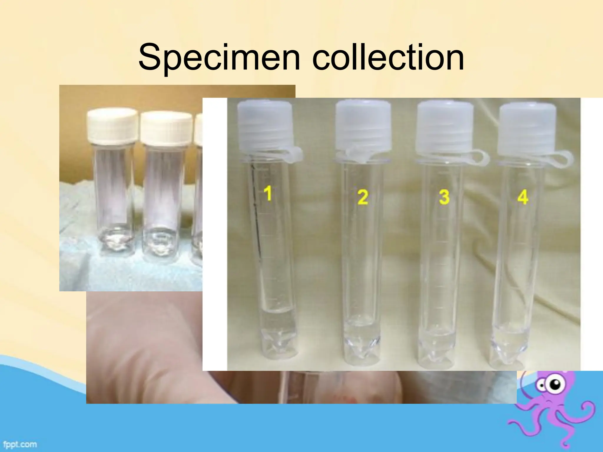
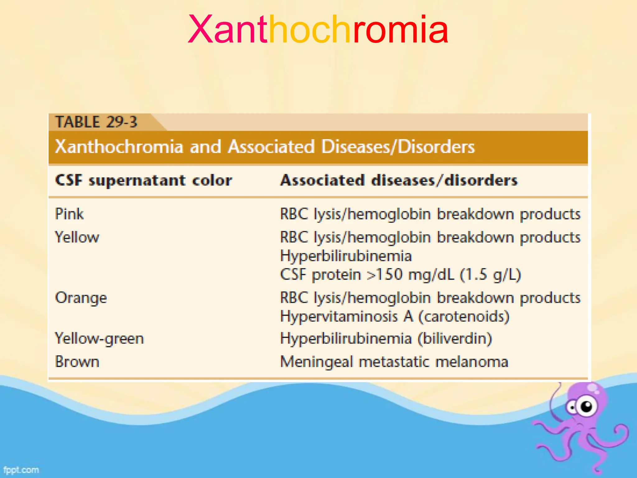
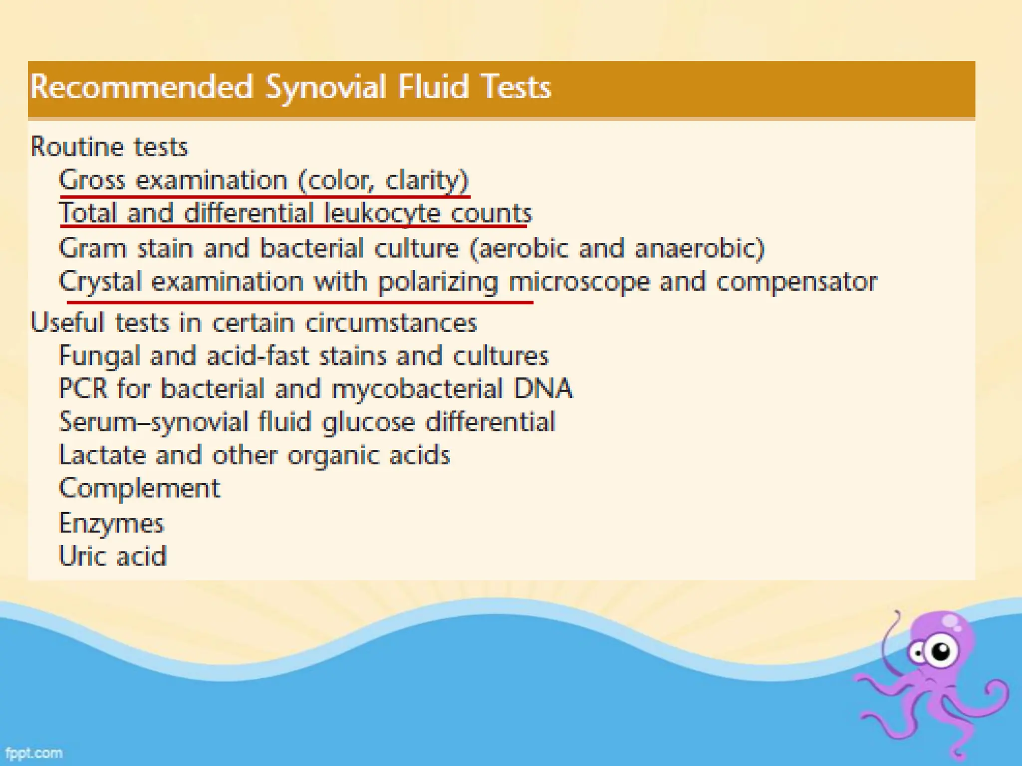
Body fluid analysis in
clinical lab
• Collection
• Turn around time
• Physician/laboratory communication
• Reliable reference values
• CSF
• Synovial fluid
• Peritoneal fluid
• Pleural fluid
• Pericardial fluid
CSF
• 10 to 60 ml……..90 to 150 ml
• Ultrafiltration and secretion
 physical support
 protective effect against sudden changes in acute
venous (respiratory and postural) and arterial blood
pressure
 excretory waste function
 maintains CNS ionic homeostasis
REASONS TO TEST
• Meningitis
• SAH
• Malignancy
• Demyelinating disease
Specimen collection
Required
Opening CSF pressure
Gross Examination
Total cell count and differential
Glucose (CSF/plasma ratio)
Protein
Optional
Cultures, gram stain, antigens, cytology
Protein electrophoresis, VDRL, D-dimers
Gross
• Crystal clear, colorless, like water
• Turbidity (WBC>200/μl or RBC>400/μl), bloody
(RBC>6000/μl)
• Viscous samples
Xanthochromia
Traumatic tap
• Decreasing amounts of
blood(Last tube will
have less)
• Clot present
SAH
• All tubes uniform
• No clot
• Hemosiderin/
hematoidin pigment
Microscopic examination
Total leucocyte count:
• 7% neutrophils with normal WBC
• WBCcorr = WBCobs −WBCadded
• WBCadded = WBC(BLD )× RBC(CSF)/ RBC(BLD)
Adults Neonates
0-5 cells/μl 0-30 cells/μl
Where to count
• a) < 200 cells are present in all nine squares, count all
nine squares. This area counted is 9 mm2.
• b) > 200 cells are present in all nine squares, then
count the four corner squares. This area counted is 4
mm2.
• c) > 200 cells are present in one square, then count
five of the squares within the center square for an
area of 0.2 mm2
N X Dilution factor
Area of total squares counted X Depth
Total cell count =
Calculation of Cell count
Correlation of cell count with cytomorphological findings is
essential.
Slide for DLC
• Cytocentrifuge, 500g for 5 min
• 2 drops of 22% albumin
• Wrights stain
Parasites
Synovial fluid
Imperfect ultra filtrate of plasma combined with
hyaluronic acid
FUNCTIONS
• Lubricate
• Provide nutrients
• Remove debris
REASONS TO TEST
• Sepsis
• Hemorrhage
• Crystal induced inflammation
Specimen collection
GROSS
• Color
• Volume
• Clarity
LOSS OF CLARITY
• Leucocytes
• Crystals
• Rice bodies
• Onchronosis
Lymphocytes:
• 15% of SF cells
• RA and other collagen disorders, chronic infections.
Monocytes & macrophages
• Most common cells present in normal SF, 65% of the
cell count.
Eosinophilia
• >2% of the leukocyte count,
• Rheumatoid arthritis, rheumatic fever, metastatic
carcinoma, Lymes disease, parasitic infection, chronic
urticaria, and angioedema & following arthrography.
Crystals
• Neutrophil predominant collection
• Intracellular crystals – pathognomic
 Monosodium urate monohydrate
 Calcium pyrophosphate dihydrate
 Apatite & basic calcium phosphates
 Calcium oxalate
 Lipids
Type of crystal Size and shape Associated conditions
Monosodium urate
(MSU)
needle-shaped rods
5–20 μm
strongly birefringent
Urate Gout
Occ in infections
Calcium
pyrophosphate
dihydrate (CPPD)
rhomboids, rods, or
rectangles 1–20 μm
birefringent
Hypomagnesemia,
Hemochromatosis,
Hyperparathyroidism &
Hypothyroidism
BCP small and
nonbirefringent
Non specific
Calcium oxalate
dihydrate
5- to 30-μm bipyramidal
octahedral “envelopes”
variable birefringence
chronic renal dialysis &
primary oxalosis, inborn
error of metabolism
Lipid crystals 1- to 20-μm spheres with
a Maltese cross
birefringent
Non specific
Can cause acute arthritis
Cholesterol crystals irregular birefringent
plates, notched corners
chronic effusions (e.g.,
TB arthritis, RA, SLE)
Crystal mimics
• Glove powder
• Talc
• Anticoagulants
• Prosthetic fragments
• Dust particles
• corticosteroids
•
Serous fluids
• Pleural
• Peritoneal
• Pericardial
Reasons to test
• Detect sepsis
• Malignancy
• Systemic disease
Specimen collection
Gross
• Transudates- usually clear
• Exudates – milky, turbid, bloody lights criteria
supernatant clear turbidity persists
cellular elements chylous or pseudochylous
centrifuge
Serous fluids
• Leucocytes
• RBC
• Mesothelial cells
• Macrophages(mononuclear phagocytes)
• Vacoulated histyocytes( can be confused with signet
ring cells)
• Bacteria
• Yeast
Mesothelial cells
• Distinguishable cell
borders
• Flat sheets
• Individual cells
• uniform
Malignant cells
• Poorly defined cell
borders
• Ball like clusters
• Cannibalism
• Non uniform
• Smooth nuclear
membrane
• No clefts
• Vacoules are limited to
cytoplasm
• Irregular nuclear
membrane
• Clefts and moulding
• Vacoules are all over
including over nuclei
DLBCL
CARCINOMA
YEAST, SPORES
Automation
Why automation
 Labour intensive
 Time consuming
 Skilled person(24x7)
 Interobserver variability
 Biohazard
Problems with automation
• Carryover
• Background counting
• Flaging of abnormal cells
• Clogging of machine
• Cytochemistry
• Immunocytochemistry
• Flow Cytometry
• Molecular studies
Ancillary Techniques
Time
delay
viability
30 min 50%
60 min 20%
90 min 10%
CSF for flow cytometry
Dux et al, J Neural Sci, 1994;121:74-78
References
• Karcher DS and McPherson RA. Cerebrospinal, Synovial, Serous Body Fluids, and
Alternative Specimens in Henry's Clinical Diagnosis and Management by Laboratory
Methods. Eds. McPherson RA and Pincus RA. Twenty Second Edition. Philadelphia, PA:
WB Saunders 2012: 480-506.
• de Graaf MT, de Jongste AHC, Kraan J, Boonstra JG, Sillevis Smitt PAE, Gratama JW.
Flow cytometric characterization of cerebrospinal fluid cells. Cytometry Part B 2011; 80B:
271–281.
• Sandhaus et al (2010) 'Automated Cerebrospinal Fluid Cell Counts Using the Sysmex XE-
5000', Am J Clin Pathol, (134), pp. 734-738.
• Grimaldi and Scopacasa (2000) 'Evaluation of the Abbott CELL-DYN 4000 Hematology
Analyzer', Am J Clin Pathol, (113), pp. 497-505.
• KRISHAN ET AL (2006) 'Detection of Tumor Cells in Body Cavity Fluids by Flow
Cytometric and Immunocytochemical Analysis', Diagnostic Cytopathology, 34(8), pp. 528-
541.

Body fluid analysis in clinically approved

  • 1.
    Body fluid analysisin clinical lab
  • 2.
    • Collection • Turnaround time • Physician/laboratory communication • Reliable reference values
  • 3.
    • CSF • Synovialfluid • Peritoneal fluid • Pleural fluid • Pericardial fluid
  • 4.
    CSF • 10 to60 ml……..90 to 150 ml • Ultrafiltration and secretion  physical support  protective effect against sudden changes in acute venous (respiratory and postural) and arterial blood pressure  excretory waste function  maintains CNS ionic homeostasis
  • 5.
    REASONS TO TEST •Meningitis • SAH • Malignancy • Demyelinating disease
  • 6.
  • 7.
    Required Opening CSF pressure GrossExamination Total cell count and differential Glucose (CSF/plasma ratio) Protein Optional Cultures, gram stain, antigens, cytology Protein electrophoresis, VDRL, D-dimers
  • 8.
    Gross • Crystal clear,colorless, like water • Turbidity (WBC>200/μl or RBC>400/μl), bloody (RBC>6000/μl) • Viscous samples
  • 9.
  • 10.
    Traumatic tap • Decreasingamounts of blood(Last tube will have less) • Clot present SAH • All tubes uniform • No clot • Hemosiderin/ hematoidin pigment
  • 11.
    Microscopic examination Total leucocytecount: • 7% neutrophils with normal WBC • WBCcorr = WBCobs −WBCadded • WBCadded = WBC(BLD )× RBC(CSF)/ RBC(BLD) Adults Neonates 0-5 cells/μl 0-30 cells/μl
  • 14.
    Where to count •a) < 200 cells are present in all nine squares, count all nine squares. This area counted is 9 mm2. • b) > 200 cells are present in all nine squares, then count the four corner squares. This area counted is 4 mm2. • c) > 200 cells are present in one square, then count five of the squares within the center square for an area of 0.2 mm2
  • 16.
    N X Dilutionfactor Area of total squares counted X Depth Total cell count = Calculation of Cell count Correlation of cell count with cytomorphological findings is essential.
  • 18.
    Slide for DLC •Cytocentrifuge, 500g for 5 min • 2 drops of 22% albumin • Wrights stain
  • 23.
  • 28.
    Synovial fluid Imperfect ultrafiltrate of plasma combined with hyaluronic acid FUNCTIONS • Lubricate • Provide nutrients • Remove debris REASONS TO TEST • Sepsis • Hemorrhage • Crystal induced inflammation
  • 29.
  • 30.
    GROSS • Color • Volume •Clarity LOSS OF CLARITY • Leucocytes • Crystals • Rice bodies • Onchronosis
  • 32.
    Lymphocytes: • 15% ofSF cells • RA and other collagen disorders, chronic infections. Monocytes & macrophages • Most common cells present in normal SF, 65% of the cell count. Eosinophilia • >2% of the leukocyte count, • Rheumatoid arthritis, rheumatic fever, metastatic carcinoma, Lymes disease, parasitic infection, chronic urticaria, and angioedema & following arthrography.
  • 33.
    Crystals • Neutrophil predominantcollection • Intracellular crystals – pathognomic  Monosodium urate monohydrate  Calcium pyrophosphate dihydrate  Apatite & basic calcium phosphates  Calcium oxalate  Lipids
  • 34.
    Type of crystalSize and shape Associated conditions Monosodium urate (MSU) needle-shaped rods 5–20 μm strongly birefringent Urate Gout Occ in infections Calcium pyrophosphate dihydrate (CPPD) rhomboids, rods, or rectangles 1–20 μm birefringent Hypomagnesemia, Hemochromatosis, Hyperparathyroidism & Hypothyroidism BCP small and nonbirefringent Non specific Calcium oxalate dihydrate 5- to 30-μm bipyramidal octahedral “envelopes” variable birefringence chronic renal dialysis & primary oxalosis, inborn error of metabolism Lipid crystals 1- to 20-μm spheres with a Maltese cross birefringent Non specific Can cause acute arthritis Cholesterol crystals irregular birefringent plates, notched corners chronic effusions (e.g., TB arthritis, RA, SLE)
  • 36.
    Crystal mimics • Glovepowder • Talc • Anticoagulants • Prosthetic fragments • Dust particles • corticosteroids •
  • 37.
    Serous fluids • Pleural •Peritoneal • Pericardial
  • 38.
    Reasons to test •Detect sepsis • Malignancy • Systemic disease
  • 39.
  • 40.
    Gross • Transudates- usuallyclear • Exudates – milky, turbid, bloody lights criteria supernatant clear turbidity persists cellular elements chylous or pseudochylous centrifuge
  • 41.
    Serous fluids • Leucocytes •RBC • Mesothelial cells • Macrophages(mononuclear phagocytes) • Vacoulated histyocytes( can be confused with signet ring cells) • Bacteria • Yeast
  • 42.
    Mesothelial cells • Distinguishablecell borders • Flat sheets • Individual cells • uniform Malignant cells • Poorly defined cell borders • Ball like clusters • Cannibalism • Non uniform
  • 43.
    • Smooth nuclear membrane •No clefts • Vacoules are limited to cytoplasm • Irregular nuclear membrane • Clefts and moulding • Vacoules are all over including over nuclei
  • 45.
  • 46.
  • 47.
  • 48.
    Automation Why automation  Labourintensive  Time consuming  Skilled person(24x7)  Interobserver variability  Biohazard
  • 49.
    Problems with automation •Carryover • Background counting • Flaging of abnormal cells • Clogging of machine
  • 50.
    • Cytochemistry • Immunocytochemistry •Flow Cytometry • Molecular studies Ancillary Techniques
  • 51.
    Time delay viability 30 min 50% 60min 20% 90 min 10% CSF for flow cytometry Dux et al, J Neural Sci, 1994;121:74-78
  • 52.
    References • Karcher DSand McPherson RA. Cerebrospinal, Synovial, Serous Body Fluids, and Alternative Specimens in Henry's Clinical Diagnosis and Management by Laboratory Methods. Eds. McPherson RA and Pincus RA. Twenty Second Edition. Philadelphia, PA: WB Saunders 2012: 480-506. • de Graaf MT, de Jongste AHC, Kraan J, Boonstra JG, Sillevis Smitt PAE, Gratama JW. Flow cytometric characterization of cerebrospinal fluid cells. Cytometry Part B 2011; 80B: 271–281. • Sandhaus et al (2010) 'Automated Cerebrospinal Fluid Cell Counts Using the Sysmex XE- 5000', Am J Clin Pathol, (134), pp. 734-738. • Grimaldi and Scopacasa (2000) 'Evaluation of the Abbott CELL-DYN 4000 Hematology Analyzer', Am J Clin Pathol, (113), pp. 497-505. • KRISHAN ET AL (2006) 'Detection of Tumor Cells in Body Cavity Fluids by Flow Cytometric and Immunocytochemical Analysis', Diagnostic Cytopathology, 34(8), pp. 528- 541.