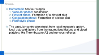
Wachemo University
NEMMCSH
Department of Pediatrics
Seminar Presentation on
Bleeding disorders
Moderator Dr. Sisay (MD, Pediatrician)
By Yishak and Yeshimebet
March 2023
Outline…
 Hemostasis
 Bleeding disorders
 hemophilia
 von willbrand disease
 Clinical approach bleeding child
 Principles of management
Hemostasis
 Hemostasis: is an active process that maintains normal blood flow
through healthy vessels but, when a vessel is damaged, rapidly
generates a platelet plug (primary hemostasis) and subsequent
thrombin clot (secondary hemostasis) at the site of vascular injury.
 The major components of the hemostatic mechanism are the platelets,
the anticoagulant proteins, the procoagulant proteins, fibrinolytic
system, and the various components of the vascular wall
 An abnormality of one element destabilizes the system, but significant
clinical symptoms often manifest only when two components are affected
Hemostasis...
 Homostasis has four stages
1. Vascular phase: constriction
2. Platelet phase: Formation of a platelet plug
3. Coagulation phase: Formation of a blood clot
4. Fibrinolytic phase
 The vascular contraction result from local myogenic spasm,
local autacoid factors from the traumatized tissues and blood
platelets like Thromboxane A2 and nervous reflexes
Platelet plug formation
Hemostasis...
 Platelet adhesion occurs when platelets bind to collagen exposed by
blood vessel damage. Most platelet adhesion is mediated through von
Willebrand factor (VWF)
 After platelets adhere to collagen, they become activated
 As platelets become activated, they express surface receptors that can
bind to fibrinogen, a plasma protein
 Activated platelets express phospholipids (platelet factor III) and
coagulation factor V, which are important in clot formation
Formation of blood clot
Hemostasis...
 The third stage of hemostasis begins
if the vasospasm & the platelet plug
are not enough to stop the bleeding
 A blood clot is a network of
threadlike protein fibers, called fibrin,
that traps blood cells, platelets, and
fluid
 The major event in the blood clot is
the formation of prothrombin activator
 .
 It is formed from the intrinsic and
extrinsic pathway
Bleeding disorders
 Bleeding is the loss of blood or blood escaping from the circulatory
system
 Bleeding can occur Internally, where blood leaks from blood
vessels inside the body or externally , either through a natural opening
such as the mouth, nose, ear, urethra, vagina or anus or through a
break in the skin
 The most common ones are epistaxis(Nose bleeding), Gum bleeding,
intracranial bleeding, rectal, menstrual bleeding, bleeding into the
joints, heavy bleeding from small cuts or dental work
Bleeding
Disorders
DIC
Clotting factor
abnrmalities
Hemophilia A
Hemophilia B
Von Willebrand's
disease
Platelet
disorders
ITP
TTP
Vascular
abnormality
Henoch-
Schönlein
Purpura
Hemophilias
 Hemophilia A (factor VIII deficiency) and hemophilia B (factor IX
deficiency) are the most common and serious congenital
coagulation factor deficiencies
 Hemophilia C is the bleeding disorder associated with reduced
levels of factor XI
 Hemophilia occurs in approximately 1 : 5,000 males, with 85%
having factor VIII deficiency and 10–15% having factor IX
deficiency
 The disease occurs predominantly in males and is inherited in an
X-linked recessive manner
Pathophysiology
Hemophilias...
 Factor VIII and IX - participate in a complex required for the activation of
factor X (tenase)
 absence impairs the ability to generate thrombin and fibrin
 delayed formation of an abnormal clot
 The clot that is formed may be friable and re-bleeding occurs during
physiological lyses of clots or with minimal trauma
Classification
Hemophilias...
 Is classified on the basis of the patient’s baseline level of clotting factor VIII or IX,
because factor levels usually correlate with the severity of bleeding symptoms
 By the WHO standard, 1 IU of each factor is defined as that amount in 1 mL of
normal plasma; thus, 100 mL of normal plasma has 100 IU/dL (100% activity) of
each factor
Classification factor level Bleeding
Severe hemophilia < 1% (< 1IU/dl) spontaneously
Moderate hemophilia 1-5% (1-5 IU/dl) mild trauma
Mild hemophilia >5% (>5 IU/dl) significant trauma
Clinical manifestations
Hemophilias...
 The symptoms of hemophilia A and B are virtually identical
 Neither factor VIII nor factor IX crosses the placenta, thus
bleeding symptoms may be present from birth or occur in the
fetus but they usually go unnoticed in the newborn
 The hallmark of hemophilic bleeding is hemarthrosis, which
may be induced by minor trauma but often occurs
spontaneously
 Easy bruising, intramuscular hematomas and hemarthroses
Clinical manifestations...
 Persistence of bleeding from minor traumatic lacerations of the
mouth
 Uncontrolled bleeding during circumcision
 Life threatening bleeding in the hemophilic patient is caused by
bleeding in to vital structures (CNS and upper air way) or by
exsanguinations (external, GI or iliopsoas hemorrhage)
Complications
Hemophilias...
 Hemophilic arthropathy: Joint contractures, limited range of
motion and chronic pain
 Intracranial bleeding(can occur without known trauma in severe
hemophilia).
 Compartment syndrome
 Airway compromise due to bleeds in the pharynx, tongue or
neck.
 Life-threatening hemorrhage due to gastrointestinal, post
traumatic or perioperative bleeds
Von Willebrand Disease
 is the most common inherited bleeding disorder, with an
estimated prevalence cited at 1 : 100 to 1 : 10,000
depending on the criteria used for diagnosis
 a quantitative or qualitative abnormality of vWF
 VWF serves to tether platelets to injured subendothelium
and as a carrier protein for factor VIII (FVIII), protecting it
from degradation in plasma
Clinical manifestations
Von Willebrand Disease...
 typically presents with mucosal bleeding, similar to that seen
with other platelet defects
 Epistaxis, easy bruising, and heavy menutrual bleeding in
women are common complaints
 Surgical bleeding, particularly with dental extractions or
adenotonsillectomy
 Severe type 3 VWD may present with joint bleeds
 men and women are equally likely to have VWD, women being
more likely to be diagnosed with VWD because of the potential
for symptoms with menorrhagia
Classifications
Von Willebrand Disease...
 Type 1: is by far the most common type, 60-80%
 partial quantitative deficiency of vWF, platelet with VWF:Ag <30 IU/dL
 Type 2: A qualitative(dysfunctional) deficiency of vWF
 Type 2A, 2B, 2N, 2M
 15% to 20% of patients
 more significant bleeding symptoms than in type 1
 Type 3: A complete quantitative deficiency of VWF
 results in a secondary deficiency of Factor VIII
 accounts for <5% of patients
 a severe bleeding disorder
Approach to a bleeding child
 History and physical examination
 Laboratory approach
History
Approach to a bleeding child...
 Clinical evaluation of a patient with bleeding symptoms begins
with taking a careful history, including the child's age, sex,
clinical presentation, personal and family medical history, and
medications
 Careful attention should be paid to the child's bleeding history,
including number and severity of bleeding episodes and
characterizing the type of bleeding
History...
 Age at symptom onset
 the median age at diagnosis is 36 months for people with mild
hemophilia, 8 months for those with moderate hemophilia, and 1 month
for those with severe hemophilia. (CDC data, United States)
 76% of men with VWD had been diagnosed by age 10, but 50% of
women with VWD were not diagnosed until after age 12.
 The peak ages for the presentation of ITP are 1–4 years of age and
adolescence, but ITP occurs throughout childhood and adolescence
History...
 Sex
 Some of the inherited hemostatic defects such as hemophilia A (FVIII
deficiency), hemophilia B (FIX deficiency), Wiskott-Aldrich syndrome
and X-linked thrombocytopenia are due to mutations on the X
chromosome manifested on males
 there is an elevated rate of diagnosis of VWD, platelet defects and FXI
deficiency in women because of menorrhagia
 All other inherited and acquired hemostatic defects occurs in both sexes
Sites of bleeding
History...
 Mucocutaneous bleeding into the skin and mucous membranes
is characteristic of disorders of platelets and their interaction
with blood vessels and may be manifested as petechiae,
ecchymoses, and/or mucosal bleeding
 includes; easy bruising with or without petechiae formation,
prolonged bleeding after tooth extraction, prolonged, frequent
nosebleeds, gum bleeding
 is usually associated with abnormalities of platelet number or
function, of platelet cofactors such as VWF, or of the vessel
wall
Sites of bleeding...
History...
 A healthy child who presents with petechiae and/or
mucocutaneous purpura in the wake of a viral infection most
likely has postinfectious immune thrombocytopenia
 Deep bleeding into soft tissue, muscle, and joints suggests the
presence of hemophilia or other disorders of coagulation
proteins
History...
 Severity and duration of bleeding
 it also help differentiating pathological bleeding from normal bleeding;
which is unusually frequent, long-lasting and severe.
 Triggering event; spontaneous vs induced with trauma
 Timing of bleeding after injury: immediate vs delayed
Family history
History...
 The family history may provide important clues about the potential inheritance of
an underlying bleeding disorder
 The presence of bleeding manifestations only in male siblings and maternal
uncles is suggestive of an X-linked recessive disorder, such as hemophilia A or
B. However, a negative family history does not exclude an inherited coagulation
disorder, as up to one-third of patients with hemophilia have a negative family
history
 Consanguinity in a family increases the risk of autosomal-recessive disorders.
 Evaluation of the bleeding history in family members by a validated bleeding
questionnaire couldbe useful for appreciating the significance of the family
bleeding history
Nutrition history
History...
 A nutrition history should be obtained to assess the likelihood
of
 vitamin K deficiency, especially if the patient also is taking broad
spectrum antibiotics,
 vitamin C deficiency, scurvy/gum bleeding and
 general malnutrition and malabsorption
Perinatal history
History...
 Superficial (bruising, petechiae)
 Vitamin K administration
 Maternal drugs
 CNS bleeding, GI bleeding, Cephalohematoma
 Unexplained anemia or hyperbilirubinemia
 Delayed cord separation, bleeding after cord separation
History...
Approach to a bleeding child...
 History of drug use; Aspirin and NSAIDs, Anticoagulants,
Antibiotics, Anticonvulsants
 History of delayed/slow wound healing
 Is there any sign of bleeding from umbilical stump, circumcision
site venepuncture?
 Are there lumps with bruising for which there is minimal
trauma?
 Was there increased bleeding during surgery or dental
procedures?
 Menstrual history in post pubertal females
 Previous personal & Family history of similar problem
Physical examination
Approach to a bleeding child...
Key words
 Petechiae (≤4 mm in diameter) are a small
red or purple spot that can appear on the
skin, conjunctiva, retina, and mucous
membranes which is caused by haemorrhage
of capillaries
 develop in areas of increased venous
pressure, such as the dependent parts of the
body
 are not found on the sole of the foot where
the vessels are protected by the strong
subcutaneous tissue
 are asymptomatic and not palpable
key words...
 Ecchymoses; lesions (>1cm in
diameter) characteristically are purple in
color and are small, multiple, and
superficial in location. They usually
develop without noticeable trauma and
do not spread into deeper tissues
 Purpura (4-10mm in diameter)
 Telangiectasias also known as spider
veins (0.5 - 1 mm in diameter), are
small dilated blood vessels that can
occur near the surface of the skin or
mucous membranes. commonly seen
on the face around the nose, cheeks
and chin
Physical examination...
 The most important determination is whether the patient
appears acutely or chronically ill, including vital signs and
growth parameters
 The nose should be examined for ulcers or anatomic bleeding
sites, and the heart should be examined for the presence of
murmurs (as occur in anemia and endocarditis).
 Joints should be examined for chronic arthropathy (as occurs in
hemophilia) or joint laxity (as occurs in Ehlers Danlos
syndrome), and the extremities are examined for thumb or
radial anomalies (thrombocytopenia–absent radius syndrome,
or Fanconi anemia).
Physical examination...
 The abdomen and lymph nodes should be examined for the
presence of hepatosplenomegaly and adenopathy
 The examination of the skin should include a search for pallor,
hematomas, petechiae, ecchymoses, telangiectasias, poor
wound healing (large or abnormal scars), lax (loose) skin, and
varicose veins (possible deep venous thrombosis)
Laboratory approach
Approach to a bleeding child...
 Guided by history
 No set of screening tests is complete and capable of detecting the
panorama of hemorrhagic disorders, but the screen should include:
 CBC
 Blood film
 Prothrombin time (PT)
 Activated Partial thromboplastin time (aPTT)
 Functional fibrinogen level or thrombin time (TT)
 VWF testing
 Liver function test
 Coombs’ test
Laboratory approach...
 CBC is performed to exclude thrombocytopenia and it also provides
information about additional cytopenias, and other WBC and RBC
abnormalities
 Peripheral blood film provides additional information regarding platelet
number, size, lumping and granularity
 Evaluation of WBC morphology allows identification of malignant
blasts, granulocyte inclusions, or other WBC abnormities
 Evaluation of RBC morphology is important to exclude a
microangiopathic process as evidenced by presence of fragmented red
blood cells, microcytosis, macrocytosis and other RBC abnormalities
Laboratory approach...
 Prothrombin Time (PT) measures the extrinsic and common pathway in
the coagulation cascade (tissue factor, VII, X, V, II, fibrinogen) and it is
sensitive to alterations in the vitamin K-dependent coagulation factors
 Activated partial thromboplastin time(aPTT) measures the intrinsic and
common pathways of coagulation and it is sensitive to deficiencies of
factors(XII, XI, IX, VIII, X, V, II, fibrinogen) and to inhibitors such as
heparin
 aPTT less sensitive than the PT to deficiencies within the common
pathway
 Results should be compared with age specific laboratory reference
intervals and are reported in seconds and/or as a percent age of a normal
control sample
Laboratory approach...
 A mixing study (patient plasma 1:1 normal plasma)
differentiates between factor deficiency (mixing corrects the PT
or aPTT) and the presence of an inhibitor (mixing does not
correct the PT or aPTT) if an abnormal PT and/or an aPTT is
identified
 Functional fibrinogen level or thrombin time (TT) measures the
thrombin-induced conversion of fibrinogen to fibrin
 A prolonged TT suggests a quantitative or qualitative
abnormality of fibrinogen or the presence of heparin in the
sample
 PT, aPTT, and TT do not screen for factor XIII deficiency
Laboratory approach...
 The bleeding time is an indirect measure of platelet number
and a more direct measure of platelet function, vascular
integrity, and platelet interaction with the vascular
subendothelium
 but is less often used now because of difficulties in
standardization
 VWF antigen and activity (ristocetin cofactor assay); these tests
measure the level and the activity of VWF for the diagnosis of
VWD
Laboratory approach...
 An isolated prolonged aPTT with normal PT can be seen in
 Hemophilia A or B
 Factor XI deficiency
 Lupus anticoagulants
 Deficiencies of factor XII, high molecular weight kininogen, and
prekallikrein
 Heparin contamination; contamination occurs most often in specimens
drawn from arterial or central venous catheters, heparin-containing
intravascular lines
Laboratory approach...
 An isolated prolonged PT with normal aPTT can be seen in
inherited factor VII deficiency and acquired factor VII inhibitors
 Combined prolongation of PT and aPTT
 In a child with bleeding symptoms but who is otherwise well
indicates an inherited disorder (rare deficiencies of factors X, V,
and II, or fibrinogen) or an acquired disorder involving multiple
pathways
 In a sick child, disorders to consider are disseminated
intravascular coagulation (DIC due to sepsis or other systemic
illness), severe liver disease, or severe vitamin K deficiency. The
factor V level can be used to distinguish between vitamin K
deficiency and liver disease or DIC (normal in Vit K deficiency)
Screening profiles
Disorder aPTT PT TT PLT Fibrin BT
Vasculopathy,
CTD
N N N N N LONG
ITP N N N LOW N LONG
PLT
dysfunction
N N N N/LOW N LONG
Hemophilia(VI
II, IX)
LONG N N N N N
VWD LONG N N N N LONG
DIC LONG LONG LONG LOW LOW LONG
HDN LONG LONG N N N LONG
Principles of management
Principles of management...
 supportive care
 ABC
 Indication for blood transfusion
 acute blood loss, when 20–30% of the total blood volume has been lost
and bleeding is continuing
 severe anaemia
 septic shock (if IV fluids are insufficient to maintain adequate circulation
and in addition to antibiotic therapy)
 to provide plasma and platelets for clotting factors, if specific blood
components are not available
 exchange transfusion in neonates with severe jaundice
Principles of management...
 Compression
 hemostatic agents
 Baby in shock (due to acute blood loss)
 Immediate blood transfusion with 20 ml/kg of whole blood
 If blood is not available use other volume expanders(N/S)
 Follow vital signs and urine out put
 Raise the level of PCV to the desired level by packed RBC
transfusion
Principles of management...
Hemophilia
 recombinant factor VIII or recombinant factor IX with the aim of
achieving to hemostatic levels, 35–50% range for mild to moderate or
100% for life threatening or major hemorrhages
 prophylaxis is the standard of care for most children with severe
hemophilia, to prevent spontaneous bleeding and early joint deformities
 desmopressin acetate an also be used to treat patients with mild
hemophilia A
 Supportive care
Von Willbrand disease
 desmopressin for type 1
 VWF-containing concentrates for type 1, and type 2
References
• Nelson Textbook of Pediatrics, 21st ed.
• Nelson Pediatric Symptom-Based Diagnosis, 2nd ed.
• Uptodate 21.3
• Pediatric Hospital Care Ethiopia, First Edition, 2010
• SickKids Handbook of Pediatric Thrombosis and Hemostasis,
2nd ed.

Bleeding Disorders.pptx

  • 1.
    Wachemo University NEMMCSH Department ofPediatrics Seminar Presentation on Bleeding disorders Moderator Dr. Sisay (MD, Pediatrician) By Yishak and Yeshimebet March 2023
  • 2.
    Outline…  Hemostasis  Bleedingdisorders  hemophilia  von willbrand disease  Clinical approach bleeding child  Principles of management
  • 3.
    Hemostasis  Hemostasis: isan active process that maintains normal blood flow through healthy vessels but, when a vessel is damaged, rapidly generates a platelet plug (primary hemostasis) and subsequent thrombin clot (secondary hemostasis) at the site of vascular injury.  The major components of the hemostatic mechanism are the platelets, the anticoagulant proteins, the procoagulant proteins, fibrinolytic system, and the various components of the vascular wall  An abnormality of one element destabilizes the system, but significant clinical symptoms often manifest only when two components are affected
  • 4.
    Hemostasis...  Homostasis hasfour stages 1. Vascular phase: constriction 2. Platelet phase: Formation of a platelet plug 3. Coagulation phase: Formation of a blood clot 4. Fibrinolytic phase  The vascular contraction result from local myogenic spasm, local autacoid factors from the traumatized tissues and blood platelets like Thromboxane A2 and nervous reflexes
  • 5.
    Platelet plug formation Hemostasis... Platelet adhesion occurs when platelets bind to collagen exposed by blood vessel damage. Most platelet adhesion is mediated through von Willebrand factor (VWF)  After platelets adhere to collagen, they become activated  As platelets become activated, they express surface receptors that can bind to fibrinogen, a plasma protein  Activated platelets express phospholipids (platelet factor III) and coagulation factor V, which are important in clot formation
  • 6.
    Formation of bloodclot Hemostasis...  The third stage of hemostasis begins if the vasospasm & the platelet plug are not enough to stop the bleeding  A blood clot is a network of threadlike protein fibers, called fibrin, that traps blood cells, platelets, and fluid  The major event in the blood clot is the formation of prothrombin activator  .  It is formed from the intrinsic and extrinsic pathway
  • 7.
    Bleeding disorders  Bleedingis the loss of blood or blood escaping from the circulatory system  Bleeding can occur Internally, where blood leaks from blood vessels inside the body or externally , either through a natural opening such as the mouth, nose, ear, urethra, vagina or anus or through a break in the skin  The most common ones are epistaxis(Nose bleeding), Gum bleeding, intracranial bleeding, rectal, menstrual bleeding, bleeding into the joints, heavy bleeding from small cuts or dental work
  • 8.
    Bleeding Disorders DIC Clotting factor abnrmalities Hemophilia A HemophiliaB Von Willebrand's disease Platelet disorders ITP TTP Vascular abnormality Henoch- Schönlein Purpura
  • 9.
    Hemophilias  Hemophilia A(factor VIII deficiency) and hemophilia B (factor IX deficiency) are the most common and serious congenital coagulation factor deficiencies  Hemophilia C is the bleeding disorder associated with reduced levels of factor XI  Hemophilia occurs in approximately 1 : 5,000 males, with 85% having factor VIII deficiency and 10–15% having factor IX deficiency  The disease occurs predominantly in males and is inherited in an X-linked recessive manner
  • 10.
    Pathophysiology Hemophilias...  Factor VIIIand IX - participate in a complex required for the activation of factor X (tenase)  absence impairs the ability to generate thrombin and fibrin  delayed formation of an abnormal clot  The clot that is formed may be friable and re-bleeding occurs during physiological lyses of clots or with minimal trauma
  • 11.
    Classification Hemophilias...  Is classifiedon the basis of the patient’s baseline level of clotting factor VIII or IX, because factor levels usually correlate with the severity of bleeding symptoms  By the WHO standard, 1 IU of each factor is defined as that amount in 1 mL of normal plasma; thus, 100 mL of normal plasma has 100 IU/dL (100% activity) of each factor Classification factor level Bleeding Severe hemophilia < 1% (< 1IU/dl) spontaneously Moderate hemophilia 1-5% (1-5 IU/dl) mild trauma Mild hemophilia >5% (>5 IU/dl) significant trauma
  • 12.
    Clinical manifestations Hemophilias...  Thesymptoms of hemophilia A and B are virtually identical  Neither factor VIII nor factor IX crosses the placenta, thus bleeding symptoms may be present from birth or occur in the fetus but they usually go unnoticed in the newborn  The hallmark of hemophilic bleeding is hemarthrosis, which may be induced by minor trauma but often occurs spontaneously  Easy bruising, intramuscular hematomas and hemarthroses
  • 13.
    Clinical manifestations...  Persistenceof bleeding from minor traumatic lacerations of the mouth  Uncontrolled bleeding during circumcision  Life threatening bleeding in the hemophilic patient is caused by bleeding in to vital structures (CNS and upper air way) or by exsanguinations (external, GI or iliopsoas hemorrhage)
  • 14.
    Complications Hemophilias...  Hemophilic arthropathy:Joint contractures, limited range of motion and chronic pain  Intracranial bleeding(can occur without known trauma in severe hemophilia).  Compartment syndrome  Airway compromise due to bleeds in the pharynx, tongue or neck.  Life-threatening hemorrhage due to gastrointestinal, post traumatic or perioperative bleeds
  • 15.
    Von Willebrand Disease is the most common inherited bleeding disorder, with an estimated prevalence cited at 1 : 100 to 1 : 10,000 depending on the criteria used for diagnosis  a quantitative or qualitative abnormality of vWF  VWF serves to tether platelets to injured subendothelium and as a carrier protein for factor VIII (FVIII), protecting it from degradation in plasma
  • 16.
    Clinical manifestations Von WillebrandDisease...  typically presents with mucosal bleeding, similar to that seen with other platelet defects  Epistaxis, easy bruising, and heavy menutrual bleeding in women are common complaints  Surgical bleeding, particularly with dental extractions or adenotonsillectomy  Severe type 3 VWD may present with joint bleeds  men and women are equally likely to have VWD, women being more likely to be diagnosed with VWD because of the potential for symptoms with menorrhagia
  • 17.
    Classifications Von Willebrand Disease... Type 1: is by far the most common type, 60-80%  partial quantitative deficiency of vWF, platelet with VWF:Ag <30 IU/dL  Type 2: A qualitative(dysfunctional) deficiency of vWF  Type 2A, 2B, 2N, 2M  15% to 20% of patients  more significant bleeding symptoms than in type 1  Type 3: A complete quantitative deficiency of VWF  results in a secondary deficiency of Factor VIII  accounts for <5% of patients  a severe bleeding disorder
  • 18.
    Approach to ableeding child  History and physical examination  Laboratory approach
  • 19.
    History Approach to ableeding child...  Clinical evaluation of a patient with bleeding symptoms begins with taking a careful history, including the child's age, sex, clinical presentation, personal and family medical history, and medications  Careful attention should be paid to the child's bleeding history, including number and severity of bleeding episodes and characterizing the type of bleeding
  • 20.
    History...  Age atsymptom onset  the median age at diagnosis is 36 months for people with mild hemophilia, 8 months for those with moderate hemophilia, and 1 month for those with severe hemophilia. (CDC data, United States)  76% of men with VWD had been diagnosed by age 10, but 50% of women with VWD were not diagnosed until after age 12.  The peak ages for the presentation of ITP are 1–4 years of age and adolescence, but ITP occurs throughout childhood and adolescence
  • 21.
    History...  Sex  Someof the inherited hemostatic defects such as hemophilia A (FVIII deficiency), hemophilia B (FIX deficiency), Wiskott-Aldrich syndrome and X-linked thrombocytopenia are due to mutations on the X chromosome manifested on males  there is an elevated rate of diagnosis of VWD, platelet defects and FXI deficiency in women because of menorrhagia  All other inherited and acquired hemostatic defects occurs in both sexes
  • 22.
    Sites of bleeding History... Mucocutaneous bleeding into the skin and mucous membranes is characteristic of disorders of platelets and their interaction with blood vessels and may be manifested as petechiae, ecchymoses, and/or mucosal bleeding  includes; easy bruising with or without petechiae formation, prolonged bleeding after tooth extraction, prolonged, frequent nosebleeds, gum bleeding  is usually associated with abnormalities of platelet number or function, of platelet cofactors such as VWF, or of the vessel wall
  • 23.
    Sites of bleeding... History... A healthy child who presents with petechiae and/or mucocutaneous purpura in the wake of a viral infection most likely has postinfectious immune thrombocytopenia  Deep bleeding into soft tissue, muscle, and joints suggests the presence of hemophilia or other disorders of coagulation proteins
  • 24.
    History...  Severity andduration of bleeding  it also help differentiating pathological bleeding from normal bleeding; which is unusually frequent, long-lasting and severe.  Triggering event; spontaneous vs induced with trauma  Timing of bleeding after injury: immediate vs delayed
  • 25.
    Family history History...  Thefamily history may provide important clues about the potential inheritance of an underlying bleeding disorder  The presence of bleeding manifestations only in male siblings and maternal uncles is suggestive of an X-linked recessive disorder, such as hemophilia A or B. However, a negative family history does not exclude an inherited coagulation disorder, as up to one-third of patients with hemophilia have a negative family history  Consanguinity in a family increases the risk of autosomal-recessive disorders.  Evaluation of the bleeding history in family members by a validated bleeding questionnaire couldbe useful for appreciating the significance of the family bleeding history
  • 26.
    Nutrition history History...  Anutrition history should be obtained to assess the likelihood of  vitamin K deficiency, especially if the patient also is taking broad spectrum antibiotics,  vitamin C deficiency, scurvy/gum bleeding and  general malnutrition and malabsorption
  • 27.
    Perinatal history History...  Superficial(bruising, petechiae)  Vitamin K administration  Maternal drugs  CNS bleeding, GI bleeding, Cephalohematoma  Unexplained anemia or hyperbilirubinemia  Delayed cord separation, bleeding after cord separation
  • 28.
    History... Approach to ableeding child...  History of drug use; Aspirin and NSAIDs, Anticoagulants, Antibiotics, Anticonvulsants  History of delayed/slow wound healing  Is there any sign of bleeding from umbilical stump, circumcision site venepuncture?  Are there lumps with bruising for which there is minimal trauma?  Was there increased bleeding during surgery or dental procedures?  Menstrual history in post pubertal females  Previous personal & Family history of similar problem
  • 29.
    Physical examination Approach toa bleeding child... Key words  Petechiae (≤4 mm in diameter) are a small red or purple spot that can appear on the skin, conjunctiva, retina, and mucous membranes which is caused by haemorrhage of capillaries  develop in areas of increased venous pressure, such as the dependent parts of the body  are not found on the sole of the foot where the vessels are protected by the strong subcutaneous tissue  are asymptomatic and not palpable
  • 30.
    key words...  Ecchymoses;lesions (>1cm in diameter) characteristically are purple in color and are small, multiple, and superficial in location. They usually develop without noticeable trauma and do not spread into deeper tissues  Purpura (4-10mm in diameter)  Telangiectasias also known as spider veins (0.5 - 1 mm in diameter), are small dilated blood vessels that can occur near the surface of the skin or mucous membranes. commonly seen on the face around the nose, cheeks and chin
  • 31.
    Physical examination...  Themost important determination is whether the patient appears acutely or chronically ill, including vital signs and growth parameters  The nose should be examined for ulcers or anatomic bleeding sites, and the heart should be examined for the presence of murmurs (as occur in anemia and endocarditis).  Joints should be examined for chronic arthropathy (as occurs in hemophilia) or joint laxity (as occurs in Ehlers Danlos syndrome), and the extremities are examined for thumb or radial anomalies (thrombocytopenia–absent radius syndrome, or Fanconi anemia).
  • 32.
    Physical examination...  Theabdomen and lymph nodes should be examined for the presence of hepatosplenomegaly and adenopathy  The examination of the skin should include a search for pallor, hematomas, petechiae, ecchymoses, telangiectasias, poor wound healing (large or abnormal scars), lax (loose) skin, and varicose veins (possible deep venous thrombosis)
  • 33.
    Laboratory approach Approach toa bleeding child...  Guided by history  No set of screening tests is complete and capable of detecting the panorama of hemorrhagic disorders, but the screen should include:  CBC  Blood film  Prothrombin time (PT)  Activated Partial thromboplastin time (aPTT)  Functional fibrinogen level or thrombin time (TT)  VWF testing  Liver function test  Coombs’ test
  • 34.
    Laboratory approach...  CBCis performed to exclude thrombocytopenia and it also provides information about additional cytopenias, and other WBC and RBC abnormalities  Peripheral blood film provides additional information regarding platelet number, size, lumping and granularity  Evaluation of WBC morphology allows identification of malignant blasts, granulocyte inclusions, or other WBC abnormities  Evaluation of RBC morphology is important to exclude a microangiopathic process as evidenced by presence of fragmented red blood cells, microcytosis, macrocytosis and other RBC abnormalities
  • 35.
    Laboratory approach...  ProthrombinTime (PT) measures the extrinsic and common pathway in the coagulation cascade (tissue factor, VII, X, V, II, fibrinogen) and it is sensitive to alterations in the vitamin K-dependent coagulation factors  Activated partial thromboplastin time(aPTT) measures the intrinsic and common pathways of coagulation and it is sensitive to deficiencies of factors(XII, XI, IX, VIII, X, V, II, fibrinogen) and to inhibitors such as heparin  aPTT less sensitive than the PT to deficiencies within the common pathway  Results should be compared with age specific laboratory reference intervals and are reported in seconds and/or as a percent age of a normal control sample
  • 36.
    Laboratory approach...  Amixing study (patient plasma 1:1 normal plasma) differentiates between factor deficiency (mixing corrects the PT or aPTT) and the presence of an inhibitor (mixing does not correct the PT or aPTT) if an abnormal PT and/or an aPTT is identified  Functional fibrinogen level or thrombin time (TT) measures the thrombin-induced conversion of fibrinogen to fibrin  A prolonged TT suggests a quantitative or qualitative abnormality of fibrinogen or the presence of heparin in the sample  PT, aPTT, and TT do not screen for factor XIII deficiency
  • 37.
    Laboratory approach...  Thebleeding time is an indirect measure of platelet number and a more direct measure of platelet function, vascular integrity, and platelet interaction with the vascular subendothelium  but is less often used now because of difficulties in standardization  VWF antigen and activity (ristocetin cofactor assay); these tests measure the level and the activity of VWF for the diagnosis of VWD
  • 38.
    Laboratory approach...  Anisolated prolonged aPTT with normal PT can be seen in  Hemophilia A or B  Factor XI deficiency  Lupus anticoagulants  Deficiencies of factor XII, high molecular weight kininogen, and prekallikrein  Heparin contamination; contamination occurs most often in specimens drawn from arterial or central venous catheters, heparin-containing intravascular lines
  • 39.
    Laboratory approach...  Anisolated prolonged PT with normal aPTT can be seen in inherited factor VII deficiency and acquired factor VII inhibitors  Combined prolongation of PT and aPTT  In a child with bleeding symptoms but who is otherwise well indicates an inherited disorder (rare deficiencies of factors X, V, and II, or fibrinogen) or an acquired disorder involving multiple pathways  In a sick child, disorders to consider are disseminated intravascular coagulation (DIC due to sepsis or other systemic illness), severe liver disease, or severe vitamin K deficiency. The factor V level can be used to distinguish between vitamin K deficiency and liver disease or DIC (normal in Vit K deficiency)
  • 41.
    Screening profiles Disorder aPTTPT TT PLT Fibrin BT Vasculopathy, CTD N N N N N LONG ITP N N N LOW N LONG PLT dysfunction N N N N/LOW N LONG Hemophilia(VI II, IX) LONG N N N N N VWD LONG N N N N LONG DIC LONG LONG LONG LOW LOW LONG HDN LONG LONG N N N LONG
  • 42.
  • 43.
    Principles of management... supportive care  ABC  Indication for blood transfusion  acute blood loss, when 20–30% of the total blood volume has been lost and bleeding is continuing  severe anaemia  septic shock (if IV fluids are insufficient to maintain adequate circulation and in addition to antibiotic therapy)  to provide plasma and platelets for clotting factors, if specific blood components are not available  exchange transfusion in neonates with severe jaundice
  • 44.
    Principles of management... Compression  hemostatic agents  Baby in shock (due to acute blood loss)  Immediate blood transfusion with 20 ml/kg of whole blood  If blood is not available use other volume expanders(N/S)  Follow vital signs and urine out put  Raise the level of PCV to the desired level by packed RBC transfusion
  • 45.
    Principles of management... Hemophilia recombinant factor VIII or recombinant factor IX with the aim of achieving to hemostatic levels, 35–50% range for mild to moderate or 100% for life threatening or major hemorrhages  prophylaxis is the standard of care for most children with severe hemophilia, to prevent spontaneous bleeding and early joint deformities  desmopressin acetate an also be used to treat patients with mild hemophilia A  Supportive care Von Willbrand disease  desmopressin for type 1  VWF-containing concentrates for type 1, and type 2
  • 46.
    References • Nelson Textbookof Pediatrics, 21st ed. • Nelson Pediatric Symptom-Based Diagnosis, 2nd ed. • Uptodate 21.3 • Pediatric Hospital Care Ethiopia, First Edition, 2010 • SickKids Handbook of Pediatric Thrombosis and Hemostasis, 2nd ed.

Editor's Notes

  • #5 Eventual growth of fibrous tissue into the blood clot to close the hole in the vessel permanently
  • #23 HSP ITP
  • #34 vWF no single test is reliable. antigen, activity with cofcator, ratio, mutimer distribution ristocetin cofactor activity
  • #36 normal PT ranges b/n 10-13 sec
  • #38 Bleeding time 4-8min
  • #39 FXI more commonly in Ashkenazi Jews, often mucocutaneous in nature Lupus, acquired inhibitors against phospholipid-protein complexes that produce a prolonged aPTT, associated with recent infections (particularly viral infections), and usually transient.
  • #46 calculated as for F8= %Desired x Kg x 0.5 for F9= %Desired x Kg x 1.3 150 µg (1 spray) for children weighing <50 kg and 300 µg (2 spray) for children weighing >50 kg VWF, adjunct therapy; antifibrinolytics