Assessment of the The Pregnant Patient by Dr. Ina.pdf
1.
HISTORY AND PHYSICAL
EXAMINATIONIN AN
OBSTETRIC PATIENT
(HOW TO CALCULATE AOG AND
ESTIMATED DATE OF DELIVERY)
INA S. IRABON, MD, FPOGS, FPSRM, FPSGE
OBSTETRICS AND GYNECOLOGY
REPRODUCTIVE ENDOCRINOLOGY AND INFERTILITY
MINIMALLY INVASIVE SURGERY
REFERENCES
■ Thompson JE.Chapter 14 The Pregnant Woman. In: Bickley
LS (ed); Bates’ Guide to Physical Examination and History
Taking, 7th edition (1999)
■ Cunningham FG, Leveno KJ, Bloom SL, Spong CY, Dashe JS,
Hoffman BL, Casey BM, Sheffield JS (eds). Williams Obstetrics
24th edition. 2014.
■ Comprehensive Gynecology 7th edition, 2017 (Lobo RA,
Gershenson DM, Lentz GM, Valea FA editors)
4.
Outline
■ Components ofan Obstetric History
■ Determining Gravidity and Parity
■ Calculating fetal age of gestation (AOG)
■ Calculating Estimated Date of Delivery (Naegele’s rule)
■ Components of an Obstetric Physical exam
Obstetric history
■ Obtainingan accurate history is important to confirm a
woman’s suspicion of pregnancy, make accurate fetal dating,
assess general health of the mother and fetus
■ Directed toward risk factors known or suspected to diminish
the health of either the woman or her developing fetus
Thompson JE. Chapter 14 The Pregnant Woman. In: Bickley LS (ed);
Bates’ Guide to Physical Examination and History Taking, 7th edition
(1999)
7.
Part II COMPREHENSIVEEVALUATION OF THE F
130
story. When the patient has completed the history of her cur-
Be culturally sensitive.
Establish rapport.
Listen and respond to the woman’s concerns (empathy).
Be nonjudgmental.
Include both verbal and nonverbal communication.
Engage the woman in discussion and treatment options
(partnership).
Convey comfort in discussing sensitive topics.
Abandon stereotypes.
Check for understanding of your explanations.
Show support by helping the woman to overcome barriers to care
and compliance with treatment.
Box 7.1 Components of Effective Physician Communication
Mendiratta V, Lentz GM. Chapter 7 History, Physical Examination, and
Preventive Health Care; In: Comprehensive Gynecology 7th edition, 2017
(Lobo RA, Gershenson DM, Lentz GM, Valea FA editors)
8.
History Outline
1. Sociodemographicdetails (Name, age, address, marital
status, occupation/Source of income)
2. Chief complaint:
examples: “regular prenatal check-up”, “abdominal pain”,
“bloody or water discharge”
3. History of present pregnancy
Examples: When amenorrhea was noted; when assisted
reproductive technique was performed; when pregnancy
test was done
9.
Components of History
3.Past Medical or Family history of chronic or genetic diseases
(Diabetes Mellitus, Hypertension, cardiac conditions, Asthma, etc)
4. Past Obstetric history (gravidity and parity, birth outcmes such as
birthweight, gender, and major complications of pregnancy, labor
or birth; history of premature birth or growth-retarded infant, etc)
5. Personal/social history (exposure to teratogenic chemicals/drugs,
toxic substances, smoking history, alcohol or illicit drugs use)
6. Menstrual history (regularity of menses, last menstrual period
(LMP)
7. Past Surgical/Gynecologic history (history of OCPs use, gyne
infections)
8. Antenatal course (symptoms of pregnancy such as nausea,
vomitting, breats tenderness, pelvic pain, fatigue, change in
urinary frequency, change in bowel habits; intake of Folic acid,
Down’s screening; previous admissions)
10.
Determining the patient’sgravidity and parity:
G_P_ (F-P-A-L)
■ Gravidity: number of times the woman has become pregnant (this
should include preterm births, ectopic pregnancies, molar
pregnancies and abortions)
■ Parity: indicates the number of pregnancies reaching viable
gestational age (> 20 wks), INCLUDING stillbirths
– The number of fetuses does not determine the parity.
– Twin pregnancy carried to viable gestational age is counted as 1
■ FPAL = F: number of fullterm babies
P: number of preterm babies
A: number or abortions, ectopic pregnancy, molar pregnancy
L: number of living children
11.
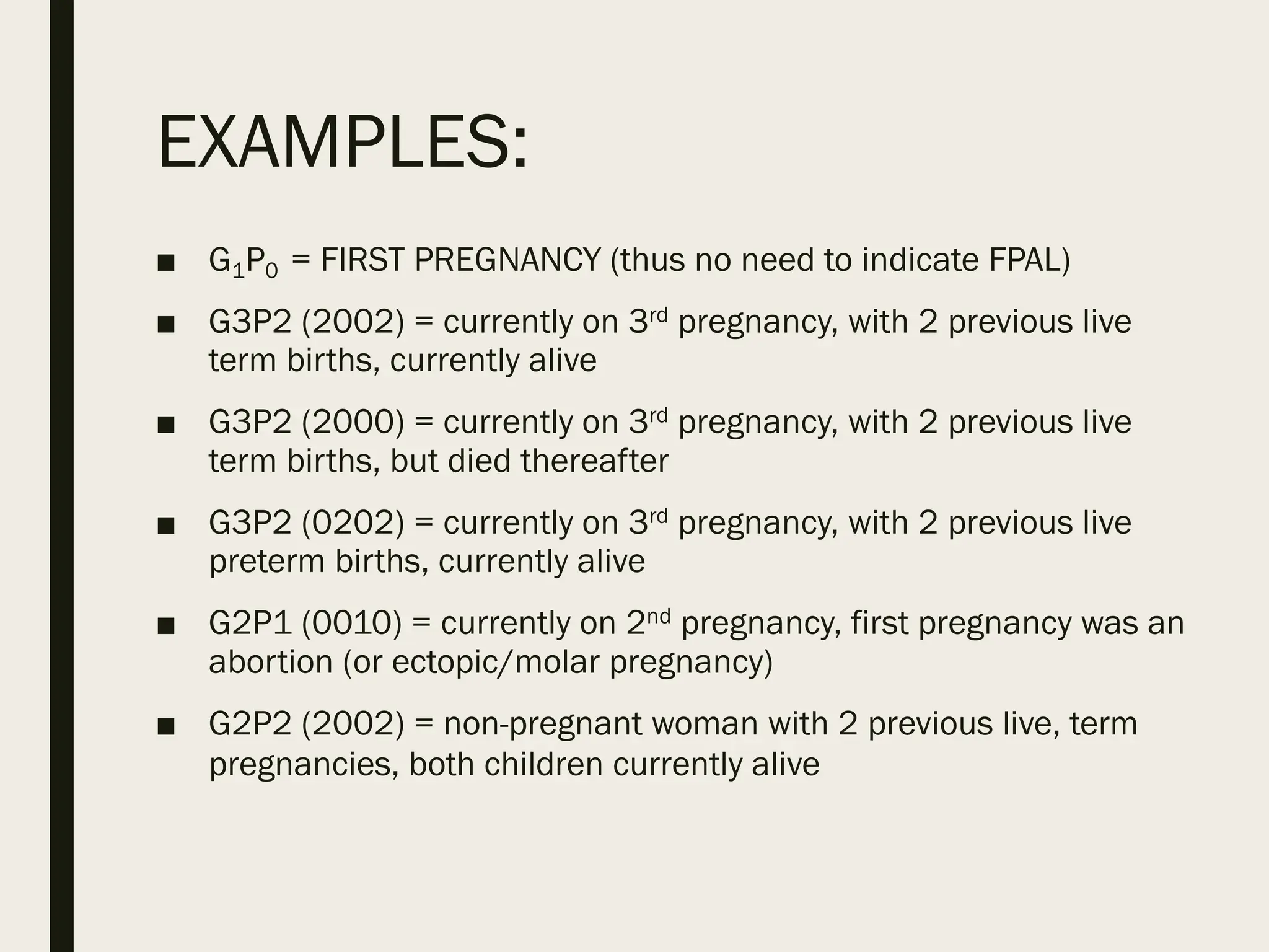
EXAMPLES:
■ G1P0 =FIRST PREGNANCY (thus no need to indicate FPAL)
■ G3P2 (2002) = currently on 3rd pregnancy, with 2 previous live
term births, currently alive
■ G3P2 (2000) = currently on 3rd pregnancy, with 2 previous live
term births, but died thereafter
■ G3P2 (0202) = currently on 3rd pregnancy, with 2 previous live
preterm births, currently alive
■ G2P1 (0010) = currently on 2nd pregnancy, first pregnancy was an
abortion (or ectopic/molar pregnancy)
■ G2P2 (2002) = non-pregnant woman with 2 previous live, term
pregnancies, both children currently alive
12.
Examples (multiple pregnancies)
■A woman currently on her 2nd pregnancy, had a previous twin
pregnancy that was carried to term, and currently alive:
G2P1 (2002)
■ A woman who just gave birth to her twin babies on her first
pregnancy:
G1P1 (2002)
13.
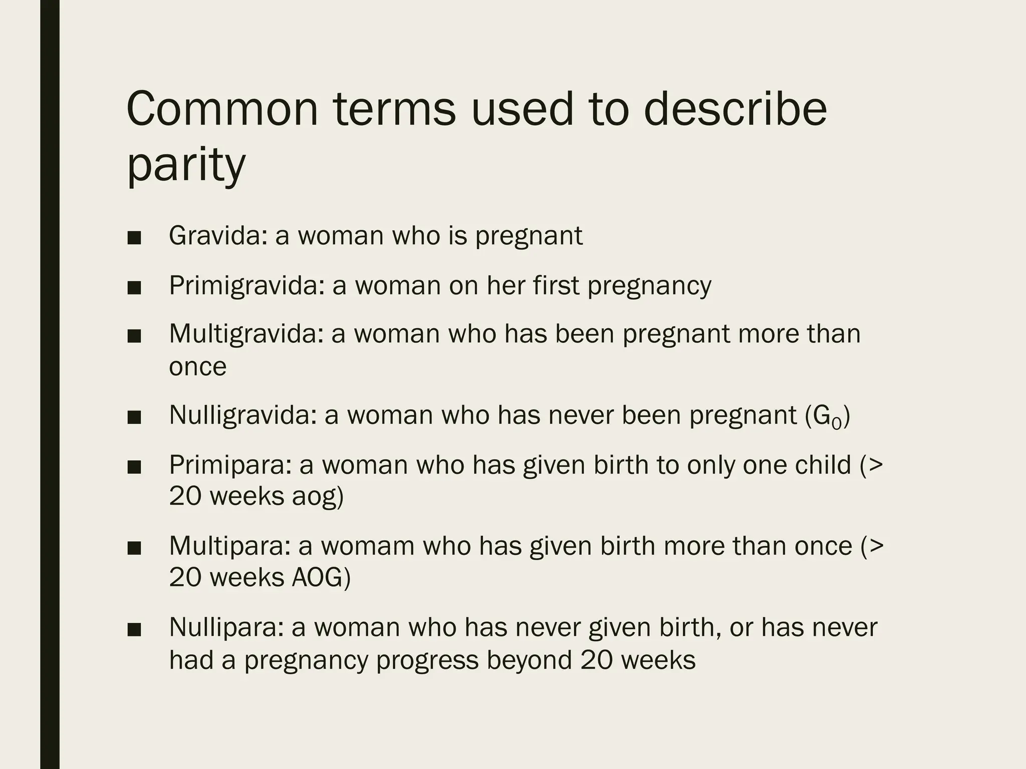
Common terms usedto describe
parity
■ Gravida: a woman who is pregnant
■ Primigravida: a woman on her first pregnancy
■ Multigravida: a woman who has been pregnant more than
once
■ Nulligravida: a woman who has never been pregnant (G0)
■ Primipara: a woman who has given birth to only one child (>
20 weeks aog)
■ Multipara: a womam who has given birth more than once (>
20 weeks AOG)
■ Nullipara: a woman who has never given birth, or has never
had a pregnancy progress beyond 20 weeks
14.
Determining fetal age
■Calculating number of weeks AOG based on LMP
■ If patient has irregular menses or does not
remember her LMP:
1. Uterine size
2. Quickening
3. First trimester ultrasound scan
15.
Calculating the ageof
gestation (AOG)
■ LMP: January 3, 2021
■ Date today: May 1, 2021
January: 31 days – 3 = 28 days
February: 28 days
March: 31 days
April: 30 days
May: 1 day
TOTAL: 118 days
118 ÷ 7 days =
16 6/7 wks
16.
Calculating the estimated
dateof delivery (EDD)
■ Naegele’s rule (using the Last Menstrual period/LMP) – used
only if patient has regular menses and is sure of her LMP
17.
Naegele’s rule
■ add7 days to the first day of the last period and
subtract 3 months, then add 1 year
■ For example:
– LMP: July 5, 2016
– EDD: July 5 + 7 days è July 12 à July 12 minus 3
months à April 12 à + 1 year à April 12, 2017
General Approach
■ Makesure to always provide comfort and sense of privacy
■ Have the needed equipment readily at hand
■ Provide gown and drapes for abdominal and pelvic exam
■ Instruct the patient to empty her bladder prior to examination
A. Positioning
Semi-sitting position with the
knees bent supported by a
pillow affords the greatest
comfort, as well as
protection from the negative
effects of the weight of the
gravid uterus on abdominal
organs and vessels
Thompson JE. Chapter 14 The Pregnant Woman. In: Bickley LS (ed); Bates’ Guide to Physical
Examination and History Taking, 7th edition (1999)
20.
■ B. Equipment
–The examiner’s hands are the “primary equipment”
for examination of the pregnant woman (should be
warmed); avoid tender areas of the body until the
end of the examination
– Speculum
– Tape measure
– Stethoscope/ fetal doppler
Thompson JE. Chapter 14 The Pregnant Woman. In: Bickley LS (ed); Bates’ Guide to Physical
Examination and History Taking, 7th edition (1999)
21.
General examination
1. Appearance(inspection
of overall health,
nutritional status.,
emotional state,
neuromuscular
coordination)
2. Weight, Height, BMI
3. Vital signs (BP, pulse
rate, temperature)
Thompson JE. Chapter 14 The Pregnant Woman. In: Bickley LS (ed); Bates’ Guide to Physical
Examination and History Taking, 7th edition (1999)
22.
Head and Neck
Skinpigmentation
changes
CHLOASMA/”MELASMA
GRAVIDARUM” -- irregular
brownish patches of varying
size appear on the face and
neck —the so-called mask of
pregnancy.
Cunningham FG, Leveno KJ, Bloom SL, Spong CY, Dashe JS, Hoffman BL, Casey
BM, Sheffield JS (eds). Williams Obstetrics 24th
edition. 2014.
23.
Head and Neck
■Hair: note texture, moisture and distribution;
dryness, oiliness and minor generalized hair loss
may be noted
■ Eyes: anemia of pregnancy may cause pallor
■ Nose: nasal congestion is common among
pregnant women; nosebleeds also common
■ Mouth: inspect gums and teeth; gingival
enlargement with bleeding is common
■ Thyroid: symmetrical enlargement may be
expected; marked enlargement is not normal
during pregnancy
Thompson JE. Chapter 14 The Pregnant Woman. In: Bickley LS (ed); Bates’ Guide to Physical
Examination and History Taking, 7th edition (1999)
24.
THORAX AND LUNGS
■Inspect thorax for pattern of breathing;
■ There are usually no abnormal physical
signs, except some women who might
experience labored breathing
Thompson JE. Chapter 14 The Pregnant Woman. In: Bickley LS (ed); Bates’ Guide to Physical
Examination and History Taking, 7th edition (1999)
25.
HEART
■ Palpate theapical impulse; In advanced
pregnancy, it may be slightly higher than
normal because of dextrorotation of the
heart due to the higher diaphragm
■ Auscultate the heart; soft blowing murmurs
are common, reflecting the increased blood
flow in normal vessels
Thompson JE. Chapter 14 The Pregnant Woman. In: Bickley LS (ed); Bates’ Guide to Physical
Examination and History Taking, 7th edition (1999)
26.
BREASTS
■ Inspect breastsand nipple for symmetry
and color; nipples and areola become
bigger and darker; Montgomery glands
prominent.
■ Compress nipples with finger and thumb à
may express colostrum from the nipples.
Thompson JE. Chapter 14 The Pregnant Woman. In: Bickley LS (ed); Bates’ Guide to Physical
Examination and History Taking, 7th edition (1999)
27.
Inspection: skin changes
■Linea Nigra : darkening of the
linea alba (midline of the
abdominal skin from xiphoid to
symphysis pubis)
■ à due to stimulation of
melanophores by increase in
melanocyte stimulating hormone
Abdomen
Cunningham FG, Leveno KJ, Bloom SL, Spong CY, Dashe JS, Hoffman BL, Casey
BM, Sheffield JS (eds). Williams Obstetrics 24th
edition. 2014.
28.
Abdomen
Skin pigmentation changes
■Striae gravidarum: “stretch marks”
■ à separation of the underlying
collagen tissue (secondary to
stretching of the abdomen) and
appear as irregular scars
■ à reddish or purplish à becomes
silvery after delivery
■ associated risk factors are weight
gain during pregnancy, younger
maternal age, and family history. Cunningham FG, Leveno KJ, Bloom SL, Spong CY,
Dashe JS, Hoffman BL, Casey BM, Sheffield JS
(eds). Williams Obstetrics 24th
edition. 2014.
29.
Skin changes
■ Occasionally,the muscles of
the abdominal walls do not
withstand the tension to
which they are subjected.
■ As a result, rectus muscles
separate in the midline,
creating diastasis recti
■ If severe, a considerable
portion of the anterior
uterine wall is covered by
only a layer of skin,
attenuated fascia, and
peritoneum to form a ventral
hernia.
Abdomen
Cunningham FG, Leveno KJ, Bloom SL, Spong
CY, Dashe JS, Hoffman BL, Casey BM, Sheffield
JS (eds). Williams Obstetrics 24th
edition. 2014.
30.
Abdomen
Skin pigmentation changes
■Spider telangieactasia : vascular stellate
marks resulting from high levels of
estrogen
■ à blanch when pressure is applied
■ à palmar erythema is an associated sign
■ Typically develops in face, neck, upper
chest and arms
Cunningham FG, Leveno KJ, Bloom SL, Spong CY, Dashe JS, Hoffman BL, Casey
BM, Sheffield JS (eds). Williams Obstetrics 24th
edition. 2014.
31.
Abdomen
Palpation: Abdominal Enlargement
■0 to 12 weeks AOG: uterus is a pelvic
organ
■ 12 weeks AOG: uterus at symphysis
pubis
■ 16 weeks AOG: midway between
symphysis pubis and umbilicus
■ 20 weeks AOG: umbilical level
■ Linear measurement from the
symphysis pubis to the uterine
fundus on an empty bladder
correlates with AOG at 16-32 weeks
(FUNDIC HEIGHT)
■ example: 20 weeks AOG = 20 cm
Cunningham FG, Leveno KJ, Bloom SL, Spong
CY, Dashe JS, Hoffman BL, Casey BM,
Sheffield JS (eds). Williams Obstetrics 24th
edition. 2014.
32.
Abdomen
Palpation
■ Perception offetal movement by the examiner
– Examiner may feel fetal movement after 24 weeks AOG
(felt by the mother around 18 weeks - ”quickening”)
■ Uterine contractility:
– abdomen feels tense or firm to the examiner, especially
if the patient is in labor, or near term (“Braxton-Hicks
contractions”)
■ Some fetal parts become palpable, espescially if mother
is non-obese
Thompson JE. Chapter 14 The Pregnant Woman. In: Bickley LS (ed); Bates’ Guide to Physical
Examination and History Taking, 7th edition (1999)
Leopold’s maneuvers
1. Leopold’smaneuver #1
(LM1)
■ “Fundal grip”
■ Uterine fundus is palpated
to detemine which fetal part
occupies the fundus
■ Fetal head should be round
and hard, ballottable
■ Breech presents as a large
nodular mass
Cunningham FG, Leveno KJ, Bloom SL, Spong CY, Dashe
JS, Hoffman BL, Casey BM, Sheffield JS (eds). Williams
Obstetrics 24th edition. 2014.
35.
Leopold’s maneuvers
2. Leopold’smaneuver #2
(LM2)
■ “Umbilical grip”
■ Palpation of paraumbilical
areas or the sides of the
uterus
■ To determine which side is
the fetal back
■ Fetal back feels like a hard,
resistant, convex structure
■ Fetal small parts feel
nodular, irregular
Cunningham FG, Leveno KJ, Bloom SL, Spong CY, Dashe
JS, Hoffman BL, Casey BM, Sheffield JS (eds). Williams
Obstetrics 24th edition. 2014.
36.
Leopold’s maneuvers
3. Leopold’smaneuver #3 (LM3)
■ “Pawlik’s grip”
■ Suprapubic palpation using
thumb and fingers just above
the symphysis pubis, to
determine fetal presentation
and station
■ the differentiation between
head and breech is made as
in LM1
■ *If presenting part is not engaged, a
movable structure can be palpated
Cunningham FG, Leveno KJ, Bloom SL, Spong CY,
Dashe JS, Hoffman BL, Casey BM, Sheffield JS (eds).
Williams Obstetrics 24th edition. 2014.
37.
Leopold’s maneuvers
4. Leopold’smaneuver #4 (LM4)
■ “Pelvic grip”
■ Palpation of the bilateral lower
quadrants to determine engagement
of the fetal presenting part
■ Fetal part is engaged: examiner’s
hands diverge
■ Fetal head is not engaged:
examiner’s hands converge
■ If fetal head is felt on same side of
the fetal small partsà fetal head is
well flexed Cunningham FG, Leveno KJ, Bloom SL, Spong CY, Dashe
JS, Hoffman BL, Casey BM, Sheffield JS (eds). Williams
Obstetrics 24th edition. 2014.
38.
Abdomen
Auscultation: Identification of
fetalheart beat; heard at fetal
back
■ FHR is usually at a range of 110-
160 bpm
■ Detected through stethoscope
at 18 weeks AOG
■ Detected though fetal Doppler at
10-12 weeks AOG
Cunningham FG, Leveno KJ, Bloom SL, Spong CY, Dashe JS, Hoffman BL, Casey BM, Sheffield JS (eds). Williams
Obstetrics 24th edition. 2014.
39.
Extremities
■ Inspect handsand legs for edema.
■ Palpate for pretibial, ankle and pedal edema
■ Physiologic edema is more common in advanced
pregnancy and in women who stand a lot.
■ Pathologic edema is often grade 3+ and often
associated with pregnancy-induced hypertension
■ Check for leg varicosities
Thompson JE. Chapter 14 The Pregnant Woman. In: Bickley LS (ed); Bates’ Guide to Physical
Examination and History Taking, 7th edition (1999)
40.
Genitalia
Inspection
■ Note hairdistribution, color, scars
■ Parous relaxation of the introitus and noticaeble
enlargement of labia and clitoris are normal
■ Scars from previous episiotomy or perineal
lacerations may be present
■ Inspect anal area for varicosities (hemorrhoids)
■ Palpate Bartholin’s and skene’s glands
■ Check for cystocoele or rectocoele
Thompson JE. Chapter 14 The Pregnant Woman. In: Bickley LS (ed); Bates’ Guide to Physical
Examination and History Taking, 7th edition (1999)
41.
Speculum exam: Changesin
the Vaginal
Mucosa
“Chadwick’s sign” – vaginal
mucosa becomes congested
and violaceous, or bluish to
purplish in color
GENITALIA
Cunningham FG, Leveno KJ, Bloom SL, Spong CY, Dashe
JS, Hoffman BL, Casey BM, Sheffield JS (eds). Williams
Obstetrics 24th edition. 2014.
42.
Speculum examination: cervical
changes
■cervical glands undergo marked
proliferation, and by the end of
pregnancy, they occupy up to
one half of the entire cervical
mass.
■ These normal pregnancy-
induced changes represent an
extension, or eversion, of the
proliferating columnar
endocervical glands.
■ This tissue tends to be red and
velvety and bleeds even with
minor trauma, such as with Pap
smear sampling.
48 Maternal Anatomy and Physiology
SECTION
2
concomitant mean Doppler velocimetry was increased eightfold.
Recall that blood flow within a vessel increases in proportion to
the fourth power of the radius. Thus, slight diameter increases
in the uterine artery produces a tremendous blood flow capac-
ity increase (Guyton, 1981). As reviewed by Mandala and Osol
(2011), the vessels that supply the uterine corpus widen and
elongate while preserving contractile function. In contrast, the
spiral arteries, which directly supply the placenta, widen but
completely lose contractility. This presumably results from endo-
vascular trophoblast invasion that destroys the intramural mus-
cular elements (Chap. 5, p. 93).
The vasodilation during pregnancy is at least in part the con-
sequence of estrogen stimulation. For example, 17β-estradiol
has been shown to promote uterine artery vasodilation and
reduce uterine vascular resistance (Sprague, 2009). Jauniaux
and colleagues (1994) found that estradiol and progesterone,
as well as relaxin, contribute to the downstream fall in vascular
resistance in women with advancing gestational age.
The downstream fall in vascular resistance leads to an accel-
eration of flow velocity and shear stress in upstream vessels. In
turn, shear stress leads to circumferential vessel growth, and
nitric oxide—a potent vasodilator—appears to play a key role
regulating this process (p. 61). Indeed, endothelial shear stress,
estrogen, placental growth factor (PlGF), and vascular endo-
thelial growth factor (VEGF)—a promoter of angiogenesis—all
augment endothelial nitric oxide synthase (eNOS) and nitric
FIGURE 4-1 Cervical eversion of pregnancy as viewed through
a colposcope. The eversion represents columnar epithelium on
the portio of the cervix. (Photograph contributed by Dr. Claudia
Werner.)
Genitalia
Cunningham FG, Leveno KJ, Bloom SL, Spong CY, Dashe
JS, Hoffman BL, Casey BM, Sheffield JS (eds). Williams
Obstetrics 24th edition. 2014.
Genitalia
■ Bimanual/internal examination
Hegar’ssign : softening of the
uterine isthmus, resulting in its
compressibility on bimanual
examination; observed by the 6th
to 8th week AOG
Goodell’s sign : cyanosis and
softening of the cervix; May occur
as early as 4 weeks AOG
Cunningham FG, Leveno KJ, Bloom SL, Spong CY, Dashe
JS, Hoffman BL, Casey BM, Sheffield JS (eds). Williams
Obstetrics 24th edition. 2014.
45.
Genitalia
Internal examination:
■ Estimatethe length of the cervix by palpating the lateral
surface of the cervix from the cervical tip to the lateral fornix.
■ Prior to 34-36 weeks, cervix should retain its normal length of
about 1.5 – 2cm
■ A shortened (“effaced”) cervix prior to 32 weeks may indicate
preterm labor
Thompson JE. Chapter 14 The Pregnant Woman. In: Bickley LS (ed); Bates’ Guide to Physical
Examination and History Taking, 7th edition (1999)
46.
Genitalia
Internal examination:
■ Noteif cervix is closed or dilated
■ If dilated, take note of the following:
– estimate the approximate size of dilatation in
centimeters
– Note the fetal station
– fetal presenting part (ex: cephalic, breech)
– Bag of waters intact?
Thompson JE. Chapter 14 The Pregnant Woman. In: Bickley LS (ed); Bates’ Guide to Physical
Examination and History Taking, 7th edition (1999)
47.
Concluding the visit
■Once the examination is completed, instruct patient to get
dressed
■ Review findings with patient
■ Answer patient’s questions
■ Advise necessary laboratory/ancillary procedures patient may
need
■ Reinforce the importance of regular prenatal check-ups
■ Record all findings in the chart/record
48.
Summary
■ Components ofan Obstetric History
■ Determining Gravidity and Parity
■ Calculating fetal age of gestation (AOG)
■ Calculating Estimated Date of Delivery (Naegele’s rule)
■ Components of an Obstetric Physical exam