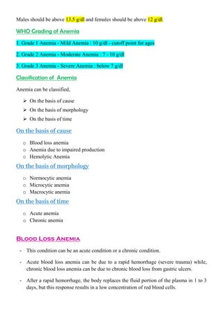
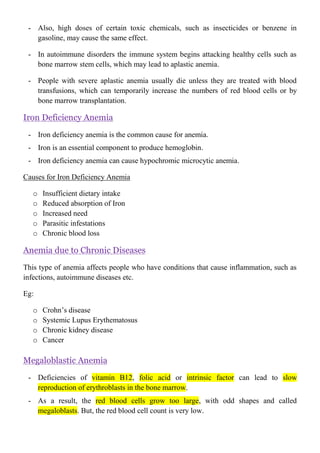
Anemia I
Learning Outcomes
 Define anemia
 Describe the different gradings of anemia
 Classify anemia types
 Describe pathophysiology of different types of anemia
What is “Anemia”?
Anemia is the deficiency of hemoglobin concentration in the blood compared to the
normal reference range.
What are the causes for anemia?
- Too few red blood cells
- Too little hemoglobin in the red blood cells
- Too little oxygen binding ability of each hemoglobin molecule due to deformity
- Increased loss of red blood cells
- Decreased production of normal red blood cells
- Increased destruction of red blood cells
- Increased physiological demand for red blood cells and iron
- Genetic factors
- Iron deficiency anemia
WHO Hemoglobin Threshold
Males should be above 13.5 g/dl and females should be above 12 g/dl.
WHO Grading of Anemia
1. Grade 1 Anemia - Mild Anemia : 10 g/dl - cutoff point for ages
2. Grade 2 Anemia - Moderate Anemia : 7 - 10 g/dl
3. Grade 3 Anemia - Severe Anemia : below 7 g/dl
Classification of Anemia
Anemia can be classified,
 On the basis of cause
 On the basis of morphology
 On the basis of time
On the basis of cause
o Blood loss anemia
o Anemia due to impaired production
o Hemolytic Anemia
On the basis of morphology
o Normocytic anemia
o Microcytic anemia
o Macrocytic anemia
On the basis of time
o Acute anemia
o Chronic anemia
Blood Loss Anemia
- This condition can be an acute condition or a chronic condition.
- Acute blood loss anemia can be due to a rapid hemorrhage (severe trauma) while,
chronic blood loss anemia can be due to chronic blood loss from gastric ulcers.
- After a rapid hemorrhage, the body replaces the fluid portion of the plasma in 1 to 3
days, but this response results in a low concentration of red blood cells.
- If a second hemorrhage does not occur, red blood cell concentration usually returns to
normal within 3 to 6 weeks.
- When chronic blood loss occurs, a person frequently cannot absorb enough iron from
the intestines to form hemoglobin as rapidly as it is lost.
- Red blood cells that are much smaller than normal and have too little hemoglobin
inside them are then produced, giving rise to microcytic, hypochromic anemia.
Anemia due to impaired production
o Aplastic anemia
o Iron deficiency anemia
o Anemia due to chronic diseases
o Megaloblastic anemia
Aplastic Anemia
- Aplastic anemia is due to lack of functioning of bone marrow.
- There may be different types of causes for aplastic anemia.
o Hereditary causes
o Acquired causes
o Idiopathic causes - Due to an unknown cause
Hereditary causes
o Fanconi anemia
o Dyskeratosis congenital
o Schwachman-Diamond syndrome
Acquired causes
o Autoimmune disorders that lead to the immune system to attack the bone marrow
o Toxic substances
o Chemotherapy or radiotherapy treatment
o Pregnancy
o Viral infections such as Epstein Barr virus, Hepatitis or HIV
o Certain medications including some antibiotics
o Cancer that affects has spread to the bone marrow
- Exposure to high dose radiation or chemotherapy for cancer treatment can damage
stem cells of the bone marrow, followed in a few weeks by anemia.
- Also, high doses of certain toxic chemicals, such as insecticides or benzene in
gasoline, may cause the same effect.
- In autoimmune disorders the immune system begins attacking healthy cells such as
bone marrow stem cells, which may lead to aplastic anemia.
- People with severe aplastic anemia usually die unless they are treated with blood
transfusions, which can temporarily increase the numbers of red blood cells or by
bone marrow transplantation.
Iron Deficiency Anemia
- Iron deficiency anemia is the common cause for anemia.
- Iron is an essential component to produce hemoglobin.
- Iron deficiency anemia can cause hypochromic microcytic anemia.
Causes for Iron Deficiency Anemia
o Insufficient dietary intake
o Reduced absorption of Iron
o Increased need
o Parasitic infestations
o Chronic blood loss
Anemia due to Chronic Diseases
This type of anemia affects people who have conditions that cause inflammation, such as
infections, autoimmune diseases etc.
Eg:
o Crohn’s disease
o Systemic Lupus Erythematosus
o Chronic kidney disease
o Cancer
Megaloblastic Anemia
- Deficiencies of vitamin B12, folic acid or intrinsic factor can lead to slow
reproduction of erythroblasts in the bone marrow.
- As a result, the red blood cells grow too large, with odd shapes and called
megaloblasts. But, the red blood cell count is very low.
- Thus, atrophy of the stomach mucosa (pernicious anemia) or loss of the entire
stomach after surgical removal can lead to megaloblastic anemia.
- Also, megaloblastic anemia develops in patients who have intestinal sprue, in which
folic acid, vitamin B12 and other vitamin B compounds are poorly absorbed.
- Because in these states the erythroblasts cannot proliferate rapidly enough to form
normal numbers of red blood cells.
- The red blood cells that are formed are mostly oversized, have bizarre shapes, and
have fragile membranes. Therefore, these cells rupture easily.
Hemolytic Anemia
- Hemolytic anemia is due to different abnormalities of the red blood cells, which can
be hereditary (genetic) or acquired.
- It makes the red blood cells fragile. So the red blood cells can be ruptured easily as
they go through the capillaries and they will have a short life span.
- But, the number of red blood cells formed may be normal or even much greater than
normal in some hemolytic diseases.
- Hemolytic anemia can be due to blood disorders, toxins or infections.
Eg:
 Hereditary spherocytosis
 Sickle cell anaemia
Hereditary Spherocytosis
- In hereditary spherocytosis, the red blood cells are very small and spherical rather
than being biconcave disks.
- These cells cannot withstand compression forces because they do not have the normal
loose, bag like cell membrane structure of the biconcave disks.
- Upon passing through the splenic pulp and some other tight vascular beds, they are
easily ruptured by even slight compression.
Sickle Cell Anemia
- In sickle cell anemia, the cells have an abnormal type of hemoglobin called
hemoglobin S, containing faulty beta chains in the hemoglobin molecule.
- When this hemoglobin is exposed to low concentrations of oxygen, it precipitates
into long crystals inside the red blood cell.
- These crystals elongate the cell and give it the appearance of a sickle rather than a
biconcave disk.
- The precipitated hemoglobin also damages the cell membrane, so the cells become
highly fragile, leading to serious anemia.
- Such patients experience a sickle cell disease, called crisis, in which low oxygen
tension in the tissues causes sickling, which leads to ruptured red blood cells, which
causes a further decrease in oxygen tension and still more sickling and red blood cell
destruction. Once the process starts, it progresses rapidly, eventuating in a serious
decrease in red blood cells within a few hours and in some cases, death.
Anemia II
Learning Outcomes
 Describe the signs and symptoms of anemia
 Describe oral manifestations of anemia
 Basic investigations used to diagnose anemia
 Describe the importance of knowledge on anemia as a dental surgeon
Symptoms and Signs of Anemia
Symptoms of Anemia
Symptoms can be minor and may go undetected.
 Feeling of weakness
 Fatigue
 General malaise
 Loss of stamina
 Poor concentration
 Dyspnea on exertion
 Pale or yellowish skin
 Irregular heartbeats
 Shortness of breath
 Dizziness or lightheadedness
 Chest pain
 Cold hands and feat
 Headaches
 Changed stool colour
 Low blood pressure
 Fainting
 Pica
 Koilonychia nails
Signs of Anemia
 Cardiac failure
 Bounding pulse
 Pallor colour (Pale skin, Mucosal linings and Nail beds)
 Jaundice in hemolytic anemia
 Bone deformities (Found in thalassemia major)
 Leg ulcers (Seen in sickle cell disease)
 Tachycardia (Fast heart rate)
 Flow murmurs
 Cardiac ventricular hypertrophy (enlargement)
Oral Manifestations of Anemia
 Oral ulcers
 Angular cheilitis
 Glossitis
 Pale tongue
Investigations for Anemia
 Complete blood count
 RBC count
 Hb level
 MCV (Mean Corpuscular Volume)
 RDW (Red blood cell Distribution Width)
 Size and shape of the RBC (Flowcytometry techniques)
 Examination of a stained blood smear using a microscope
- To get a blood cell count we can use,
 Automatic analyzers
 Manual hemocytometer
Packed Cell Volume (PCV) / Hematocrit (HCT)
 Packed cell volume is the proportion of the volume of blood sample that is occupied by
the red cells.
 It is represented as a percentage.
 There are white blood cells, plasma, platelets other than red blood cells.
 When we need to define the exact proportion of the red blood cells we can use packed
cell volume.
 It can be determined by manual and automated techniques.
 The normal reference range for PCV is 38-47.7%.
MCV (Mean Corpuscular Volume)
 Mean corpuscular volume is the volume of a single red cell.
 It is a kind of a laboratory value that measures the average size and volume of a single
red blood cell.
 It allows classification as either,
1. Microcytic anemia (MCV below normal range)
2. Normocytic anemia (MCV within normal range)
3. Macrocytic anemia (MCV above normal range)
 In automated systems MCV is measured directly, but in semi-automated counters
MCV has to be calculated.
 The unit of MCV is expressed in fl (ficoliter).
 The normal range of MCV is 80 - 95 fl.
Red blood cell distribution width (RDW or RCDW)
 Red cell distribution width reflects the range of red cell sizes measured within the
sample. It’s a measure of the variations of the red blood cells.
 Usually, the size of the red blood cell is 6 - 8 micrometers. The normal reference range
for RDW is 11 - 15%.
 It means if there is a wide range of sizes and shapes of red blood cells in the sample,
red cell distribution width is high.
 If all the red blood cells are in the same size and shape, red cell distribution width is
low.
 RDW is useful in,
- Early classification of anaemias (Becomes abnormal earlier in nutritional deficiency
anaemias than any of the other red cell parameters).
- Identifying red cell fragmentation, agglutination or dimorphic cell population.
Anisocytosis
 In anisocytosis the patient's red blood cells are of unequal size.
Flowcytometry
 Flowcytometry (FC) is a technique used to detect and measure physical and chemical
characteristics of a population of cells.
Mean Corpuscular Hemoglobin (MCH)
 Mean corpuscular haemoglobin is the amount of haemoglobin in each red blood cell.
 The unit of MCH is expressed in picograms (pg).
 The normal reference range in MCH is 27 - 31 pg.
Mean Corpuscular Hemoglobin Concentration (MCHC)
 Mean corpuscular haemoglobin concentration is the percentage of haemoglobin in 100
ml of packed red blood cells.
 The unit of MCHC is expressed in g/dl.
 The normal range of MCHC is 32 - 38 g/dl.
Blood Pictures
Examples for normocytic anemia
 Anemia of chronic diseases
 Hemolytic anemia
 Anemia of acute hemorrhage
 Aplastic anemia
 Renal disease
 Malignancy
Examples for microcytic anemia
 Iron deficiency anemia
 Thalassemia
 Anemia of chronic diseases
 Sideroblastic anemia
Examples for macrocytic anemia
 Vitamin B12 deficiency
 Folate deficiency
 Liver disease
 Alcoholism
 Reticulocytosis
 Drugs
A patient with severe anemia suddenly had an acute cardiac failure after doing
some jogging in the morning. Explain the physiological basis behind this
phenomena?
- The viscosity of the blood depends largely on the blood concentration of red blood
cells.
- In persons with severe anemia, the blood viscosity may fall to as low as 1.5 times that
of water rather than the normal value of about 3.
- This change decreases the resistance to blood flow in the peripheral blood vessels, so
far greater than normal quantities of blood flow through the tissues and return to the
heart, thereby greatly increasing cardiac output.
- Hypoxia resulting from diminished transport of oxygen by the blood causes the
peripheral tissue blood vessels to dilate, allowing a further increase in the return of
blood to the heart and increasing the cardiac output to a still higher level.
- Thus, one of the major effects of anemia is greatly increased cardiac output, as well as
increased pumping workload on the heart.
- The increased cardiac output in persons with anemia partially offsets the reduced
oxygen carrying effect of the anemia because even though each unit quantity of blood
carries only small quantities of oxygen, the rate of blood flow may be increased
enough that almost normal quantities of oxygen are actually delivered to the tissues.
- However, when a person with anemia begins to exercise, the heart is not capable of
pumping much greater quantities of blood than it is already pumping.
- Consequently, during exercise, which greatly increases tissue demand for oxygen,
extreme tissue hypoxia and acute cardiac failure can be resulted.

Anemia Basic Lecture Note Prepared by a Student.pdf

  • 1.
    Anemia I Learning Outcomes Define anemia  Describe the different gradings of anemia  Classify anemia types  Describe pathophysiology of different types of anemia What is “Anemia”? Anemia is the deficiency of hemoglobin concentration in the blood compared to the normal reference range. What are the causes for anemia? - Too few red blood cells - Too little hemoglobin in the red blood cells - Too little oxygen binding ability of each hemoglobin molecule due to deformity - Increased loss of red blood cells - Decreased production of normal red blood cells - Increased destruction of red blood cells - Increased physiological demand for red blood cells and iron - Genetic factors - Iron deficiency anemia WHO Hemoglobin Threshold
  • 2.
    Males should beabove 13.5 g/dl and females should be above 12 g/dl. WHO Grading of Anemia 1. Grade 1 Anemia - Mild Anemia : 10 g/dl - cutoff point for ages 2. Grade 2 Anemia - Moderate Anemia : 7 - 10 g/dl 3. Grade 3 Anemia - Severe Anemia : below 7 g/dl Classification of Anemia Anemia can be classified,  On the basis of cause  On the basis of morphology  On the basis of time On the basis of cause o Blood loss anemia o Anemia due to impaired production o Hemolytic Anemia On the basis of morphology o Normocytic anemia o Microcytic anemia o Macrocytic anemia On the basis of time o Acute anemia o Chronic anemia Blood Loss Anemia - This condition can be an acute condition or a chronic condition. - Acute blood loss anemia can be due to a rapid hemorrhage (severe trauma) while, chronic blood loss anemia can be due to chronic blood loss from gastric ulcers. - After a rapid hemorrhage, the body replaces the fluid portion of the plasma in 1 to 3 days, but this response results in a low concentration of red blood cells.
  • 3.
    - If asecond hemorrhage does not occur, red blood cell concentration usually returns to normal within 3 to 6 weeks. - When chronic blood loss occurs, a person frequently cannot absorb enough iron from the intestines to form hemoglobin as rapidly as it is lost. - Red blood cells that are much smaller than normal and have too little hemoglobin inside them are then produced, giving rise to microcytic, hypochromic anemia. Anemia due to impaired production o Aplastic anemia o Iron deficiency anemia o Anemia due to chronic diseases o Megaloblastic anemia Aplastic Anemia - Aplastic anemia is due to lack of functioning of bone marrow. - There may be different types of causes for aplastic anemia. o Hereditary causes o Acquired causes o Idiopathic causes - Due to an unknown cause Hereditary causes o Fanconi anemia o Dyskeratosis congenital o Schwachman-Diamond syndrome Acquired causes o Autoimmune disorders that lead to the immune system to attack the bone marrow o Toxic substances o Chemotherapy or radiotherapy treatment o Pregnancy o Viral infections such as Epstein Barr virus, Hepatitis or HIV o Certain medications including some antibiotics o Cancer that affects has spread to the bone marrow - Exposure to high dose radiation or chemotherapy for cancer treatment can damage stem cells of the bone marrow, followed in a few weeks by anemia.
  • 4.
    - Also, highdoses of certain toxic chemicals, such as insecticides or benzene in gasoline, may cause the same effect. - In autoimmune disorders the immune system begins attacking healthy cells such as bone marrow stem cells, which may lead to aplastic anemia. - People with severe aplastic anemia usually die unless they are treated with blood transfusions, which can temporarily increase the numbers of red blood cells or by bone marrow transplantation. Iron Deficiency Anemia - Iron deficiency anemia is the common cause for anemia. - Iron is an essential component to produce hemoglobin. - Iron deficiency anemia can cause hypochromic microcytic anemia. Causes for Iron Deficiency Anemia o Insufficient dietary intake o Reduced absorption of Iron o Increased need o Parasitic infestations o Chronic blood loss Anemia due to Chronic Diseases This type of anemia affects people who have conditions that cause inflammation, such as infections, autoimmune diseases etc. Eg: o Crohn’s disease o Systemic Lupus Erythematosus o Chronic kidney disease o Cancer Megaloblastic Anemia - Deficiencies of vitamin B12, folic acid or intrinsic factor can lead to slow reproduction of erythroblasts in the bone marrow. - As a result, the red blood cells grow too large, with odd shapes and called megaloblasts. But, the red blood cell count is very low.
  • 5.
    - Thus, atrophyof the stomach mucosa (pernicious anemia) or loss of the entire stomach after surgical removal can lead to megaloblastic anemia. - Also, megaloblastic anemia develops in patients who have intestinal sprue, in which folic acid, vitamin B12 and other vitamin B compounds are poorly absorbed. - Because in these states the erythroblasts cannot proliferate rapidly enough to form normal numbers of red blood cells. - The red blood cells that are formed are mostly oversized, have bizarre shapes, and have fragile membranes. Therefore, these cells rupture easily. Hemolytic Anemia - Hemolytic anemia is due to different abnormalities of the red blood cells, which can be hereditary (genetic) or acquired. - It makes the red blood cells fragile. So the red blood cells can be ruptured easily as they go through the capillaries and they will have a short life span. - But, the number of red blood cells formed may be normal or even much greater than normal in some hemolytic diseases. - Hemolytic anemia can be due to blood disorders, toxins or infections. Eg:  Hereditary spherocytosis  Sickle cell anaemia Hereditary Spherocytosis - In hereditary spherocytosis, the red blood cells are very small and spherical rather than being biconcave disks. - These cells cannot withstand compression forces because they do not have the normal loose, bag like cell membrane structure of the biconcave disks. - Upon passing through the splenic pulp and some other tight vascular beds, they are easily ruptured by even slight compression. Sickle Cell Anemia - In sickle cell anemia, the cells have an abnormal type of hemoglobin called hemoglobin S, containing faulty beta chains in the hemoglobin molecule. - When this hemoglobin is exposed to low concentrations of oxygen, it precipitates into long crystals inside the red blood cell.
  • 6.
    - These crystalselongate the cell and give it the appearance of a sickle rather than a biconcave disk. - The precipitated hemoglobin also damages the cell membrane, so the cells become highly fragile, leading to serious anemia. - Such patients experience a sickle cell disease, called crisis, in which low oxygen tension in the tissues causes sickling, which leads to ruptured red blood cells, which causes a further decrease in oxygen tension and still more sickling and red blood cell destruction. Once the process starts, it progresses rapidly, eventuating in a serious decrease in red blood cells within a few hours and in some cases, death. Anemia II Learning Outcomes  Describe the signs and symptoms of anemia  Describe oral manifestations of anemia  Basic investigations used to diagnose anemia  Describe the importance of knowledge on anemia as a dental surgeon Symptoms and Signs of Anemia Symptoms of Anemia Symptoms can be minor and may go undetected.  Feeling of weakness  Fatigue  General malaise  Loss of stamina  Poor concentration  Dyspnea on exertion  Pale or yellowish skin  Irregular heartbeats  Shortness of breath  Dizziness or lightheadedness  Chest pain  Cold hands and feat  Headaches  Changed stool colour  Low blood pressure  Fainting
  • 7.
     Pica  Koilonychianails Signs of Anemia  Cardiac failure  Bounding pulse  Pallor colour (Pale skin, Mucosal linings and Nail beds)  Jaundice in hemolytic anemia  Bone deformities (Found in thalassemia major)  Leg ulcers (Seen in sickle cell disease)  Tachycardia (Fast heart rate)  Flow murmurs  Cardiac ventricular hypertrophy (enlargement) Oral Manifestations of Anemia  Oral ulcers  Angular cheilitis  Glossitis  Pale tongue Investigations for Anemia  Complete blood count  RBC count  Hb level  MCV (Mean Corpuscular Volume)  RDW (Red blood cell Distribution Width)  Size and shape of the RBC (Flowcytometry techniques)  Examination of a stained blood smear using a microscope
  • 8.
    - To geta blood cell count we can use,  Automatic analyzers  Manual hemocytometer Packed Cell Volume (PCV) / Hematocrit (HCT)  Packed cell volume is the proportion of the volume of blood sample that is occupied by the red cells.  It is represented as a percentage.  There are white blood cells, plasma, platelets other than red blood cells.  When we need to define the exact proportion of the red blood cells we can use packed cell volume.  It can be determined by manual and automated techniques.  The normal reference range for PCV is 38-47.7%. MCV (Mean Corpuscular Volume)  Mean corpuscular volume is the volume of a single red cell.  It is a kind of a laboratory value that measures the average size and volume of a single red blood cell.
  • 9.
     It allowsclassification as either, 1. Microcytic anemia (MCV below normal range) 2. Normocytic anemia (MCV within normal range) 3. Macrocytic anemia (MCV above normal range)  In automated systems MCV is measured directly, but in semi-automated counters MCV has to be calculated.  The unit of MCV is expressed in fl (ficoliter).  The normal range of MCV is 80 - 95 fl. Red blood cell distribution width (RDW or RCDW)  Red cell distribution width reflects the range of red cell sizes measured within the sample. It’s a measure of the variations of the red blood cells.  Usually, the size of the red blood cell is 6 - 8 micrometers. The normal reference range for RDW is 11 - 15%.  It means if there is a wide range of sizes and shapes of red blood cells in the sample, red cell distribution width is high.  If all the red blood cells are in the same size and shape, red cell distribution width is low.  RDW is useful in, - Early classification of anaemias (Becomes abnormal earlier in nutritional deficiency anaemias than any of the other red cell parameters). - Identifying red cell fragmentation, agglutination or dimorphic cell population. Anisocytosis  In anisocytosis the patient's red blood cells are of unequal size.
  • 10.
    Flowcytometry  Flowcytometry (FC)is a technique used to detect and measure physical and chemical characteristics of a population of cells. Mean Corpuscular Hemoglobin (MCH)  Mean corpuscular haemoglobin is the amount of haemoglobin in each red blood cell.  The unit of MCH is expressed in picograms (pg).  The normal reference range in MCH is 27 - 31 pg. Mean Corpuscular Hemoglobin Concentration (MCHC)  Mean corpuscular haemoglobin concentration is the percentage of haemoglobin in 100 ml of packed red blood cells.  The unit of MCHC is expressed in g/dl.  The normal range of MCHC is 32 - 38 g/dl. Blood Pictures
  • 11.
    Examples for normocyticanemia  Anemia of chronic diseases  Hemolytic anemia  Anemia of acute hemorrhage  Aplastic anemia  Renal disease  Malignancy Examples for microcytic anemia  Iron deficiency anemia  Thalassemia  Anemia of chronic diseases  Sideroblastic anemia Examples for macrocytic anemia  Vitamin B12 deficiency  Folate deficiency  Liver disease  Alcoholism  Reticulocytosis  Drugs A patient with severe anemia suddenly had an acute cardiac failure after doing some jogging in the morning. Explain the physiological basis behind this phenomena? - The viscosity of the blood depends largely on the blood concentration of red blood cells. - In persons with severe anemia, the blood viscosity may fall to as low as 1.5 times that of water rather than the normal value of about 3. - This change decreases the resistance to blood flow in the peripheral blood vessels, so far greater than normal quantities of blood flow through the tissues and return to the heart, thereby greatly increasing cardiac output. - Hypoxia resulting from diminished transport of oxygen by the blood causes the peripheral tissue blood vessels to dilate, allowing a further increase in the return of blood to the heart and increasing the cardiac output to a still higher level. - Thus, one of the major effects of anemia is greatly increased cardiac output, as well as increased pumping workload on the heart. - The increased cardiac output in persons with anemia partially offsets the reduced oxygen carrying effect of the anemia because even though each unit quantity of blood
  • 12.
    carries only smallquantities of oxygen, the rate of blood flow may be increased enough that almost normal quantities of oxygen are actually delivered to the tissues. - However, when a person with anemia begins to exercise, the heart is not capable of pumping much greater quantities of blood than it is already pumping. - Consequently, during exercise, which greatly increases tissue demand for oxygen, extreme tissue hypoxia and acute cardiac failure can be resulted.