2. 2
INTRODUCTION
Ayurveda is the first systematically written record of medicine in the world and
incorporating all aspect of human life.
The main aim of Ayurveda is being to provide guidelines for maintenance and
promotion of health as well as prevention and then treatment of diseases.
SROTAS :-
Achaarya Charak has described 13 Srotas, In female there is one more
i.e. Artavwaha Srotas in Garbha prakaran,where as Acharya Sushrut has
told 11 srotas but they are pair in number.
Acharya Charaka described Srotas as the channels for transportation and
transformation of dhatu.
ANNAVAHA SROTAS :-
Annavaha Srotas is one of the type of srotasa described in all important samhita,
it can be considered to Gastro Intestinal Tract except colon from modern view.
Mahsrotas and Kostha are describe synonymous to Annavaha Srotas. The
pakwashaya (colon) and further
part of alimentary tract are included in Purishvaha srotasa.
3. 3
COMPONENTS OF ANNAVAHA SROTAS
AMASHAYA :-
Acharya Charak described location of amashaya in between nabhi (umbilicus)
and stana (nipples). It perform the function of pachan (digestion) of all type of
food material.
Amashaya is divided into two parts, Urdhva and AdhoAmashya is
considered as Kshudrantantra and agnyasaya.
GRAHANI AND PITTADHARAKALA :-
Sushruta described that grahani is located between amashaya (stomach)
and pakwashaya (large intestine) and it is the site of pittadharakala.
Acharya Charaka described grahani as a seat of agni and it is
called so because of holding up the food.
This description of grahani indicate it to be whole of the
small intestine including duodenum, jejunum and ilium.
Pittadharakala is stated to cover that part of the gastro-intestinal
tract which is described as grahani, is related with digestion of
food.Ultimately the separation of Sara and Kitta takes place by
Pittadharakala[19] it is the site of antaragni.
4. 4
YAKRIT :-
In Ayurveda, according to Charaka and Sushruta it has
been described as Kosthanga, origin of Yakrit is described from Matrija Bhava.
In Ayurveda, yakrit is considered as root and place of Rakta and Raktavaha
srotas. It means store house of blood.
AGNI :-
The vital role of the Agni for the existence of human life has been
appreciated by the Acharyas of Ayurveda.
The main function of Agni in the body is the breakdown or to disintegrate the
food into their simplest possible components making it suitable for absorption
and utilization by the body.
THE PROCESS OF PACHAN :-
Pranavayu takes anna to the koshtha.Saman vayu intensify the agni. The
pachana takes place in the presence of Agni.Along with Bhutagni and
Dhatwagni especially, Jatharagni.
The pachak pitta which is responsible for splitting-up of the ingested food materials
may be compared with the enzymes like secretin, cholecystokinin, enterokinase,
lipase, amylase, invertase, enterogastrone, etc.
5. 5
VIPAKA :-
In Ayurveda, Ahara is digested by the action of Jatharagni leads to formation
of Ahara rasa and rest is known as vipaka.
It is of 3 types :
1)Madhura Vipaka :- Madhura and Lavana Rasa.
2)Amla Vipaka :- Amla Rasa.
3)Katu Vipaka :- Katu, Tikta and Kashaya Rasa.
STAGES OF PACHAN :-
The Madhura bhava is the stage of digestion carried out by bodhaka-kapha, i.e.
salivary digestion and also kledaka-kapha in the fundus portion of the stomach.
The second avasthapaka starts in the stomach and here the food partially
digests. Charaka has described this kind of food as the ‘Vidagdha-Aharara’ i.e.
Pakwapakwam (partially digested food). After the food enters in the duodenum
(the first part of the Grahani) and stimulates the Brunner’s glands through which
numbers of internal secretions are secreted. Bile and pancreatic juices are also
poured in the duodenum to carry-out further digestion of the partially digested
acid-chyme. So in Awasthapaka ingested food of any rasa which will be
transformed into the sweet taste and leads to the kapha formation and gradually
it takes the form of Amla and Katu-bhava, step by step it leads to the formation
of pitta and vata, respectively.
6. OBJECTIVES
• By the end of this lecture the student
should be able to:
• Describe the anatomy of the esophagus; extent,
length, parts, strictures, relations, blood & nerve
supply and lymphatics.
• Describe the anatomy of the stomach; location,
shape, parts, relations, blood & nerve supply and
lymphatics.
6
7. 7
The abdominal cavity is
divided into 9
compartments:
by:
2 vertical and
2 horizontal planes
Vertical planes:
2 Midclavicular lines.
Horizontal plane:
Subcostal (L3) and
intertubercular (L5) lines.
INTRODUCTIO
N
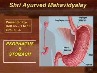
8. ESOPHAGUS
• It is a tubular structure about 25
cm long.
• It begins as the continuation of
the pharynx at the level of the
6th cervical vertebra.
• It pierces the diaphragm at the
level of the 10th thoracic
vertebra to join the stomach.
• It is formed of 3 parts:
• 1- Cervical.
• 2- Thoracic.
• 3- Abdominal.
Abdominal
thoracic
Cervical
8
9. 9
RELATIONS OF
CERVICAL PART
• Posteriorly:
• Cervical vertebral.
• Laterally:
• Lobes of thyroid
gland.
• Anteriorly:
• Trachea
• Recurrent laryngeal
nerves.
10. 10
THORACIC PART
• In the thorax, it descends
downward and to the left
through superior and then the
posterior mediastinum
• At the level of the sternal
angle, the aortic arch
pushes the esophagus again
to the midline.
12. 12
POSTERIOR
RELATIONS
1. Bodies of the
thoracic
vertebrae.
2. Thoracic duct.
3. Azygos vein.
4. Right posterior
intercostal
arteries.
5. Descending
thoracic aorta (at
the lower end).
Thoracic part
13. LATERAL
RELATIONS
• On the Right side:
1. Right mediastinal
pleura.
2. Terminal part of the
azygos vein.
• On the Left side:
1. Left mediastinal pleura.
2. Left subclavian artery.
3. Aortic arch.
4. Thoracic duct.
13
14. ESOPHAGEAL
CONSTRICTIONS
• The esophagus has 3 anatomic
constrictions.
• The first is at the junction with
the pharynx.
• The second is at the crossing
with the aortic arch and the
left main bronchus.
• The third is at the junction
with the stomach.
• They have a considerable
clinical importance.
• Why?
15. ESOPHAGEAL
STRICTURES
1. They may cause difficulties in
passing an esophagoscope or
gastroscope.
2. In case of swallowing of caustic
liquids (mostly in children), this
is where the burning is the worst
and strictures develop.
3. The esophageal strictures are a
common site of esophageal
carcinoma.
4. What is the importance of the
scale in this picture?
16. 16
ARTERIAL
SUPPLY
• Upper third is
supplied by the
inferior thyroid
artery.
• The middle third
by the thoracic
aorta.
• The lower third
by the left
gastric artery.
17. 17
VENOUS
DRAINAGE
• The upper third
drains in into the
inferior thyroid
veins.
• The middle third
into the azygos
veins.
• The lower third
into the left
gastric vein,
which is a
tributary of the
portal vein.
18. By Prof. Saeed Abuel Makarem 18
LYMPH
DRAINAGE
• The upper third
is drained in the
deep cervical
nodes.
• The middle third
is drained into
the superior
and inferior
mediastinal
nodes.
• The lower third
is drained in the
celiac lymph
nodes in the
abdomen.
19. By Prof. Saeed Abuel Makarem 19
NERVE SUPPLY
• It is supplied by
sympathetic fibers from
the sympathetic
trunks.
• The parasympathetic
supply comes form the
vagus nerves.
• Inferior to the roots of
the lungs, the vagus
nerves join the
sympathetic fibers to
form the esophageal
plexus.
• The left vagus lies
anterior to the
esophagus.
• The right vagus lies
posterior to it.
20. LOCATION
• The stomach is the
most dilated part of
the alimentary canal.
• It is located in the
upper part of the
abdomen.
• It extends from
beneath the left
costal region to the
epigastric and
umbilical regions.
• Most of the stomach
is protected by the
lower ribs.
• It is roughly J-
shaped.
STOMACH
21. 21
PARTS
2 Orifices:
• Cardiac orifice
• Pyloric orifice
2 Borders:
• Greater curvature
• Lesser curvature
2 Surfaces:
• Anterior surface
• Posterior surface
3 Parts:
• Fundus
• Body
• Pylorus:
The pylorus is subdivided
into 3 parts
• Pyloric antrum
• Pyloric canal
• Pyloric sphincter
22. 22
CARDIAC ORIFICE • It is the site of the
gastro- esophageal
sphincter.
• It is a physiological
rather than an
anatomical, sphincter.
• Consists of circular
layer of smooth
muscle (under vagal &
hormonal control).
• Function:
• Prevents esophageal
regurgitation (reflux)
24. 24
BODY
• Extends from:
– The level of the
fundus
– to
– The level of
Incisura angularis
• Incisura
angularis:
• a constant notch
on the lesser
curvature
25. 25
LESSER CURVATURE
• Forms the right
border of the
stomach.
• Extends from
the cardiac
orifice to the
pylorus.
• Attached to the
liver by the
lesser
omentum.
26. 26
GREATER CURVATURE • Forms the left
border of the
stomach.
• Extends from
the cardiac
orifice to the
pylorus.
• Its upper part is
attached to the
spleen by
gastrosplenic
ligament
• Its lower part is
attached to the
transverse colon
by the greater
omentum.
27. PYLORIC ANTRUM AND PYLORUS
• The pyloric antrum
extends from Incisura
angularis to the pylorus
• The pylorus is a tubular
part of the stomach
• It lies in the
transpyloric plane (L1)
• It has a thick muscular
end called pyloric
sphincter.
• The cavity of the pylorus
is the pyloric canal.
27
29. 29
POSTERIOR RELATIONS
1. Diaphragm
2. Left Kidney
3. Left suprarenal
gland
4. Pancreas
5. Trasverse
mesocolon
6. Splenic flexure of
colon
7. Splenic artery
All these structures
form the stomach
bed
30. 30
ARTERIES
• Left gastric artery:
• It is a branch of
celiac artery.
– Ascends along
the lesser
curvature.
• Right gastric
artery:
From the hepatic
artery of celiac.
– Runs to the left
along the lesser
curvature.
31. 31
ARTERIES
• Short gastric arteries –
arise from the splenic
artery.
– Pass in the
gastrosplenic
ligament.
• Left gastroepiploic
artery:
from splenic artery
– Pass in the
gastrosplenic
ligament.
• Right gastroepiploic
artery:
• from the
gastroduodenal artery
of hepatic .
– Passes to the left
along the greater
curvature.
32. 32
VEINS
• All of them drain into the portal circulation.
• The right and left gastric veins drain directly in the portal vein.
• The short gastric veins and the left gastroepiploic vein join the
splenic vein.
• The right gastroepiploic vein drain in the superior mesenteric vein.
33. 33
LYMPH DRAINAGE
• The lymph vessels
follow the arteries.
• They first drain to the:
– Left and right
gastric nodes
– Left and right
gastroepiploic
nodes and the
– Short gastric
nodes
• Ultimately, all the
lymph from the
stomach is collected
at the celiac nodes.
34. 34
NERVE
SUPPL
Y
• Sympathetic fibers are derived from the celiac plexus.
• Parasympathetic fibers from both vagi.
• Anterior vagal trunk:
– Formed from the left vagus
– Supply the anterior surface of the stomach
– Gives off a hepatic branch and from it, a branch to the
pylorus.
• Posterior vagal trunk:
– Formed from the right vagus
– Supply the posterior surface of the stomach
– Gives off a large branch to the celiac and the superior
mesenteric plexuses.
35. 35
Cardiac orifice lies deep to the left
7th costal cartilage 2.5 cm. from
the sternum ,(T10).
Pyloric orifice lies on transpyloric
plane (L1), 1 cm. to the right of the
middle line.
Lesser curvature:
A curved line, concave to the right
joining these 2 points.
The fundus:
Reaches to the left 5th intercostal
space (the point of the apex of the
heart).
Greater curvature:
A curved line drawn from the cardiac
orifice to the summit of the fundus,
then downward and to the left, finally
turning medial toward to the pyloric
orifice, passing through the
intersection of the left lateral with the
transpyloric line.
SURFAC ANATOMY OF
THE STOMACH