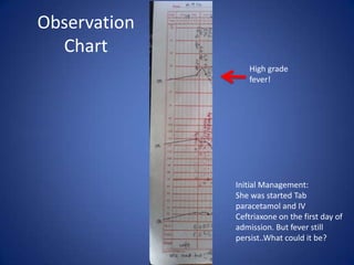
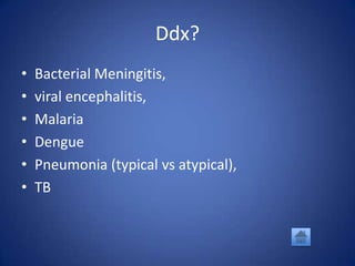
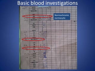
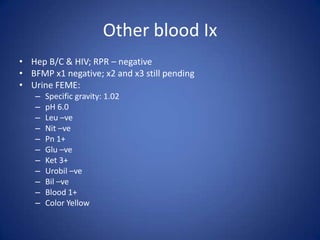
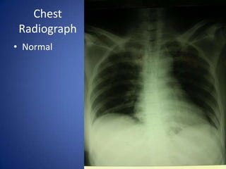
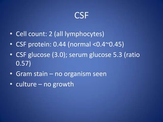
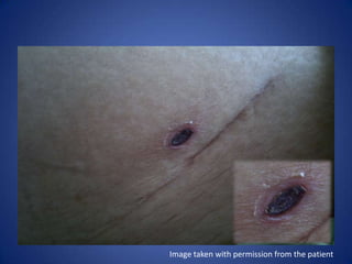
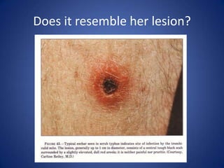
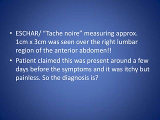
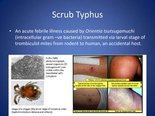
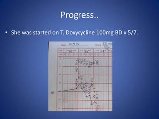
An interesting fever..
    Lee Chiang Sheng
Mdm B, 51 years old Ibanese
• Known history of hypertension under
  treatment
• Previously well.
• Was admitted on the 20th September with the
  chief complain
  – fever, chills, rigor x 1/52
• Fever started after returning from work in the afternoon.
• Was associated with
   – nausea and vomiting x 1 episode (non blood/billous stained,
     only food particles; non projectile),
   – generalized body weakness,
   – photophobia,
   – headache (mainly frontal area with ?radiation to the neck)
   – LOA
• No LOW, night sweats, no SOB/cough/chest pain, weakness
  of the limbs, seizure, visual changes, syncopal attack, no
  dysuria/hematuria/frequency, no rash.
  No PV discharge/ bleed
• Father claimed that ever since she was sick, she has
  become withdrawn from her family; ?personality change.
• No reversal of sleeping pattern.
• Has not returned to work ever since.
Past Medical History
• Hypertension
  – Taking T. Methyldopa 250mg TDS
  – Regular follow up at community clinic


           Past Surgical History
• History of appendicectomy many years back.
Family History
• Married with 5 children.
• Husband passed away d/t UGIB.
• No family members with fever.
Social History
• Denies any recent sexual contact/partners
• Social drinker (drinks rice wine)
• Non-smoker/ non IVDU
On Examination
• GCS full, Pink, conscious but slightly drowsy
  looking lady, not on any oxygen therapy/IV drip,
  not breathless with a RR of around 20 bpm, not in
  pain/cyanosed.

• Vitals:
   –   BP 116/70,
   –   Temp 38.3C,
   –   RR 20 bpm,
   –   SpO2 99% under room air
• Hand: Warm and sweaty hands. No clubbing,
  CRT <2s, no splinter hemorrhages, no nail fold
  vasculitis, no Osler nodes/Janeway lesions.
• Pulse: 80 bpm, regular rhythm, good volume,
  equal on both sides, good caliber
• Arm: No injection marks seen, no bruising
• Face: conjunctival pallor, no scleral icterus,
  cornea arcus
• Dental hygiene: good; tongue: moist
• Neck: no regional lymphadenopathy
• Praecordium: DRNM; good air entry with
  vesicular breath sounds/no adventitious
  sound

• Abdomen: not distended, Soft non tender; no
  hepato/splenomegaly; kidney not ballotable,
  bowel sounds present; Murphy’s punch –ve

• Lower limb: No swelling/tender calf.
• Neuro Exam: essentially normal with no focal
  neurological deficits; no cranial nerve palsies,
  Neck stiffness.
Observation
  Chart
                 High grade
                 fever!


              Initial Management:
              She was started Tab
              paracetamol and IV
              Ceftriaxone on the first day of
              admission. But fever still
              persist..What could it be?
WAIT! Don’t proceed to the
   next slide yet. Reflect
through the case and think..
Any further history and examination you’d like
to do? What is your differential diagnosis? And
      what investigations will you order?
Further Social History..
• No history of travel/ jungle trekking/
  swimming in river
• Works as a farmer in a paddy field (owned by
  the family)
• Lives in a village, near a rubber plantation.

  *Inguinal region:
  ++ inguinal lymphadenopathy
Ddx?
•   Bacterial Meningitis,
•   viral encephalitis,
•   Malaria
•   Dengue
•   Pneumonia (typical vs atypical),
•   TB
Basic blood investigations
                Normochromic
                normocytic
Other blood Ix
• Hep B/C & HIV; RPR – negative
• BFMP x1 negative; x2 and x3 still pending
• Urine FEME:
   –   Specific gravity: 1.02
   –   pH 6.0
   –   Leu –ve
   –   Nit –ve
   –   Pn 1+
   –   Glu –ve
   –   Ket 3+
   –   Urobil –ve
   –   Bil –ve
   –   Blood 1+
   –   Color Yellow
• ECG: normal sinus rhythm with no ischemic
  changes
• ECHO: essentially normal findings; EF 68%, no
  regional wall motion abnormality, no
  apical/mural thrombus
Chest
 Radiograph
• Normal
Essentially
 normal
CSF
• Cell count: 2 (all lymphocytes)
• CSF protein: 0.44 (normal <0.4~0.45)
• CSF glucose (3.0); serum glucose 5.3 (ratio
  0.57)
• Gram stain – no organism seen
• culture – no growth
So, what do you think you’ve missed?

 Fortunately, this lady was seen by one of the
physician on call and he meticulously examined
   her and found something unusual on her
                   abdomen..

           Do you know what is it?
Image taken with permission from the patient
Does it resemble her lesion?
• ESCHAR/ “Tache noire” measuring approx.
  1cm x 3cm was seen over the right lumbar
  region of the anterior abdomen!!
• Patient claimed this was present around a few
  days before the symptoms and it was itchy but
  painless. So the diagnosis is?
Scrub Typhus
• An acute febrile illness caused by Orientia tsutsugamuchi
  (intracellular gram –ve bacteria) transmitted via larval stage of
  trombiculid mites from rodent to human, an accidental host.
• The initial lesion of scrub typhus is a papule that
  develops at the site of the inoculation and it
  subsequently forms an eschar. Approximately 1 to 2
  weeks after the development of the initial lesion, an
  abrupt onset of high fever, headache, and
  disseminated papulomacular skin rash develops.
• The diagnosis of scrub typhus has been based on the
  assessment of the titer of antibody in serum samples
  that are obtained during the acute and convalescent
  phases of the illness. However, it takes several weeks
  to confirm the diagnosis by serologic testing to
  establish a fourfold or greater titer increase.
Distribution of Eschar
• The reported incidence of eschar in most studies
  varies from 46-85%. Irons et al reported that
  eschar is usually located in warm, damp areas
  where pressure from clothing occurs.
• Thus perineum, inguinal region, axilla,
  underneath the breasts are common areas.
Management
• Diagnosis of scrub typhus is mainly clinical
  especially in patients with characteristic
  symptoms and signs.
• Antibiotic therapy should begin promptly
  – Doxycycline and chloramphenicol are commonly
    used.
  – In Doxycycline resistant scrub typhus,
    azithromycin and rifampicin have been effective.
  – Diagnosis can be confirmed via lab tests (usually
    >1 week needed)
Complication
Since it is a vasculitic process, it is capable of
causing multiple organ involvement.
   • Renal: azotemia/proteinuria
   • Hematologic: leukopenia, leukocytosis,
     thrombocytopenia
   • Hepatic: elevated liver enzymes
   • Metabolic: hypoalbuminemia/electrolyte abnormalities
     (e.g. hypoNa)
   • Heart: myocarditis/pericarditis
   • CNS: aseptic meningitis/ meningoencephalitis
Progress..
• She was started on T. Doxycycline 100mg BD x 5/7.
Learning Pearls
• Always enquire about patient’s SOCIAL
  HISTORY as it may give a hint to the
  underlying problem!
• Always be meticulous and thorough in your
  examination especially in patients with fever
  without a known source.
Thank you and Happy New Year!

An interesting fever

  • 1.
    An interesting fever.. Lee Chiang Sheng
  • 3.
    Mdm B, 51years old Ibanese • Known history of hypertension under treatment • Previously well. • Was admitted on the 20th September with the chief complain – fever, chills, rigor x 1/52
  • 4.
    • Fever startedafter returning from work in the afternoon. • Was associated with – nausea and vomiting x 1 episode (non blood/billous stained, only food particles; non projectile), – generalized body weakness, – photophobia, – headache (mainly frontal area with ?radiation to the neck) – LOA • No LOW, night sweats, no SOB/cough/chest pain, weakness of the limbs, seizure, visual changes, syncopal attack, no dysuria/hematuria/frequency, no rash. No PV discharge/ bleed • Father claimed that ever since she was sick, she has become withdrawn from her family; ?personality change. • No reversal of sleeping pattern. • Has not returned to work ever since.
  • 5.
    Past Medical History •Hypertension – Taking T. Methyldopa 250mg TDS – Regular follow up at community clinic Past Surgical History • History of appendicectomy many years back.
  • 6.
    Family History • Marriedwith 5 children. • Husband passed away d/t UGIB. • No family members with fever.
  • 7.
    Social History • Deniesany recent sexual contact/partners • Social drinker (drinks rice wine) • Non-smoker/ non IVDU
  • 8.
    On Examination • GCSfull, Pink, conscious but slightly drowsy looking lady, not on any oxygen therapy/IV drip, not breathless with a RR of around 20 bpm, not in pain/cyanosed. • Vitals: – BP 116/70, – Temp 38.3C, – RR 20 bpm, – SpO2 99% under room air
  • 9.
    • Hand: Warmand sweaty hands. No clubbing, CRT <2s, no splinter hemorrhages, no nail fold vasculitis, no Osler nodes/Janeway lesions. • Pulse: 80 bpm, regular rhythm, good volume, equal on both sides, good caliber • Arm: No injection marks seen, no bruising
  • 10.
    • Face: conjunctivalpallor, no scleral icterus, cornea arcus • Dental hygiene: good; tongue: moist • Neck: no regional lymphadenopathy
  • 11.
    • Praecordium: DRNM;good air entry with vesicular breath sounds/no adventitious sound • Abdomen: not distended, Soft non tender; no hepato/splenomegaly; kidney not ballotable, bowel sounds present; Murphy’s punch –ve • Lower limb: No swelling/tender calf.
  • 12.
    • Neuro Exam:essentially normal with no focal neurological deficits; no cranial nerve palsies, Neck stiffness.
  • 13.
    Observation Chart High grade fever! Initial Management: She was started Tab paracetamol and IV Ceftriaxone on the first day of admission. But fever still persist..What could it be?
  • 14.
    WAIT! Don’t proceedto the next slide yet. Reflect through the case and think.. Any further history and examination you’d like to do? What is your differential diagnosis? And what investigations will you order?
  • 15.
    Further Social History.. •No history of travel/ jungle trekking/ swimming in river • Works as a farmer in a paddy field (owned by the family) • Lives in a village, near a rubber plantation. *Inguinal region: ++ inguinal lymphadenopathy
  • 16.
    Ddx? • Bacterial Meningitis, • viral encephalitis, • Malaria • Dengue • Pneumonia (typical vs atypical), • TB
  • 17.
    Basic blood investigations Normochromic normocytic
  • 18.
    Other blood Ix •Hep B/C & HIV; RPR – negative • BFMP x1 negative; x2 and x3 still pending • Urine FEME: – Specific gravity: 1.02 – pH 6.0 – Leu –ve – Nit –ve – Pn 1+ – Glu –ve – Ket 3+ – Urobil –ve – Bil –ve – Blood 1+ – Color Yellow
  • 19.
    • ECG: normalsinus rhythm with no ischemic changes • ECHO: essentially normal findings; EF 68%, no regional wall motion abnormality, no apical/mural thrombus
  • 20.
  • 21.
  • 22.
    CSF • Cell count:2 (all lymphocytes) • CSF protein: 0.44 (normal <0.4~0.45) • CSF glucose (3.0); serum glucose 5.3 (ratio 0.57) • Gram stain – no organism seen • culture – no growth
  • 23.
    So, what doyou think you’ve missed? Fortunately, this lady was seen by one of the physician on call and he meticulously examined her and found something unusual on her abdomen.. Do you know what is it?
  • 24.
    Image taken withpermission from the patient
  • 25.
    Does it resembleher lesion?
  • 26.
    • ESCHAR/ “Tachenoire” measuring approx. 1cm x 3cm was seen over the right lumbar region of the anterior abdomen!! • Patient claimed this was present around a few days before the symptoms and it was itchy but painless. So the diagnosis is?
  • 27.
    Scrub Typhus • Anacute febrile illness caused by Orientia tsutsugamuchi (intracellular gram –ve bacteria) transmitted via larval stage of trombiculid mites from rodent to human, an accidental host.
  • 28.
    • The initiallesion of scrub typhus is a papule that develops at the site of the inoculation and it subsequently forms an eschar. Approximately 1 to 2 weeks after the development of the initial lesion, an abrupt onset of high fever, headache, and disseminated papulomacular skin rash develops. • The diagnosis of scrub typhus has been based on the assessment of the titer of antibody in serum samples that are obtained during the acute and convalescent phases of the illness. However, it takes several weeks to confirm the diagnosis by serologic testing to establish a fourfold or greater titer increase.
  • 29.
    Distribution of Eschar •The reported incidence of eschar in most studies varies from 46-85%. Irons et al reported that eschar is usually located in warm, damp areas where pressure from clothing occurs. • Thus perineum, inguinal region, axilla, underneath the breasts are common areas.
  • 30.
    Management • Diagnosis ofscrub typhus is mainly clinical especially in patients with characteristic symptoms and signs. • Antibiotic therapy should begin promptly – Doxycycline and chloramphenicol are commonly used. – In Doxycycline resistant scrub typhus, azithromycin and rifampicin have been effective. – Diagnosis can be confirmed via lab tests (usually >1 week needed)
  • 31.
    Complication Since it isa vasculitic process, it is capable of causing multiple organ involvement. • Renal: azotemia/proteinuria • Hematologic: leukopenia, leukocytosis, thrombocytopenia • Hepatic: elevated liver enzymes • Metabolic: hypoalbuminemia/electrolyte abnormalities (e.g. hypoNa) • Heart: myocarditis/pericarditis • CNS: aseptic meningitis/ meningoencephalitis
  • 32.
    Progress.. • She wasstarted on T. Doxycycline 100mg BD x 5/7.
  • 33.
    Learning Pearls • Alwaysenquire about patient’s SOCIAL HISTORY as it may give a hint to the underlying problem! • Always be meticulous and thorough in your examination especially in patients with fever without a known source.
  • 34.
    Thank you andHappy New Year!

Editor's Notes

  • #23 Pandy test – developed in 1910, to detect elevated levels of proteins (mainly globulins)  reagen used is phenol; basically 1 drop of CSF sample is added to 1 ml of Pandy’s solution. If CSF turns turbid/ bluish white streak elevated levels of globulin (+vepandy)Positive Pandy’s test may indicate: DMBrain tumorEncapsulated brain abscessSpinal cord tumorMSGBSAcute purulent meningitisGranulomatous meningitisCarcinomatous meningitisSyphillisGBSCushing’s dzCTDUremiaMyxedemaCerebral hemorrhage