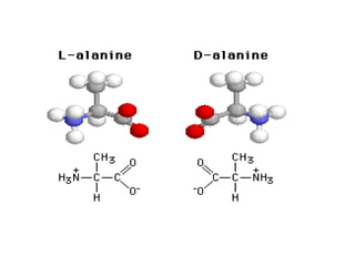
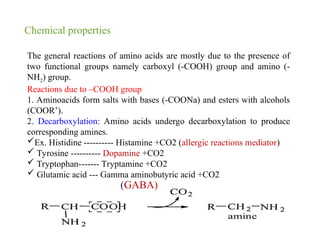
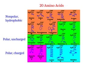
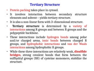
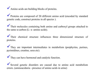
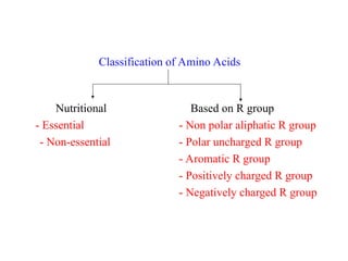
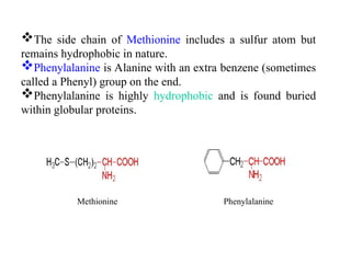
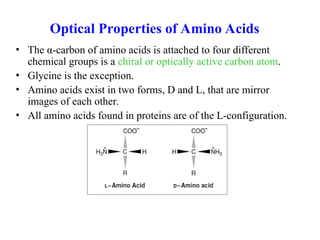
Amino acids are the fundamental building blocks of proteins, characterized by their unique structures involving amino and carboxyl groups attached to a central carbon. They play crucial roles in various biological functions including enzymatic activity and structural support, and are classified based on their side chains into categories such as essential, non-essential, polar, and non-polar amino acids. The document also delves into the chemical properties, classifications, and behavior of amino acids in physiological conditions, highlighting their importance in metabolism and genetic disorders.

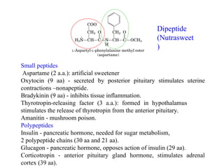
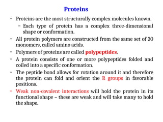
![The Henderson hasselbalch equation for acid is :-
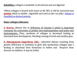
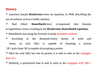
pH = pKa + log [ Aˉ ]
[HA]
Here, pKa= -log(Ka)
where Ka is the acid dissociation constant, that is
pKa= -log [H3O+
][A-
]
[HA]
for the non-specific Brønsted acid-base reaction:
HA + H20 A-
+ H3O+
( Acid ) ( Conjugate base )](https://image.slidesharecdn.com/aminoacidsandproperties-240811181322-eaf1e3ae/85/Aminoacids-properties-and-classification-32-320.jpg)
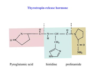
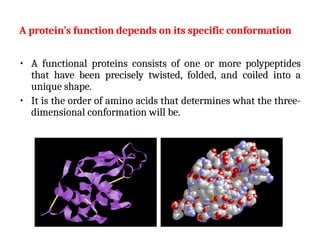
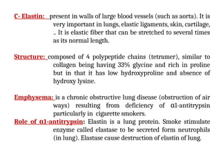
![The Henderson Hasselbalch Equation for base is :
pOH = pKb + log [ BH+
]
-*[B]
where BH+
denotes the conjugate acid of the corresponding
base B.
B + H2O+
BH + OH-
(Base ) (Conjugate acid)](https://image.slidesharecdn.com/aminoacidsandproperties-240811181322-eaf1e3ae/85/Aminoacids-properties-and-classification-33-320.jpg)
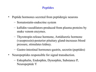
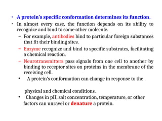
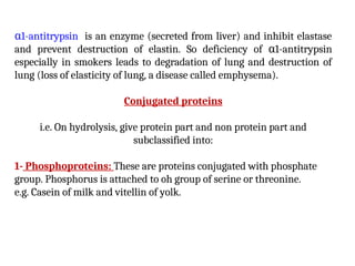
![The dissociation of an acid can be described by an
equilibrium expression
HA + H2O H3O+
+ A-
Consider the case of acetic acid (CH3COOH) and acetate anion
.
(CH3COO-
): CH3COOH + H2O CH3COO-
+ H3O+
Acetate is the conjugate base of acetic acid. Acetic acid and
acetate are a conjugate acid/base pair. We can describe this
relationship with an equilibrium constant:
Ka = [H3O+
][A-
]
[HA]](https://image.slidesharecdn.com/aminoacidsandproperties-240811181322-eaf1e3ae/85/Aminoacids-properties-and-classification-35-320.jpg)
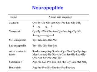
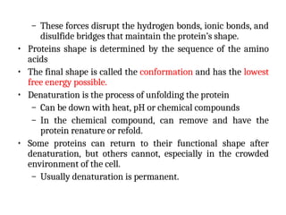
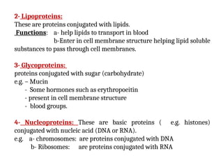
![Taking the negative log of both sides of the equation gives
-logKa = -log [H3O+
][A-
]
[HA]
or, -logKa = -log [H3O+
] + (-log [A-
] )
[HA]
By definition,
pKa = -logKa and pH = -log[H3O+], so
pka=pH – log [A-]
[HA]
This equation can then be rearranged to give the
Henderson-Hasselbalch equation:
pH = pKa + log [A-] = pKa + log [conjugate base]
[HA] [acid]](https://image.slidesharecdn.com/aminoacidsandproperties-240811181322-eaf1e3ae/85/Aminoacids-properties-and-classification-36-320.jpg)