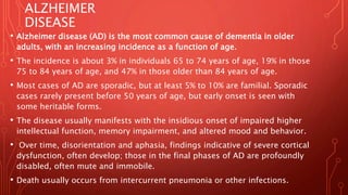
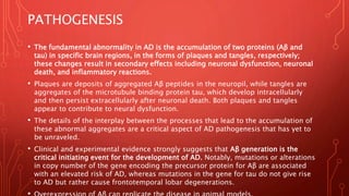
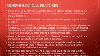
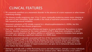
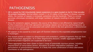
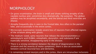
ALZHEIMER DISEASE
PARKINSON DISEASE
PRION DISEASE
• Neurodegenerative diseases are characterized by the progressive loss of
neurons, typically affecting groups of neurons with functional
interconnections.
Different diseases tend to involve particular neural systems and therefore
have relatively stereotypic presenting signs and symptoms.
• • Diseases that involve the hippocampus and associated cortices present
with cognitive changes, often including disturbances of memory, behavior,
and language.
With time these progress to dementia, as occurs with Alzheimer disease.
• • Diseases that affect the basal ganglia manifest as movement disorders;
these may be hypokinetic, as with Parkinson disease, or hyperkinetic, as
with Huntington disease.
• • Diseases that affect the cerebellum or its input and output circuitry result
in ataxia, as seen in the spinocerebellar ataxias.
• A pathologic process shared by most neurodegenerative diseases is
the accumulation of protein aggregates, which serve as histologic
hallmarks of specific disorders.
• Aggregates may arise because of mutations that alter the protein’s
conformation or that disrupt pathways involved in processing or
clearance of the proteins.
• In other situations, there may be a subtle imbalance between protein
synthesis and clearance (due to genetic, environmental, or stochastic
factors) that allows gradual accumulation of proteins.
• The aggregates often are resistant to degradation by normal cellular
proteases, accumulate within cells, elicit an inflammatory response,
and may be directly toxic to neurons. As is evident from the same
proteins may be present as aggregates in multiple diseases.
• The clinical phenotype of the neurodegenerative disease is determined
more by the distribution of the aggregates than by the nature of the
aggregating protein.
• Two other features are common to many neurodegenerative diseases:
• • There is experimental evidence that many of the protein aggregates
that accumulate in neurons affected in these diseases appear to be
capable of spreading to healthy neurons. Thus, aggregates can seed
the development of more aggregates, and the disease process can
spread, like prions. However, there is no evidence for transmission
from affected to healthy individuals.
• • Activation of the innate immune system is a common feature of
neurodegenerative diseases. The importance of this interaction
between the brain and the immune system has been further
strengthened by the identification of genes that confer risk for
diseases (e.g., TREM2 for Alzheimer disease) that encode components
of immune regulatory pathways.
ALZHEIMER
DISEASE
• Alzheimer disease (AD) is the most common cause of dementia in older
adults, with an increasing incidence as a function of age.
• The incidence is about 3% in individuals 65 to 74 years of age, 19% in those
75 to 84 years of age, and 47% in those older than 84 years of age.
• Most cases of AD are sporadic, but at least 5% to 10% are familial. Sporadic
cases rarely present before 50 years of age, but early onset is seen with
some heritable forms.
• The disease usually manifests with the insidious onset of impaired higher
intellectual function, memory impairment, and altered mood and behavior.
• Over time, disorientation and aphasia, findings indicative of severe cortical
dysfunction, often develop; those in the final phases of AD are profoundly
disabled, often mute and immobile.
• Death usually occurs from intercurrent pneumonia or other infections.
PATHOGENESIS
• The fundamental abnormality in AD is the accumulation of two proteins (Aβ and
tau) in specific brain regions, in the forms of plaques and tangles, respectively;
these changes result in secondary effects including neuronal dysfunction, neuronal
death, and inflammatory reactions.
• Plaques are deposits of aggregated Aβ peptides in the neuropil, while tangles are
aggregates of the microtubule binding protein tau, which develop intracellularly
and then persist extracellularly after neuronal death. Both plaques and tangles
appear to contribute to neural dysfunction.
• The details of the interplay between the processes that lead to the accumulation of
these abnormal aggregates are a critical aspect of AD pathogenesis that has yet to
be unraveled.
• Clinical and experimental evidence strongly suggests that Aβ generation is the
critical initiating event for the development of AD. Notably, mutations or alterations
in copy number of the gene encoding the precursor protein for Aβ are associated
with an elevated risk of AD, whereas mutations in the gene for tau do not give rise
to AD but rather cause frontotemporal lobar degenerations.
• The pathogenesis of AD involves Aβ and Tau deposits, as well as other risk factors
and inflammatory reactions.
• • Role of Aβ. Aβ is created when the transmembrane protein amyloid precursor
protein (APP) is sequentially cleaved by the enzymes β-amyloid–converting enzyme
(BACE) and γ-secretase (Fig. 23.24). APP also can be cleaved by α-secretase and γ-
secretase, liberating a different peptide that is nonpathogenic. Mutations in APP or
in components of γ-secretase (encoded by the presenilin-1 or presenilin-2 gene)
lead to familial AD by increasing the rate at which Aβ, particularly its most
aggregation-prone form, is generated.
• The APP gene is located on chromosome 21, and the risk for AD also is higher in
those with an extra copy of the APP gene, such as patients with trisomy 21 (Down
syndrome) and individuals with small interstitial duplications of APP, presumably
because this too leads to greater Aβ generation.
• Once generated, Aβ is highly prone to aggregation; it first forms small oligomers,
and these eventually propagate into large aggregates and fibrils.
• It is these aggregates that deposit in the brain and are visible as plaques.
• There is evidence that these oligomers decrease the number of synapses present
and alter the function of those that remain so that the cellular processes felt to
underlie learning and memory are disrupted.
• Role of tau. Because neurofibrillary tangles contain the tau protein, there has been much
interest in the role of this protein in AD. Tau is a microtubule-associated protein present in
axons in association with the microtubular network.
• With the development of tangles in AD, tau shifts to a somatic-dendritic distribution,
becomes hyperphosphorylated, and loses the ability to bind to microtubules.
• The formation of tangles is an important component of AD, but the mechanism of tangle
injury to neurons remains poorly understood.
• Two pathways have been suggested;
1) aggregates of tau protein elicit a stress response, which persists and eventually leads to cell
death; and
2), the microtubule stabilizing function of tau protein is lost, leading to neuronal toxicity and
death.
• It has been shown that tau aggregates can be passed across synapses from one neuron to the
next; this may underlie some of the spread of lesions across the brain.
• • Other genetic risk factors. The genetic locus on chromosome 19 that encodes
apolipoprotein E (ApoE) has a strong influence on the risk for developing AD.
• Three alleles have been identified (ε2, ε3, and ε4) based on two amino acid polymorphisms.
The dosage of the ε4 allele increases the risk for AD.
• This ApoE isoform promotes Aβ generation and deposition, although the mechanisms have
not been established.
• Overall, this locus hasbeen estimated to convey about one fourth of the risk for development of
late-onset AD. Genome-wide association studies have identified multiple other loci that
contribute to the risk for AD, but the roles of the encoded proteins in disease pathogenesis are
not established.
• • Role of inflammation. Both genetic and histologic studies have indicated that the innate
immune system responds to Aβ and tau. Deposits of Aβ elicit an inflammatory response from
microglia and astrocytes.
• This response probably assists in the clearance of the aggregated peptide, but also may
stimulate the secretion of mediators that cause neuronal injury over time.
• • Basis for cognitive impairment. Deposits of Aβ and tangles begin to appear in the brain well in
advance of cognitive impairment.
• While there remains disagreement regarding the best correlate of dementia in individuals with
AD, the presence of a large burden of plaques and tangles is strongly associated with severe
cognitive dysfunction.
• The number of neurofibrillary tangles correlates better with the degree of dementia than does
the number of neuritic plaques.
• Biochemical markers that have been correlated with the degree of dementia include amyloid
burden.
MORPHOLOGICAL FEATURES
• Brains involved by AD show a variable degree of cortical atrophy, resulting in a
widening of the cerebral sulci that is most pronounced in the frontal, temporal,
and parietal lobes.
• The atrophy produces a compensatory ventricular enlargement (hydrocephalus
ex vacuo).
• At the microscopic level, AD is diagnosed by the presence of neuritic plaques (an
extracellular lesion) and neurofibrillary tangles (an intracellular lesion) Neuritic
plaques are focal, spherical collections of dilated, tortuous, processes derived
from dystrophic neurites, often around a central amyloid core .
• Neuritic plaques range in size from 20 to 200 μm in diameter; microglial cells
and reactive astrocytes are present at their periphery.
• Plaques can be found in the hippocampus and amygdala as well as in the
neocortex, although there is relative sparing of primary motor and sensory
cortices until late in the disease course.
• The amyloid core contains Aβ . Aβ deposits also can be found that lack the
surrounding neuritic reaction, termed diffuse plaques; these are found in the
• Neurofibrillary tangles are bundles of paired helical filaments visible as
basophilic fibrillary structures in the cytoplasm of the neurons that
displace or encircle the nucleus; tangles can persist after neurons die,
becoming a form of extracellular pathology.
• They are commonly found in cortical neurons, especially in the
entorhinal cortex, as well as in the pyramidal cells of the hippocampus,
the amygdala, the basal forebrain, and the raphe nuclei. A major
component of paired helical filaments is
• hyperphosphorylated tau .
• In individuals harboring autosomal dominant mutations that cause AD,
deposition of Aβ and the formation of tangles precede the emergence of
cognitive impairment by as much as 15 to 20 years.
• For this reason, the current diagnostic criteria consider the burden and
distribution of amyloid deposits, tangles, and neuritic plaques—a
constellation known as Alzheimer disease neuropathologic changes.
• The staging of each of these processes, which has a fairly consistent
pattern across individuals, is then used to assess the likelihood that the
PARKINSON DISEASE
• Parkinson disease (PD) is a neurodegenerative disease marked by a
hypokinetic movement disorder that is caused by loss of dopaminergic
neurons from the substantia nigra.
• Parkinsonism is a clinical syndrome characterized by tremor, rigidity,
bradykinesia, and instability.
• These types of motor disturbances may be seen in a range of diseases that
damage dopaminergic neurons, which project from the substantia nigra to
the striatum and are involved in control of motor activity.
• Parkinsonism can be induced by drugs such as dopamine antagonists or
toxins that selectively injure dopaminergic neurons.
• Among the neurodegenerative diseases, most cases of parkinsonism are
caused by PD, which is associated with characteristic neuronal inclusions
PATHOGENESI
S
• PD is associated with protein accumulation and aggregation, mitochondrial
abnormalities, and neuronal loss in the substantia nigra and elsewhere in the brain.
Based on the genetics of PD, it appears that abnormal protein and organelle
clearance due to defects in autophagy and lysosomal degradation have a pathogenic
role in the disease.
• One clue and diagnostic feature of the disease is the Lewy body, a characteristic
inclusion containing α-synuclein, a protein involved in synaptic transmission.
• While PD in most cases is sporadic, point mutations and duplications of the gene
encoding α-synuclein cause autosomal dominant PD.
• Synuclein aggregates are cleared by autophagy and several mutations associated
with PD are in genes whose products (LRRK2, Parkin, others) all appear to have roles
in endosomal trafficking pathways implicated in autophagy.
• It also has been demonstrated that heterozygosity for the Gaucher disease–causing
mutation in glucocerebrosidase is a risk factor for PD.
• Glucocerebrosidase is a lysosomal enzyme, yet another clue suggesting that
abnormal turnover of cellular constituents somehow sets the stage for the
MORPHOLOGICAL FEATURES
• A typical gross finding at autopsy is pallor of the substantia nigra
and locus ceruleus. Microscopic features include
• loss of the pigmented, catecholaminergic neurons in these
• regions associated with gliosis. Lewy bodies (see Fig. 23.27C)
• may be found in those neurons that remain. These are single or
• multiple, cytoplasmic, eosinophilic, round to elongated inclusions.
• On ultrastructural examination, Lewy bodies consist of fine filaments,
• composed of α-synuclein and other proteins, including
neurofilaments and ubiquitin.
CONT
• The other major histologic finding is the presence of Lewy
neurites, dystrophic neurites that also contain aggregated α-
synuclein. Immunohistochemical staining for α-synuclein
highlights more subtle Lewy bodies and Lewy neurites in many
brain regions outside of the substantia nigra and in non-
dopaminergic neurons, including regions of the medulla, the
pons, the amygdala, and the cerebral cortex.
• Eventually they appear in the subcortical areas and the cerebral
cortex. With involvement of the cerebral cortex, there is typically
dementia present in addition to the movement disorder.
CLINICAL FEATURES
• PD commonly manifests as a movement disorder in the absence of a toxic exposure or other known
underlying etiology.
• The disease usually progresses over 10 to 15 years, eventually producing severe motor slowing to
the point of near immobility. Death usually is the result of aspiration pneumonia or trauma from
falls caused by postural instability.
• Movement symptoms of PD initially respond to L-dihydroxyphenylalanine (L-DOPA), but this
treatment does not slow disease progression.
• Over time, L-DOPA becomes less effective and begins to cause problematic fluctuations in motor
function. Another treatment for the motor symptoms of PD is deep brain stimulation, in which
electrodes are implanted in the globus pallidus or subthalamic nucleus to modulate basal ganglia
circuitry, often allowing a significant reduction in L-DOPA dose.
• While the movement disorder associated with loss of the nigrostriatal dopaminergic pathway is an
important feature of PD, it is clear that the disease has more extensive clinical and pathologic
manifestations. Lesions in the brain stem (in the dorsal motor nucleus of the vagus and in the
reticular formation), in advance of nigral involvement, can give rise to behavioral sleep disorder
often before the motor problems.
• Dementia, typically with a mildly fluctuating course and hallucinations, emerges in many individuals
with PD and is attributable to involvement of the cerebral cortex. When dementia arises within 1
HUNTINGTON DISEASE
• Huntington disease (HD) is an autosomal dominant movement
disorder associated with degeneration of the striatum (caudate and
putamen).
• The disorder is characterized by involuntary jerky movements of all
parts of the body; writhing movements of the extremities are typical.
• The disease is relentlessly progressive, resulting in death after an
average course of about 15 years. Early cognitive symptoms include
forgetfulness and thought and affective disorders, and there may be
a progression to severe dementia. As a part of these early behavioral
changes, HD carries an increased risk for suicide.
PATHOGENESIS
• HD is caused by CAG trinucleotide repeat expansions in a gene located on 4p16.3 that encodes
the protein huntingtin. Normal alleles contain 11 to 34 copies of the repeat; in disease-causing
alleles, the number of repeats is increased, sometimes into the hundreds. There is a strong
genotype-phenotype correlation, with larger numbers of repeats resulting in earlier-onset
disease.
• Once the symptoms appear, however, the course of the illness is not affected by repeat length.
Further expansions of the CAG (glutamine-encoding) repeats occur during spermatogenesis, so
paternal transmission may be associated with earlier onset in the next generation, a phenomenon
referred to as anticipation .
• HD appears to be caused by a toxic gain-of-function related to the expanded polyglutamine tract
in huntingtin .
• The mutant protein is subject to ubiquitination and proteolysis, yielding fragments that can form
large intranuclear aggregates. As in other degenerative diseases, smaller aggregates of the
abnormal protein fragments are suspected to be toxic.
• These aggregates have been shown to have a range of potentially injurious actions, including
sequestration of transcription factors, disruption of protein degradation pathways, and
perturbation of mitochondrial function. It is likely that some combination of these aberrations
contributes to HD pathogenesis.
MORPHOLOGY
• On gross examination, the brain is small and shows striking atrophy of the
caudate nucleus and, sometimes less dramatically, the putamen. The globus
pallidus may be atrophied secondarily, and the lateral and third ventricles are
dilated.
• Atrophy frequently also is seen in the frontal lobe, less often in the parietal
lobe, and occasionally in the entire cortex.
• Microscopic examination reveals severe loss of neurons from affected regions
of the striatum along with gliosis.
• The medium-sized, spiny neurons that release the neurotransmitters γ-
aminobutyric acid (GABA), enkephalin, dynorphin, and substance P are
especially sensitive, disappearing early in the disease.
• There is a strong correlation between the degree of degeneration in the
striatum and the severity of motor symptoms; there is also an association
between cortical neuronal loss and dementia.
• In remaining striatal neurons and in the cortex, there are intranuclear inclusions
PRION
DISEASES
• Prion diseases are a group of infectious diseases in which the causative
agent is an abnormal form of a cellular protein.
• These include sporadic, familial, iatrogenic, and variant forms of
Creutzfeldt-Jakob disease (CJD), as well as animal diseases such as
scrapie in sheep and bovine spongiform encephalopathy in cattle (“mad
cow disease”).
• The causative protein, termed prion protein (PrP), may undergo a
conformational change from its normal shape (PrPc) to an abnormal
conformation called PrPsc (sc for scrapie).
• PrP normally is rich in α-helices, but PrPsc has a high content of β-
sheets, a characteristic that makes it resistant to proteolysis (hence an
alternative term for the pathogenic form, PrPres—i.e., protease-
resistant). More important, when PrPsc physically interacts with PrP
• Over time, this self-amplifying process leads to the accumulation
of a high burden of pathogenic PrPsc molecules in the brain.
• Certain mutations in the gene encoding PrPc (PRNP) accelerate the
rate of spontaneous conformational change; these variants are
associated with early-onset familial forms of prion disease
(familial Creutzfeldt-Jakob disease [fCJD]).
• PrPc also may change its conformation spontaneously (but at an
extremely low rate), accounting for sporadic cases of prion
disease (sporadic Creutzfeldt-Jakob disease [sCJD]).
• Accumulation of PrPsc in neural tissue seems to be the cause of
cell injury, but the mechanisms underlying the cytopathic changes
and eventual neuronal death are still unknown.
CREUTZFELDT-JAKOB DISEASE
(CJD)
• CJD is a rapidly progressive dementing illness, with a typical
duration from first onset of subtle changes in memory and behavior
to death in only 7 months.
• It is sporadic in approximately 85% of cases and has a worldwide
annual incidence of about 1 per million. While commonly affecting
individuals older than 70 years of age, familial forms caused by
mutations in PRNP may present in younger individuals. In keeping
with the infectious nature of PrPsc, there are well-established cases
of iatrogenic transmission by contaminated deep implantation
electrodes and human growth hormone preparations.
MORPHOLOGICAL FEATURES
• The progression to death in CJD usually is so rapid that there is little, if
any, macroscopic evidence of brain atrophy.
• On microscopic examination, the pathognomonic finding is a spongiform
transformation of the cerebral cortex and deep gray matter structures
(caudate, putamen); this multifocal process results in the uneven
formation of small, apparently empty, microscopic vacuoles of varying
sizes within the neuropil (the eosinophilic regions in grey matter that
contain dendrites, axons, and synapses) and sometimes in the perikaryon
of neurons.
• In advanced cases, there is severe neuronal loss, reactive gliosis, and
sometimes expansion of the vacuolated areas into cystlike spaces
(“status spongiosus”). No inflammatory infiltrate is present.
Immunohistochemical staining demonstrates the presence of proteinase
VARIANT CREUTZFELDT-JAKOB
DISEASE
• Starting in 1995, cases of a CJD-like illness appeared in the United Kingdom.
• The neuropathologic findings and molecular features of these new cases were
similar to those of CJD, suggesting a close relationship between the two illnesses,
yet this new disorder differed from typical CJD in several important respects: the
disease affected young adults; behavioral disorders figured prominently in early
disease stages; and the neurologic syndrome progressed somewhat more slowly
than typical CJD.
• Multiple lines of evidence indicate that this new disease, termed variant
Creutzfeldt-Jakob disease (vCJD) is a consequence of exposure to the prion
disease of cattle, called bovine spongiform encephalopathy. Also, there is now
documentation of transmission by blood transfusion.
• This variant form has a similar pathologic appearance to that in other types of
CJD, with spongiform change and absence of inflammation.
• In vCJD, however, there are abundant cortical amyloid plaques, surrounded by the
spongiform change
THANK YOU

Alzheimer disease.pptx

  • 1.
  • 2.
    • Neurodegenerative diseasesare characterized by the progressive loss of neurons, typically affecting groups of neurons with functional interconnections. Different diseases tend to involve particular neural systems and therefore have relatively stereotypic presenting signs and symptoms. • • Diseases that involve the hippocampus and associated cortices present with cognitive changes, often including disturbances of memory, behavior, and language. With time these progress to dementia, as occurs with Alzheimer disease. • • Diseases that affect the basal ganglia manifest as movement disorders; these may be hypokinetic, as with Parkinson disease, or hyperkinetic, as with Huntington disease. • • Diseases that affect the cerebellum or its input and output circuitry result in ataxia, as seen in the spinocerebellar ataxias.
  • 3.
    • A pathologicprocess shared by most neurodegenerative diseases is the accumulation of protein aggregates, which serve as histologic hallmarks of specific disorders. • Aggregates may arise because of mutations that alter the protein’s conformation or that disrupt pathways involved in processing or clearance of the proteins. • In other situations, there may be a subtle imbalance between protein synthesis and clearance (due to genetic, environmental, or stochastic factors) that allows gradual accumulation of proteins. • The aggregates often are resistant to degradation by normal cellular proteases, accumulate within cells, elicit an inflammatory response, and may be directly toxic to neurons. As is evident from the same proteins may be present as aggregates in multiple diseases. • The clinical phenotype of the neurodegenerative disease is determined more by the distribution of the aggregates than by the nature of the aggregating protein.
  • 4.
    • Two otherfeatures are common to many neurodegenerative diseases: • • There is experimental evidence that many of the protein aggregates that accumulate in neurons affected in these diseases appear to be capable of spreading to healthy neurons. Thus, aggregates can seed the development of more aggregates, and the disease process can spread, like prions. However, there is no evidence for transmission from affected to healthy individuals. • • Activation of the innate immune system is a common feature of neurodegenerative diseases. The importance of this interaction between the brain and the immune system has been further strengthened by the identification of genes that confer risk for diseases (e.g., TREM2 for Alzheimer disease) that encode components of immune regulatory pathways.
  • 6.
    ALZHEIMER DISEASE • Alzheimer disease(AD) is the most common cause of dementia in older adults, with an increasing incidence as a function of age. • The incidence is about 3% in individuals 65 to 74 years of age, 19% in those 75 to 84 years of age, and 47% in those older than 84 years of age. • Most cases of AD are sporadic, but at least 5% to 10% are familial. Sporadic cases rarely present before 50 years of age, but early onset is seen with some heritable forms. • The disease usually manifests with the insidious onset of impaired higher intellectual function, memory impairment, and altered mood and behavior. • Over time, disorientation and aphasia, findings indicative of severe cortical dysfunction, often develop; those in the final phases of AD are profoundly disabled, often mute and immobile. • Death usually occurs from intercurrent pneumonia or other infections.
  • 7.
    PATHOGENESIS • The fundamentalabnormality in AD is the accumulation of two proteins (Aβ and tau) in specific brain regions, in the forms of plaques and tangles, respectively; these changes result in secondary effects including neuronal dysfunction, neuronal death, and inflammatory reactions. • Plaques are deposits of aggregated Aβ peptides in the neuropil, while tangles are aggregates of the microtubule binding protein tau, which develop intracellularly and then persist extracellularly after neuronal death. Both plaques and tangles appear to contribute to neural dysfunction. • The details of the interplay between the processes that lead to the accumulation of these abnormal aggregates are a critical aspect of AD pathogenesis that has yet to be unraveled. • Clinical and experimental evidence strongly suggests that Aβ generation is the critical initiating event for the development of AD. Notably, mutations or alterations in copy number of the gene encoding the precursor protein for Aβ are associated with an elevated risk of AD, whereas mutations in the gene for tau do not give rise to AD but rather cause frontotemporal lobar degenerations.
  • 8.
    • The pathogenesisof AD involves Aβ and Tau deposits, as well as other risk factors and inflammatory reactions. • • Role of Aβ. Aβ is created when the transmembrane protein amyloid precursor protein (APP) is sequentially cleaved by the enzymes β-amyloid–converting enzyme (BACE) and γ-secretase (Fig. 23.24). APP also can be cleaved by α-secretase and γ- secretase, liberating a different peptide that is nonpathogenic. Mutations in APP or in components of γ-secretase (encoded by the presenilin-1 or presenilin-2 gene) lead to familial AD by increasing the rate at which Aβ, particularly its most aggregation-prone form, is generated. • The APP gene is located on chromosome 21, and the risk for AD also is higher in those with an extra copy of the APP gene, such as patients with trisomy 21 (Down syndrome) and individuals with small interstitial duplications of APP, presumably because this too leads to greater Aβ generation. • Once generated, Aβ is highly prone to aggregation; it first forms small oligomers, and these eventually propagate into large aggregates and fibrils. • It is these aggregates that deposit in the brain and are visible as plaques. • There is evidence that these oligomers decrease the number of synapses present and alter the function of those that remain so that the cellular processes felt to underlie learning and memory are disrupted.
  • 9.
    • Role oftau. Because neurofibrillary tangles contain the tau protein, there has been much interest in the role of this protein in AD. Tau is a microtubule-associated protein present in axons in association with the microtubular network. • With the development of tangles in AD, tau shifts to a somatic-dendritic distribution, becomes hyperphosphorylated, and loses the ability to bind to microtubules. • The formation of tangles is an important component of AD, but the mechanism of tangle injury to neurons remains poorly understood. • Two pathways have been suggested; 1) aggregates of tau protein elicit a stress response, which persists and eventually leads to cell death; and 2), the microtubule stabilizing function of tau protein is lost, leading to neuronal toxicity and death. • It has been shown that tau aggregates can be passed across synapses from one neuron to the next; this may underlie some of the spread of lesions across the brain. • • Other genetic risk factors. The genetic locus on chromosome 19 that encodes apolipoprotein E (ApoE) has a strong influence on the risk for developing AD. • Three alleles have been identified (ε2, ε3, and ε4) based on two amino acid polymorphisms. The dosage of the ε4 allele increases the risk for AD. • This ApoE isoform promotes Aβ generation and deposition, although the mechanisms have not been established.
  • 10.
    • Overall, thislocus hasbeen estimated to convey about one fourth of the risk for development of late-onset AD. Genome-wide association studies have identified multiple other loci that contribute to the risk for AD, but the roles of the encoded proteins in disease pathogenesis are not established. • • Role of inflammation. Both genetic and histologic studies have indicated that the innate immune system responds to Aβ and tau. Deposits of Aβ elicit an inflammatory response from microglia and astrocytes. • This response probably assists in the clearance of the aggregated peptide, but also may stimulate the secretion of mediators that cause neuronal injury over time. • • Basis for cognitive impairment. Deposits of Aβ and tangles begin to appear in the brain well in advance of cognitive impairment. • While there remains disagreement regarding the best correlate of dementia in individuals with AD, the presence of a large burden of plaques and tangles is strongly associated with severe cognitive dysfunction. • The number of neurofibrillary tangles correlates better with the degree of dementia than does the number of neuritic plaques. • Biochemical markers that have been correlated with the degree of dementia include amyloid burden.
  • 11.
    MORPHOLOGICAL FEATURES • Brainsinvolved by AD show a variable degree of cortical atrophy, resulting in a widening of the cerebral sulci that is most pronounced in the frontal, temporal, and parietal lobes. • The atrophy produces a compensatory ventricular enlargement (hydrocephalus ex vacuo). • At the microscopic level, AD is diagnosed by the presence of neuritic plaques (an extracellular lesion) and neurofibrillary tangles (an intracellular lesion) Neuritic plaques are focal, spherical collections of dilated, tortuous, processes derived from dystrophic neurites, often around a central amyloid core . • Neuritic plaques range in size from 20 to 200 μm in diameter; microglial cells and reactive astrocytes are present at their periphery. • Plaques can be found in the hippocampus and amygdala as well as in the neocortex, although there is relative sparing of primary motor and sensory cortices until late in the disease course. • The amyloid core contains Aβ . Aβ deposits also can be found that lack the surrounding neuritic reaction, termed diffuse plaques; these are found in the
  • 12.
    • Neurofibrillary tanglesare bundles of paired helical filaments visible as basophilic fibrillary structures in the cytoplasm of the neurons that displace or encircle the nucleus; tangles can persist after neurons die, becoming a form of extracellular pathology. • They are commonly found in cortical neurons, especially in the entorhinal cortex, as well as in the pyramidal cells of the hippocampus, the amygdala, the basal forebrain, and the raphe nuclei. A major component of paired helical filaments is • hyperphosphorylated tau . • In individuals harboring autosomal dominant mutations that cause AD, deposition of Aβ and the formation of tangles precede the emergence of cognitive impairment by as much as 15 to 20 years. • For this reason, the current diagnostic criteria consider the burden and distribution of amyloid deposits, tangles, and neuritic plaques—a constellation known as Alzheimer disease neuropathologic changes. • The staging of each of these processes, which has a fairly consistent pattern across individuals, is then used to assess the likelihood that the
  • 13.
    PARKINSON DISEASE • Parkinsondisease (PD) is a neurodegenerative disease marked by a hypokinetic movement disorder that is caused by loss of dopaminergic neurons from the substantia nigra. • Parkinsonism is a clinical syndrome characterized by tremor, rigidity, bradykinesia, and instability. • These types of motor disturbances may be seen in a range of diseases that damage dopaminergic neurons, which project from the substantia nigra to the striatum and are involved in control of motor activity. • Parkinsonism can be induced by drugs such as dopamine antagonists or toxins that selectively injure dopaminergic neurons. • Among the neurodegenerative diseases, most cases of parkinsonism are caused by PD, which is associated with characteristic neuronal inclusions
  • 14.
    PATHOGENESI S • PD isassociated with protein accumulation and aggregation, mitochondrial abnormalities, and neuronal loss in the substantia nigra and elsewhere in the brain. Based on the genetics of PD, it appears that abnormal protein and organelle clearance due to defects in autophagy and lysosomal degradation have a pathogenic role in the disease. • One clue and diagnostic feature of the disease is the Lewy body, a characteristic inclusion containing α-synuclein, a protein involved in synaptic transmission. • While PD in most cases is sporadic, point mutations and duplications of the gene encoding α-synuclein cause autosomal dominant PD. • Synuclein aggregates are cleared by autophagy and several mutations associated with PD are in genes whose products (LRRK2, Parkin, others) all appear to have roles in endosomal trafficking pathways implicated in autophagy. • It also has been demonstrated that heterozygosity for the Gaucher disease–causing mutation in glucocerebrosidase is a risk factor for PD. • Glucocerebrosidase is a lysosomal enzyme, yet another clue suggesting that abnormal turnover of cellular constituents somehow sets the stage for the
  • 15.
    MORPHOLOGICAL FEATURES • Atypical gross finding at autopsy is pallor of the substantia nigra and locus ceruleus. Microscopic features include • loss of the pigmented, catecholaminergic neurons in these • regions associated with gliosis. Lewy bodies (see Fig. 23.27C) • may be found in those neurons that remain. These are single or • multiple, cytoplasmic, eosinophilic, round to elongated inclusions. • On ultrastructural examination, Lewy bodies consist of fine filaments, • composed of α-synuclein and other proteins, including neurofilaments and ubiquitin.
  • 16.
    CONT • The othermajor histologic finding is the presence of Lewy neurites, dystrophic neurites that also contain aggregated α- synuclein. Immunohistochemical staining for α-synuclein highlights more subtle Lewy bodies and Lewy neurites in many brain regions outside of the substantia nigra and in non- dopaminergic neurons, including regions of the medulla, the pons, the amygdala, and the cerebral cortex. • Eventually they appear in the subcortical areas and the cerebral cortex. With involvement of the cerebral cortex, there is typically dementia present in addition to the movement disorder.
  • 17.
    CLINICAL FEATURES • PDcommonly manifests as a movement disorder in the absence of a toxic exposure or other known underlying etiology. • The disease usually progresses over 10 to 15 years, eventually producing severe motor slowing to the point of near immobility. Death usually is the result of aspiration pneumonia or trauma from falls caused by postural instability. • Movement symptoms of PD initially respond to L-dihydroxyphenylalanine (L-DOPA), but this treatment does not slow disease progression. • Over time, L-DOPA becomes less effective and begins to cause problematic fluctuations in motor function. Another treatment for the motor symptoms of PD is deep brain stimulation, in which electrodes are implanted in the globus pallidus or subthalamic nucleus to modulate basal ganglia circuitry, often allowing a significant reduction in L-DOPA dose. • While the movement disorder associated with loss of the nigrostriatal dopaminergic pathway is an important feature of PD, it is clear that the disease has more extensive clinical and pathologic manifestations. Lesions in the brain stem (in the dorsal motor nucleus of the vagus and in the reticular formation), in advance of nigral involvement, can give rise to behavioral sleep disorder often before the motor problems. • Dementia, typically with a mildly fluctuating course and hallucinations, emerges in many individuals with PD and is attributable to involvement of the cerebral cortex. When dementia arises within 1
  • 18.
    HUNTINGTON DISEASE • Huntingtondisease (HD) is an autosomal dominant movement disorder associated with degeneration of the striatum (caudate and putamen). • The disorder is characterized by involuntary jerky movements of all parts of the body; writhing movements of the extremities are typical. • The disease is relentlessly progressive, resulting in death after an average course of about 15 years. Early cognitive symptoms include forgetfulness and thought and affective disorders, and there may be a progression to severe dementia. As a part of these early behavioral changes, HD carries an increased risk for suicide.
  • 19.
    PATHOGENESIS • HD iscaused by CAG trinucleotide repeat expansions in a gene located on 4p16.3 that encodes the protein huntingtin. Normal alleles contain 11 to 34 copies of the repeat; in disease-causing alleles, the number of repeats is increased, sometimes into the hundreds. There is a strong genotype-phenotype correlation, with larger numbers of repeats resulting in earlier-onset disease. • Once the symptoms appear, however, the course of the illness is not affected by repeat length. Further expansions of the CAG (glutamine-encoding) repeats occur during spermatogenesis, so paternal transmission may be associated with earlier onset in the next generation, a phenomenon referred to as anticipation . • HD appears to be caused by a toxic gain-of-function related to the expanded polyglutamine tract in huntingtin . • The mutant protein is subject to ubiquitination and proteolysis, yielding fragments that can form large intranuclear aggregates. As in other degenerative diseases, smaller aggregates of the abnormal protein fragments are suspected to be toxic. • These aggregates have been shown to have a range of potentially injurious actions, including sequestration of transcription factors, disruption of protein degradation pathways, and perturbation of mitochondrial function. It is likely that some combination of these aberrations contributes to HD pathogenesis.
  • 20.
    MORPHOLOGY • On grossexamination, the brain is small and shows striking atrophy of the caudate nucleus and, sometimes less dramatically, the putamen. The globus pallidus may be atrophied secondarily, and the lateral and third ventricles are dilated. • Atrophy frequently also is seen in the frontal lobe, less often in the parietal lobe, and occasionally in the entire cortex. • Microscopic examination reveals severe loss of neurons from affected regions of the striatum along with gliosis. • The medium-sized, spiny neurons that release the neurotransmitters γ- aminobutyric acid (GABA), enkephalin, dynorphin, and substance P are especially sensitive, disappearing early in the disease. • There is a strong correlation between the degree of degeneration in the striatum and the severity of motor symptoms; there is also an association between cortical neuronal loss and dementia. • In remaining striatal neurons and in the cortex, there are intranuclear inclusions
  • 21.
    PRION DISEASES • Prion diseasesare a group of infectious diseases in which the causative agent is an abnormal form of a cellular protein. • These include sporadic, familial, iatrogenic, and variant forms of Creutzfeldt-Jakob disease (CJD), as well as animal diseases such as scrapie in sheep and bovine spongiform encephalopathy in cattle (“mad cow disease”). • The causative protein, termed prion protein (PrP), may undergo a conformational change from its normal shape (PrPc) to an abnormal conformation called PrPsc (sc for scrapie). • PrP normally is rich in α-helices, but PrPsc has a high content of β- sheets, a characteristic that makes it resistant to proteolysis (hence an alternative term for the pathogenic form, PrPres—i.e., protease- resistant). More important, when PrPsc physically interacts with PrP
  • 22.
    • Over time,this self-amplifying process leads to the accumulation of a high burden of pathogenic PrPsc molecules in the brain. • Certain mutations in the gene encoding PrPc (PRNP) accelerate the rate of spontaneous conformational change; these variants are associated with early-onset familial forms of prion disease (familial Creutzfeldt-Jakob disease [fCJD]). • PrPc also may change its conformation spontaneously (but at an extremely low rate), accounting for sporadic cases of prion disease (sporadic Creutzfeldt-Jakob disease [sCJD]). • Accumulation of PrPsc in neural tissue seems to be the cause of cell injury, but the mechanisms underlying the cytopathic changes and eventual neuronal death are still unknown.
  • 23.
    CREUTZFELDT-JAKOB DISEASE (CJD) • CJDis a rapidly progressive dementing illness, with a typical duration from first onset of subtle changes in memory and behavior to death in only 7 months. • It is sporadic in approximately 85% of cases and has a worldwide annual incidence of about 1 per million. While commonly affecting individuals older than 70 years of age, familial forms caused by mutations in PRNP may present in younger individuals. In keeping with the infectious nature of PrPsc, there are well-established cases of iatrogenic transmission by contaminated deep implantation electrodes and human growth hormone preparations.
  • 24.
    MORPHOLOGICAL FEATURES • Theprogression to death in CJD usually is so rapid that there is little, if any, macroscopic evidence of brain atrophy. • On microscopic examination, the pathognomonic finding is a spongiform transformation of the cerebral cortex and deep gray matter structures (caudate, putamen); this multifocal process results in the uneven formation of small, apparently empty, microscopic vacuoles of varying sizes within the neuropil (the eosinophilic regions in grey matter that contain dendrites, axons, and synapses) and sometimes in the perikaryon of neurons. • In advanced cases, there is severe neuronal loss, reactive gliosis, and sometimes expansion of the vacuolated areas into cystlike spaces (“status spongiosus”). No inflammatory infiltrate is present. Immunohistochemical staining demonstrates the presence of proteinase
  • 25.
    VARIANT CREUTZFELDT-JAKOB DISEASE • Startingin 1995, cases of a CJD-like illness appeared in the United Kingdom. • The neuropathologic findings and molecular features of these new cases were similar to those of CJD, suggesting a close relationship between the two illnesses, yet this new disorder differed from typical CJD in several important respects: the disease affected young adults; behavioral disorders figured prominently in early disease stages; and the neurologic syndrome progressed somewhat more slowly than typical CJD. • Multiple lines of evidence indicate that this new disease, termed variant Creutzfeldt-Jakob disease (vCJD) is a consequence of exposure to the prion disease of cattle, called bovine spongiform encephalopathy. Also, there is now documentation of transmission by blood transfusion. • This variant form has a similar pathologic appearance to that in other types of CJD, with spongiform change and absence of inflammation. • In vCJD, however, there are abundant cortical amyloid plaques, surrounded by the spongiform change
  • 26.