Downloaded 33 times

![at seven different wavelengths [300 nm, 415 nm, 455 nm, CSS
(Crime Scene Search), 515 nm, 535 nm, 555 nm] while wearing
three different filtering goggles (red, orange, yellow). Areas of
fluorescence were documented independently (i.e., post-trauma
alternate light body maps) for days 1, 7, and 14.
Identification and Location of Skin Lesions
The dimensions of all findings were measured using a stan-
dard metric measuring tape. On the white light examination, the
investigator labeled all findings that were identifiable (i.e., hem-
angioma, scar, bruise, birthmark, tattoo). On the alternate light
source examination, the investigators measured the width and
length of each area of fluorescence. The location of all skin
lesions was documented using the standard metric measuring
tape by measuring the distances of each lesion from the most
distal antecubital fossa crease and from the most proximal wrist
crease. This technique was used to locate all lesions under white
light and all areas of fluorescence under ALS. Findings were
recorded on separate but identical body maps as described
above. Upon completion, each body map was placed in a sepa-
rate prelabeled envelope with the corresponding study subject
number on it. Bruises were not reapplied at any time during the
study period. New body maps were used for all examinations, so
all investigators remained blinded to prior findings including
their own.
Definitions
Positive Fluorescence—An area on the ventral surface of the
arm whereby fluorescence was identified by study examiners
under ALS. This includes true-positive subclinical bruises and
false positives (substances or objects that fluoresce that are
not bruises).
Subclinical Bruise—A case forearm that fluoresced under ALS
yet had no visualized bruise under the white light exam con-
ducted on the same day. This includes only true positives.
True-positive subclinical bruise—a subclinical bruise seen under
ALS within 0.5 cm of the inflicted trauma by both observers,
but not under white light in the same location.
False-positive subclinical bruise—an area of positive fluores-
cence seen under ALS by both observers, but not under white
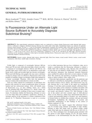
TABLE 1––Performance measures of alternate light source vs. visible white
light in examining subclinical bruises. (N = 118)
Day 1 Day 7 Day 14
(95% CI) (95% CI) (95% CI)
Alternative light source
Sensitivity 76.8%
(63.6, 87.0)
69.6%
(55.9, 81.2)
60.7%
(46.8, 73.5)
Specificity 51.6%
(38.6, 64.5)
59.7%
(46.5, 72.0)
53.2%
(40.1, 66.0)
Positive predictive
value
58.9%
(46.8, 70.3)
60.9%
(47.9, 72.9)
54.0%
(40.9, 66.6)
Negative predictive
value
71.1%
(55.7, 83.6)
68.5%
(54.5, 80.5)
60.0%
(45.9, 73.0)
Visible white light
Sensitivity 69.6%
(55.9, 81.2)
60.0%
(45.9%, 73.0%)
32.1%
(20.0%, 46.3%)
Specificity 71.0%
(58.1, 81.8)
81.4%
(69.1, 90.3)
86.9%
(75.8, 94.2)
Positive predictive
value
68.4%
(54.8, 80.1)
75.0%
(59.7, 86.8)
68.0%
(46.5, 85.1)
Negative Predictive
Value
72.1%
(59.2, 82.9)
68.6%
(56.4, 79.2)
59.6%
(48.6, 70.0)
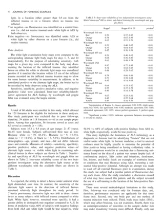
TABLE 2––Sensitivity and specificity of Mini-Crimescopeâ
400 in detecting subclinical bruising (by wavelength and goggle filter)
Day 1 (%) Day 7 (%) Day 14 (%)
Sensitivity Specificity Sensitivity Specificity Sensitivity Specificity
Wavelength 300 nm
Red 75.0 51.6 69.6 61.3 51.8 54.8
Orange 76.8 51.6 69.6 61.3 53.8 54.8
Yellow 76.8 51.6 67.9 64.5 50.0 53.2
Wavelength 415 nm
Red 75.0 51.6 69.6 59.7 57.1 54.8
Orange 76.8 51.6 69.6 59.7 57.1 54.8
Yellow 76.8 51.6 67.9 62.9 57.8 53.2
Wavelength 455 nm
Red 75.0 51.6 69.6 59.7 57.1 54.8
Orange 76.8 51.6 69.6 59.7 57.1 54.8
Yellow 76.8 51.6 67.9 62.9 51.8 53.2
Wavelength CSS
Red 75.0 51.6 69.6 61.3 58.9 54.8
Orange 76.8 51.6 67.9 61.3 50.0 56.5
Yellow 39.3 79.0 26.8 87.1 8.9 87.1
Wavelength 515 nm
Red 75.0 51.6 67.9 61.3 58.9 54.8
Orange 73.2 54.8 64.3 69.4 42.9 61.3
Yellow 23.2 83.9 16.1 95.2 5.4 95.2
Wavelength 535 nm
Red 75.0 51.6 67.9 62.9 57.1 54.8
Orange 58.9 74.2 33.9 82.3 25.0 74.2
Yellow 19.6 90.3 8.9 98.4 3.6 96.8
Wavelength 555 nm
Red 73.2 51.6 67.9 62.9 57.1 45.2
Orange 37.5 77.4 26.8 90.3 19.6 75.8
Yellow 17.9 90.3 8.9 98.4 3.6 96.8
Bolded areas represent filters and wavelengths indicated by the manufacturer to be optimal in the diagnosis of subclinical bruising at different stages.
LOMBARDI ET AL. . ALS TO DIAGNOSE SUBCLINICAL BRUISING 3](https://image.slidesharecdn.com/alsarticle-150314100335-conversion-gate01/85/Alternate-Light-Source-Usage-to-Detect-Bruising-Is-This-Evidence-Based-Practice-3-320.jpg)


This study evaluated the effectiveness of fluorescence under alternate light sources (ALS) for diagnosing subclinical bruising on randomly selected forearms. While ALS showed higher sensitivity than white light in detecting these bruises, its low specificity raises concerns about false positives, suggesting it is not a reliable standalone diagnostic tool for subclinical bruising. Evidence indicates that without further confirmation through chemical analysis, the interpretation of ALS fluorescence may lead to over-interpretation in forensic settings.


