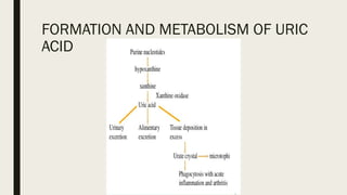
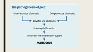
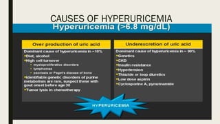
Acute Gout
SLO
■ To describe:
■ aetiology
■ clinical features
■ investigations
■ treatment of;
■ Acute Gout
Introduction:
■ Gout is characterized by intermittent painful inflammatory joint attacks, resulting
from crystallization of excessive uric acid because of hyperurecimia.
■ Aetiology: Gout is increasingly common among older individuals. Approximately 13%
of patients over the age of 80 years are affected.
■ Gout is typically associated with multiple comorbidities (hypertension, obesity,
diabetes mellitus, chronic kidney disease, vascular disease, dyslipidemia, and
nephrolithiasis) that may complicate treatment.
Pathology:
■ Uric acid is the end product of purine metabolism in humans. Free purines arise
from nucleic acid breakdown during cell turnover, from adenosine triphosphate
metabolism, and through dietary intake. Xanthineoxidase is the terminal enzyme in
human uric acid synthesis. In contrast to most mammals, humans lack the enzyme
uricase, which processes urate to the more soluble allantoin .
■ At physiologic conditions (pH, 7.4), uric acid exists primarily as its ionized form,
urate; therefore, hyperuricemia technically refers to elevated serum urate. The
saturation concentration of urate is 6.8 mg/dL , with monosodium urate crystals
unlikely to form at concentrations below this level; hyperuricemia is therefore
defined based on this physiologic parameter. Urate is cleared by the kidneys via
both glomerular filtration and active secretion in PCT.
Pathology
■ Lesch–Nyhan syndrome (LNS), is a rare inherited disorder caused by a deficiency of
the enzyme hypoxanthine-guanine phosphoribosyltransferase(HGPRT), produced by
mutations in the HPRT gene located on the X chromosome. The HGPRT deficiency
causes a build-up of uric acid in all body fluids. The combination of increased synthesis
and decreased utilization of purines leads to high levels of uric acid production. This
results in both hyperuricemia and hyperuricosuria, associated with severe gout.
■ Neurological signs include poor muscle control and moderate intellectual disability.
These complications usually appear in the first year of life. Beginning in the second
year of life, a particularly striking feature of LNS is self-mutilating behaviors,
characterized by lip and finger biting.
FORMATION AND METABOLISM OF URIC
ACID
CAUSES OF HYPERURICEMIA
CLINICAL FEATURES
SYMPTOMS OF GOUT:-
■ Podagra (initial joint manifestation in 50% of gout cases and eventually involved in
90%). The metatarsophaleangeal joint of the great toe is the commonest site.
■ Arthritis in other sites – In gout, the instep, ankle, wrist, finger joints, and knee.
■ Monoarticular involvement most commonly, though polyarticular acute flares are
not rare, and many different joints may be involved simultaneously or in rapid
succession
■ In gout, attacks that begin abruptly and typically reach maximum intensity within 8-
12 hours.
PHYSICAL FINDINGS MAY INCLUDE THE FOLLOWING:-
■ Involvement of a single (most common) or multiple joints
■ Signs of inflammation – Swelling, warmth, erythema (sometimes resembling
cellulitis), and tenderness
■ Fever (also consider infectious arthritis)
■ Tophi in soft tissues (helix of the ear, fingers, toes, prepatellar bursa, olecranon) are
seen in chronic gout.
■ Eye involvement – Tophi, crystal-containing conjunctival nodules, band keratopathy,
blurred vision, anterior uveitis (rare), scleritis
PODAGRA
COMPLICATIONS OF GOUT:-
■ Severe degenerative arthritis
■ Secondary infections
■ Urate or uric acid nephropathy
■ Increased susceptibility to infection
■ Urate nephropathy
■ Renal stones
■ Nerve or spinal cord impingement
■ Fractures in joints with tophaceous gout
INVESTIGATIONS
SYNOVIAL FLUID ASPIRATE:-
■ A drop of SF should be examined promptly under routine light and polarizing light microscopy.
SF is best examined while fresh. If MSU crystals cannot be identified on the wet preparation
after 5 to 10 minutes, the remaining SF should be centrifuged and the pellet examined.
■ MSU (Mono sodium urate) crystals are needle-shaped and approximately 2 to 20 mm long. They
exhibit strong negative birefringence under polarized light. They appear yellow when they are
parallel to the axis of polarized light and blue when lying perpendicular to the same axis.
■
■ Synovial fluid leukocyte count should also be measured: gout fluid is typically inflammatory
(2000'μL to >100,000 μL with neutrophils >50%.
MSU crystals under polarizing
microscopy.
■ Serum uric acid measurement (though hyperuricemia is not diagnostic of gout) a
value of > 6.8 mg/dL is abnormal.
■ 24-hour urinary uric acid evaluation
■ Blood studies (including white blood cells [WBCs, triglyceride, high-density
lipoprotein, glucose, and renal and liver function tests)
■
Investigations:
X RAY:-
■ Typical well-defined, "punched out," periarticular erosions with overhanging edges are not seen
radiographically until 6 to 12 years after the initial acute attack.
■ Magnetic Resonance Imaging (MRI). MRI is a useful method of determining the extent of disease in
tophaceous gout and may provide information regarding the patterns of deposition and spread of MSU
crystals.
■ Ultrasonography is a more reliable, noninvasive method for diagnosing gout.Ultrasonographic investigation
can detect deposition of MSU crystals on cartilaginous surfaces, as well as tophaceous material and
typical erosions.
TREATMENT
■ NSAIDS: Commonly used NSAIDs during an acute gout attack include ibuprofen 800 mg three to four
times daily or indomethacin 25 to 50 mg four times daily. Treatment should be discontinued when
symptoms resolve.
■ Colchicine: Intravenous colchicine is associated with serious toxicities and side effects, so it should be
used as an oral formulation only.
■ High dose oral colchicine (1.2 mg followed by 0.6 mg every hour for 6 doses) is generally poorly tolerated
because of GI side effects. Lower doses are much better received and may be used in combination with
NSAIDs.
■ Corticosteroids: In patients with contraindications to NSAID use, corticosteroids are the next choice.
Corticosteroids can be administered as an injection into the effected joint (intra-articular steroids) or
given systemically (orally, such as prednisone). Intra-articular steriods are useful if only one or two joints
are affected and the treating physician is proficient in injecting those joints.
■ Oral corticosteroids can be used starting at 30-40 mg daily tapering over 10-14 days.
URICOSURIC DRUGS
■ Probenecid may be given to patients with decreased clearance of uric acid by the kidney and normal renal
function. In general its use should be limited to patients under the age of 60.
■ Probenecid acts by inhibiting reabsorption of uric acid in the proximal tubules of the kidney.
■ Starting dose is at 500 mg to 1000 mg daily and increased to 1500 mg to 2000 mg as needed.
■ Probenecid may precipitate renal stone formation and good oral hydration should be encouraged.
■ Probenecid is contraindicated in patients with renal stones (including calcium and uric acid stones) and in
patients with urate nephropathy.
XANTHINE OXIDASE INHIBITOR
■ Allopurinol is a Xanthine oxidase inhibitor. It is well tolerated, inexpensive, and commonly used uric acid
lowering agent.
■
■ Allopurinol can be started at doses as low as 100 mg daily (100 mg qod if creatinine clearance < 10 cc/min)
and titrated by 100 mg every 10-14 days to achieve a serum uric acid level of 4-5 mg/dl.
■
■ Liver tests, blood counts, and renal function and should be monitored while on therapy.
■
■ Toxicites include rash, hepatoxicity, bone marrow suppression and severe hypersensitivity reactions.
■
■ Medication interactions can occur with allopurinol, warfarin, and theophylline and levels should be monitored.
Febuxostat
 Febuxostat is a xanthine oxidase inhibitor approved for the treatment of hyperuricemia in gout.
 It has demonstrated a dose-dependent decrease in serum uric acid (daily doses 40-80 mg/day).
 Its efficacy has been demonstrated in patients with mild or moderate renal impairment and gout.
 It can cause abnormalities in liver function tests and routine monitoring of bloodwork is recommended.
 Similar to allopurinol, there are interactions of febuxostat with azathioprine, 6MP, and theophylline.
PEGLOTICASE
■ Uricase is an enzyme that converts poorly soluable urate (uric acid) to the more soluable
allantoin (excreted in the urine). Uricase is present in most mammals, so mammals with
uricase do not develop gout.
■ However, humans and some primates lack uricase (because of evoluationary gene inactivation)
and lack the ability to make uric acid more soluable and hence, have gout.
■ Pegloticase is a porcine uricase which is approved for the treatment of gout in patients who
have failed conventional therapy.
■ Pegloticase is administered by intravenous infusion every 2 weeks.
■ Patients should be treated prophylactically for allergic reactions to the infusion with steroids
and anti-histamines and monitored closely for the development of an infusion reaction.
■ Caution should be used in prescribing this treatment in patients with a known cardiac history.
■ Thank you

Acute Gout Pathophysiology and Management

  • 1.
  • 2.
    SLO ■ To describe: ■aetiology ■ clinical features ■ investigations ■ treatment of; ■ Acute Gout
  • 3.
    Introduction: ■ Gout ischaracterized by intermittent painful inflammatory joint attacks, resulting from crystallization of excessive uric acid because of hyperurecimia. ■ Aetiology: Gout is increasingly common among older individuals. Approximately 13% of patients over the age of 80 years are affected. ■ Gout is typically associated with multiple comorbidities (hypertension, obesity, diabetes mellitus, chronic kidney disease, vascular disease, dyslipidemia, and nephrolithiasis) that may complicate treatment.
  • 4.
    Pathology: ■ Uric acidis the end product of purine metabolism in humans. Free purines arise from nucleic acid breakdown during cell turnover, from adenosine triphosphate metabolism, and through dietary intake. Xanthineoxidase is the terminal enzyme in human uric acid synthesis. In contrast to most mammals, humans lack the enzyme uricase, which processes urate to the more soluble allantoin . ■ At physiologic conditions (pH, 7.4), uric acid exists primarily as its ionized form, urate; therefore, hyperuricemia technically refers to elevated serum urate. The saturation concentration of urate is 6.8 mg/dL , with monosodium urate crystals unlikely to form at concentrations below this level; hyperuricemia is therefore defined based on this physiologic parameter. Urate is cleared by the kidneys via both glomerular filtration and active secretion in PCT.
  • 5.
    Pathology ■ Lesch–Nyhan syndrome(LNS), is a rare inherited disorder caused by a deficiency of the enzyme hypoxanthine-guanine phosphoribosyltransferase(HGPRT), produced by mutations in the HPRT gene located on the X chromosome. The HGPRT deficiency causes a build-up of uric acid in all body fluids. The combination of increased synthesis and decreased utilization of purines leads to high levels of uric acid production. This results in both hyperuricemia and hyperuricosuria, associated with severe gout. ■ Neurological signs include poor muscle control and moderate intellectual disability. These complications usually appear in the first year of life. Beginning in the second year of life, a particularly striking feature of LNS is self-mutilating behaviors, characterized by lip and finger biting.
  • 6.
  • 8.
  • 9.
    CLINICAL FEATURES SYMPTOMS OFGOUT:- ■ Podagra (initial joint manifestation in 50% of gout cases and eventually involved in 90%). The metatarsophaleangeal joint of the great toe is the commonest site. ■ Arthritis in other sites – In gout, the instep, ankle, wrist, finger joints, and knee. ■ Monoarticular involvement most commonly, though polyarticular acute flares are not rare, and many different joints may be involved simultaneously or in rapid succession ■ In gout, attacks that begin abruptly and typically reach maximum intensity within 8- 12 hours.
  • 10.
    PHYSICAL FINDINGS MAYINCLUDE THE FOLLOWING:- ■ Involvement of a single (most common) or multiple joints ■ Signs of inflammation – Swelling, warmth, erythema (sometimes resembling cellulitis), and tenderness ■ Fever (also consider infectious arthritis) ■ Tophi in soft tissues (helix of the ear, fingers, toes, prepatellar bursa, olecranon) are seen in chronic gout. ■ Eye involvement – Tophi, crystal-containing conjunctival nodules, band keratopathy, blurred vision, anterior uveitis (rare), scleritis
  • 11.
  • 12.
    COMPLICATIONS OF GOUT:- ■Severe degenerative arthritis ■ Secondary infections ■ Urate or uric acid nephropathy ■ Increased susceptibility to infection ■ Urate nephropathy ■ Renal stones ■ Nerve or spinal cord impingement ■ Fractures in joints with tophaceous gout
  • 13.
    INVESTIGATIONS SYNOVIAL FLUID ASPIRATE:- ■A drop of SF should be examined promptly under routine light and polarizing light microscopy. SF is best examined while fresh. If MSU crystals cannot be identified on the wet preparation after 5 to 10 minutes, the remaining SF should be centrifuged and the pellet examined. ■ MSU (Mono sodium urate) crystals are needle-shaped and approximately 2 to 20 mm long. They exhibit strong negative birefringence under polarized light. They appear yellow when they are parallel to the axis of polarized light and blue when lying perpendicular to the same axis. ■ ■ Synovial fluid leukocyte count should also be measured: gout fluid is typically inflammatory (2000'μL to >100,000 μL with neutrophils >50%.
  • 14.
    MSU crystals underpolarizing microscopy. ■ Serum uric acid measurement (though hyperuricemia is not diagnostic of gout) a value of > 6.8 mg/dL is abnormal. ■ 24-hour urinary uric acid evaluation ■ Blood studies (including white blood cells [WBCs, triglyceride, high-density lipoprotein, glucose, and renal and liver function tests) ■
  • 15.
    Investigations: X RAY:- ■ Typicalwell-defined, "punched out," periarticular erosions with overhanging edges are not seen radiographically until 6 to 12 years after the initial acute attack. ■ Magnetic Resonance Imaging (MRI). MRI is a useful method of determining the extent of disease in tophaceous gout and may provide information regarding the patterns of deposition and spread of MSU crystals. ■ Ultrasonography is a more reliable, noninvasive method for diagnosing gout.Ultrasonographic investigation can detect deposition of MSU crystals on cartilaginous surfaces, as well as tophaceous material and typical erosions.
  • 17.
    TREATMENT ■ NSAIDS: Commonlyused NSAIDs during an acute gout attack include ibuprofen 800 mg three to four times daily or indomethacin 25 to 50 mg four times daily. Treatment should be discontinued when symptoms resolve. ■ Colchicine: Intravenous colchicine is associated with serious toxicities and side effects, so it should be used as an oral formulation only. ■ High dose oral colchicine (1.2 mg followed by 0.6 mg every hour for 6 doses) is generally poorly tolerated because of GI side effects. Lower doses are much better received and may be used in combination with NSAIDs. ■ Corticosteroids: In patients with contraindications to NSAID use, corticosteroids are the next choice. Corticosteroids can be administered as an injection into the effected joint (intra-articular steroids) or given systemically (orally, such as prednisone). Intra-articular steriods are useful if only one or two joints are affected and the treating physician is proficient in injecting those joints. ■ Oral corticosteroids can be used starting at 30-40 mg daily tapering over 10-14 days.
  • 18.
    URICOSURIC DRUGS ■ Probenecidmay be given to patients with decreased clearance of uric acid by the kidney and normal renal function. In general its use should be limited to patients under the age of 60. ■ Probenecid acts by inhibiting reabsorption of uric acid in the proximal tubules of the kidney. ■ Starting dose is at 500 mg to 1000 mg daily and increased to 1500 mg to 2000 mg as needed. ■ Probenecid may precipitate renal stone formation and good oral hydration should be encouraged. ■ Probenecid is contraindicated in patients with renal stones (including calcium and uric acid stones) and in patients with urate nephropathy.
  • 19.
    XANTHINE OXIDASE INHIBITOR ■Allopurinol is a Xanthine oxidase inhibitor. It is well tolerated, inexpensive, and commonly used uric acid lowering agent. ■ ■ Allopurinol can be started at doses as low as 100 mg daily (100 mg qod if creatinine clearance < 10 cc/min) and titrated by 100 mg every 10-14 days to achieve a serum uric acid level of 4-5 mg/dl. ■ ■ Liver tests, blood counts, and renal function and should be monitored while on therapy. ■ ■ Toxicites include rash, hepatoxicity, bone marrow suppression and severe hypersensitivity reactions. ■ ■ Medication interactions can occur with allopurinol, warfarin, and theophylline and levels should be monitored.
  • 20.
    Febuxostat  Febuxostat isa xanthine oxidase inhibitor approved for the treatment of hyperuricemia in gout.  It has demonstrated a dose-dependent decrease in serum uric acid (daily doses 40-80 mg/day).  Its efficacy has been demonstrated in patients with mild or moderate renal impairment and gout.  It can cause abnormalities in liver function tests and routine monitoring of bloodwork is recommended.  Similar to allopurinol, there are interactions of febuxostat with azathioprine, 6MP, and theophylline.
  • 21.
    PEGLOTICASE ■ Uricase isan enzyme that converts poorly soluable urate (uric acid) to the more soluable allantoin (excreted in the urine). Uricase is present in most mammals, so mammals with uricase do not develop gout. ■ However, humans and some primates lack uricase (because of evoluationary gene inactivation) and lack the ability to make uric acid more soluable and hence, have gout. ■ Pegloticase is a porcine uricase which is approved for the treatment of gout in patients who have failed conventional therapy. ■ Pegloticase is administered by intravenous infusion every 2 weeks. ■ Patients should be treated prophylactically for allergic reactions to the infusion with steroids and anti-histamines and monitored closely for the development of an infusion reaction. ■ Caution should be used in prescribing this treatment in patients with a known cardiac history.
  • 22.