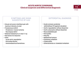
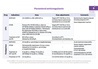
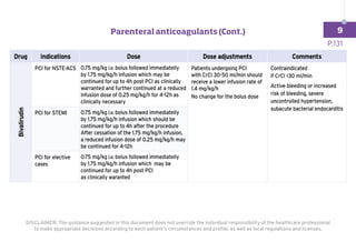
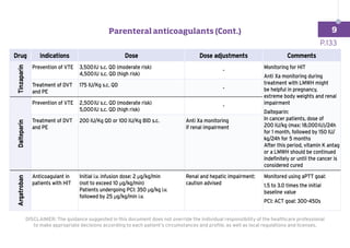
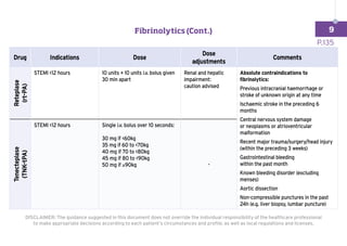
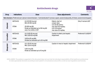
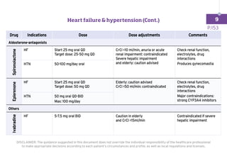
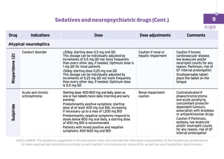
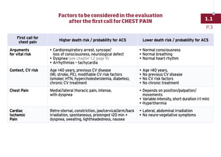
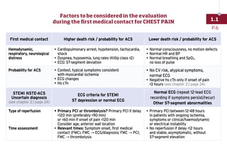
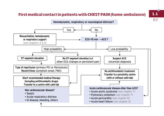
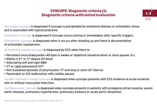
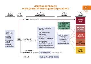
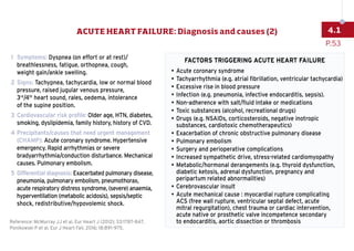
The initial assessment of patients presenting with chest pain involves considering the presentation, ECG, and troponin levels to determine the likelihood of acute coronary syndrome. A higher likelihood is present with factors like cardiorespiratory arrest, syncope, dyspnea, arrhythmias, age over 40, previous cardiovascular disease or risk factors. A lower likelihood is suggested by factors like age under 40, no prior disease or risk factors, and chest pain that is variable, short-lived or position-dependent. The assessment guides whether the diagnosis is likely to be cardiac ischemia, noncardiac chest pain, or another cause.


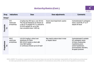
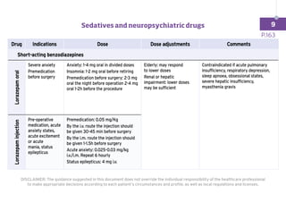
![INSTRUMENTATION INVESTIGATIONS:
Intravenous line (peripheral/central) and BP monitoring (arterial line in shock
and severe ventilatory/gas-exchange disturbances)
Laboratory measures
• Cardiac markers (troponin, BNP/NT-proBNP/MR-proANP)
•
Complete blood count, electrolytes, creatinine, urea, glucose,
inflammation, TSH
•
Consider arterial or venous blood gases, lactate, D-dimer
(suspicion of acute pulmonary embolism)
Standard 12-lead ECG
• Rhythm, rate, conduction times?
• Signs of ischemia/myocardial infarction? Hypertrophy?
Echocardiography
a) Immediately in haemodynamically unstable patients
b)
Within 48 hours when cardiac structure and function are either not known or
may have changed since previous studies
Ventricular function (systolic and diastolic)? Estimated left-and right-side filling
pressures? Lung ultrasound? Presence of valve dysfunction (severe stenosis/
insufficiency)? Pericardial tamponade?
ACTIONS:
Rule in/out
acute heart failure
as cause of symptoms
and signs
Determine
clinical profile
Start as soon as
possible treatment of
both heart failure and
the factors identified
as triggers
Establish cause
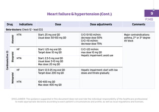
C - CIRCULATION*
HR (bradycardia [60/min], normal [60-100/min], tachycardia [100/min]), rhythm (regular, irregular), SBP (very low
[90 mmHg], low, normal [110-140 mmHg], high [140 mmHg]), and elevated jugular pressure should be checked.
P.56
4.1
ACUTE HEART FAILURE: Initial diagnosis (CDE)](https://image.slidesharecdn.com/acca-toolkit2018-220822064208-c4d36a8a/85/ACCA-TOOLKIT_2018-pdf-66-320.jpg)