Background
• To defineabnormal labor, a definition of normal labor must be
understood and accepted.
• Normal labor is defined as uterine contractions that result in
progressive dilation and effacement of the cervix.
• By following thousands of labors resulting in uncomplicated
vaginal deliveries, time limits and progress milestones have
been identified that define normal labor.
• Failure to meet these milestones defines abnormal labor,
which suggests an increased risk of an unfavorable outcome.
Thus, abnormal labor alerts the obstetrician to consider
alternative methods for a successful delivery that minimize
risks to both the mother and the infant.
3.
INCIDENCE
• Over thelast quarter of a century, the cesarean
section rate in the United States has risen to
approximately 35% of deliveries done each year.
• The Caesarean section rate in the developed world is
consistently over 20%, with one of the key causes
being prolonged or dysfunctional labor (30%),
particularly in nulliparous women.
• Dystocia is currently the most common indication for
primary cesarean section, and is about three times
more common than either non reassuring fetal
status or malpresentation.
4.
Abnormal labor andDystocia
• Normal labor is the presence of uterine
contractions of sufficient intensity ,frequency
and duration to bring about progressive
effacement and dilation of the cervix and
descent of the fetus
5.
• Dystocia isdefined as difficult labor or childbirth/Slow
progress of labor /failure to progress
• Abnormal labor, dystocia, and failure to progress :
• Terms used to describe a difficult labor pattern that
deviates from that observed in the majority of women
who have spontaneous vaginal deliveries
• It may be associated with abnormalities involving:
• Abnormalities of the Passage
• Abnormalities of the Passenger
• Abnormalities of the Powers
• or a combination of these factors
6.
• Second stage:
Timefrom complete cervical dilatation to expulsion of the fetus
• Third stage:
Time from expulsion of the fetus to expulsion of the placenta
latent
Active
Acceleration Phase
Maximum slope
Deceleration phase
• First stage:
Time from the onset of labor until complete cervical dilatation
Expected
Primipara Multiparous
Latent Stage1 <20 hrs. <14 hrs.
Active Stage 1 <5-6 hrs. <4-5 hrs.
Rate of Cervical dilatation 1.0-12.2 cm /hr 1.2-1.5 cm /hr.
Stage 2 <2 hrs. <1 hrs.
Stage 3 30 minutes
11.
0
2
4
6
8
10
12
2 4 68 10 12 14 16
Latent phase Active phase
2nd
stage
1st stage
max slope
acceleration
dec
Time (hours)
Cervical dilatation
(cm)
Friedman labor curve in nulliparous
12.
ABNORMAL PATTERNS OFLABOR
• The progress of labor is evaluated primarily
through estimates of cervical dilatation and
descent of the fetal presenting part. Normal
labor patterns in primigravidas and multiparas
have been described in detail by Friedman
and others.
13.
• Protraction disorders:refer to slower-than-normal
labor progress.
• Arrest disorders: refer to complete cessation of
progress.
Protraction and arrest disorders may occur in both the first and second stage of
labor
It is important to emphasize that the rates of cervical change listed in Table 1 are
two standard deviations from the mean and thereby used to define abnormal;
they do not represent the mean or median rates.
CLASSIFICATION – Of Labor Abnormalities:
14.
• Friedman describedfour abnormal patterns of
labor:
(1)prolonged latent phase,
(2) protraction disorders (protracted active-
phase dilatation and protracted descent),
(3) arrest disorders (prolonged deceleration
phase, secondary arrest of dilatation, arrest of
descent, and failure of descent), and
(4) precipitate labor disorders.
15.
ETIOLOGY OF PROTRACTIONAND ARREST
DISORDERS :
Abnormal labor can be the result of one or more
abnormalities:
o The cervix.
o The uterus.
o The maternal pelvis.
o The Fetus (i.e., power, passenger, or pelvis).
16.
• INCIDENCE –In one large series, the
incidence or protraction or arrest disorders in
the first stage of labor was 13 percent ,
second stage abnormalities appeared to be as
common .
17.
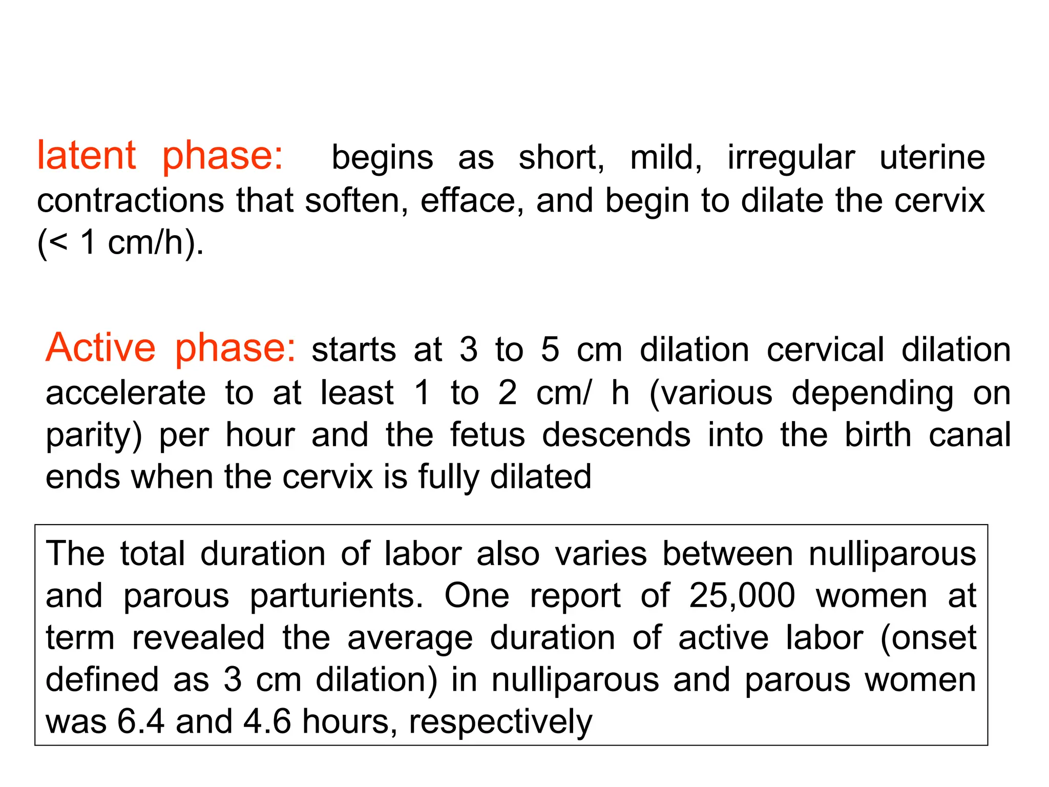
latent phase: beginsas short, mild, irregular uterine
contractions that soften, efface, and begin to dilate the cervix
(< 1 cm/h).
Active phase: starts at 3 to 5 cm dilation cervical dilation
accelerate to at least 1 to 2 cm/ h (various depending on
parity) per hour and the fetus descends into the birth canal
ends when the cervix is fully dilated
The total duration of labor also varies between nulliparous
and parous parturients. One report of 25,000 women at
term revealed the average duration of active labor (onset
defined as 3 cm dilation) in nulliparous and parous women
was 6.4 and 4.6 hours, respectively
18.
Prolonged Latent Phase
•An abnormally long latent phase is defined patient is in labor
and remains <3 cm dilated for 20 hours for the nullipara and
14 hours for the multiparous woman .Occur in 4-6%
• Prolonged latent phase is responsible for 30 % abnormalities
in nulliparas and over 50 % of abnormalities in multiparous
women
19.
Prolonged Latent Phase
Causes
•Anesthesia administered too early
• labor beginning with an unfavorable cervix.
• Irregular contraction
- hypotonic
- hypertonic
Ddx – False Labor
21.
Prolonged Latent Phase
Treatmentoptions:
• therapeutic rest with sedation and hydration.
• active management of labor.
• 85% of patients spontaneously enter the active phase of labor.
• Ten percent of patients will have been in false labor, and may
be allowed to return home to await the onset of true labor if
fetal status is reassuring.
• In the remaining 5% of patients, uterine contractions remain
ineffective in producing dilatation; in the absence of any
contraindication, active stimulation of labor with oxytocin
infusion may be effective in terminating the latent phase
22.
Prolong Active Phase
•Protracted cervical dilatation in the active phase of labor
• Protracted descent of the fetus constitute the protraction disorders.
• Protracted active-phase dilatation is characterized by an abnormally slow
rate of dilatation in the active phase, ie, less than 1.2 cm/h in nulliparas or
less than 1.5 cm/h in multiparas.
• Protracted descent of the fetus is characterized by a rate of descent under
1 cm/h in nulliparas or under 2 cm/h in multiparas.
• The second stage of labor, which normally averages 20 minutes for parous
women and 50 minutes in nulliparous women, is protracted when it
exceeds 2 hours in nulliparas or 1 hour in multiparas, or 3 and 2 hours
respectively in the presence of conduction anesthesia.
23.
Prolong Active Phase
•Patient is in labor and have cervical changes of <1.2 cm
/hr(primiparas) or <1.5 cm /hr (multiparas)
Causes
-Passenger –Macrosomia ,abnormal orientation
- Pelvis – Inadequate bony pelvic anatomy-
History may be important –compare current fetal size to fetal size of previous
pregnancies .If CPD with similar or smaller babies ,then it is probably the case
here .If no CPR with larger babied CPD unlikely
- Power-Dysfunctional or inadequate contraction
Management –depends on the tonicity of the contraction
- Hypotonic-IV Oxytocin
- Hypertonic – Morphine sedation
- Eutonic - Emergency Caesarean section
24.
It refersto uterine activity that is either not sufficiently
strong or not appropriately coordinated to dilate the
cervix and expel the fetus.
Is the most common cause of protraction or arrest
disorders in the first stage of labor.
It occurs in 3 to 8 percent of parturients and can be
quantified as uterine contraction pressures less than 200
Montevideo units.
Hypocontractile uterine activity
25.
Arrest Active Phase
•Patient is in labor and has no cervical change
for 2-3 hrs.
• Management is same as prolonged active
phase
26.
• Causes:
• About50% of patients with arrest disorders
demonstrate fetopelvic disproportion.
• various fetal malpositions (eg, occiput
posterior, occiput transverse, face, or brow).
• inappropriately administered anesthesia, or
excessive sedation.
• If fetopelvic disproportion is established,
cesarean section is done.
• If fetopelvic disproportion is not present and
uterine activity is less than optimal, oxytocin
stimulation is generally effective in producing
further progress.
28.
The median durationvaries in nulliparous and multiparous
women is 50 and 20 minutes, respectively.
The upper limit of duration associated with a normal
perinatal outcome had been defined as two hours ( but
was subsequently lengthened)
Other factors may affect its duration:
Epidural analgesia, duration of the first stage, parity,
maternal size, birth weight, and station at complete
dilation.
THE SECOND STAGE
The normal duration of 2nd
stage of labor should be based upon parity and
presence of regional anesthesia, with no intervention as long as the fetal heart
rate pattern is normal and some degree of progress is observed.
29.
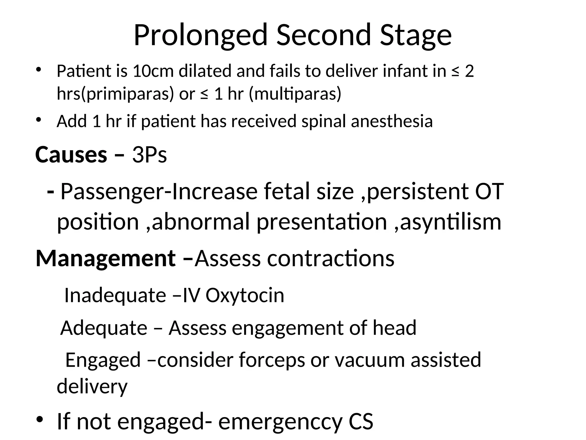
Prolonged Second Stage
•Patient is 10cm dilated and fails to deliver infant in ≤ 2
hrs(primiparas) or ≤ 1 hr (multiparas)
• Add 1 hr if patient has received spinal anesthesia
Causes – 3Ps
- Passenger-Increase fetal size ,persistent OT
position ,abnormal presentation ,asyntilism
Management –Assess contractions
Inadequate –IV Oxytocin
Adequate – Assess engagement of head
Engaged –consider forceps or vacuum assisted
delivery
• If not engaged- emergenccy CS
Quantitatives Assessment:
- Palpation.
-External tocodynamometry.
- Internal uterine pressure catheters.
95 % of women in labor will have 3-5 contractions per 10 minutes.
Quantifying assessment:
The Montevideo units (i.e., the peak strength of contractions in
mmHg measured by an internal monitor multiplied by their frequency
per 10 minutes)
90 % of women in spontaneous active labor achieved contractile
activity > 200 Montevideo units (in 40 % reaches 300 units).
Normal uterine activity
33.
Role of Epiduralanalgesia:
Dystocia due to cephalopelvic disproportion (Relative or
Absolute) :
• This diagnosis is currently based upon slow or arrested labor during the active
phase.
• Absolute: true disparity between fetal and maternal pelvic dimensions.
• Relative: due to fetal malposition (e.g., extended or asynclitic fetal head) or
malpresentation (mentum posterior, brow), rather than a.
Causes of Dystocia
Dystocia due to malposition:
5 % of cephalic presenting fetuses experience malposition with persistent occiput
posterior (OP) position or transverse arrest.
35.
• The underlyingpathogenesis of protracted
labor is probably multifactorial.
• Fetopelvic disproportion.
• minor malpositions such as occiput posterior.
• improperly administered conduction
anesthesia.
• excessive sedation.
• pelvic tumors obstructing the birth canal.
36.
• Precipitate LaborDisorders
• Precipitate dilatation occurs if cervical dilation
occurs at a rate of 5 or more centimeters per
hour in a primipara or at 10 cm or more per
hour in a multipara. Precipitate descent
occurs with descent of the fetal presenting
part of 5 cm or more per hour in primparas
and 10 cm or more per hour in multiparas.
37.
• Causes:
• 1-extremelystrong uterine contractions
• 2-low birth canal resistance.
• abnormal contractions may be associated with
administration of oxytocin and abruptio placentae.
• If oxytocin administration is the cause of abnormal
contractions, it may simply be stopped. The problem
typically resolves in less than 5 minutes.
• If excessive uterine activity is associated with fetal heart
rate abnormalities, and this pattern persists despite
discontinuation of oxytocin, a b-mimetic such as
terbutaline or ritodrine can be given and magnesium
sulfate also
• Lacerations of the birth canal are common.
• maternal amniotic fluid embolism.
• predisposing to postpartum hemorrhage.
• Perinatal mortality is increased secondary to hypoxia,
possible intracranial hemorrhage, and risks associated with
unattended delivery.
38.
• PATHOGENESIS &TREATMENT
• --Abnormalities of the Passage
• Causes:
• bony abnormalities (pelvic dystocia).
• soft tissue obstruction of the birth canal.
• abnormal placental location.
• Pelvic dystocia, is the most common cause of passage
abnormalities.
• The etiology and diagnosis of pelvic abnormalities
begins with the shape, classification, and clinical
assessment of the adult female pelvis..
• Ultrasound, magnetic resonance imaging (MRI), and x-
rays have been used to investigate pelvic size and
shape for evidence of pelvic contraction obstructing the
normal progress of labor.
39.
• Inlet contractionis suspected if the anteroposterior
diameter of the pelvis is less than 10 cm, the transverse
diameter is under 12 cm, or both.
• floating vertex presentation with no descent during
labor,
• abnormal presentation,
• prolapsed cord or extremity.
• considerable molding of the fetal head,
• caput succedaneum formation,
• and prolonged rupture of the membranes.
• If allowed to continue, abnormal thinning of the lower
uterine segment may occur, with development of a
Bandl's retraction ring, or even frank uterine rupture.
• Cesarean section is the treatment of choice in true inlet
contraction.
40.
• X-ray pelvimetryhas now fallen into limited use.
• Clinical pelvimetry has been largely used in the routine
evaluation of most obstetric patients.
• The diagnosis of fetopelvic disproportion has generally
become a diagnosis of exclusion, after fetal factors and
uterine dysfunction have been ruled out.
• However, x-ray pelvimetry retains a role in the
evaluation of a pelvis for the feasibility of vaginal
breech delivery and in the assessment of gross bony
distortion such as previous pelvic fracture or rachitic
deformity.
• Contractions of the pelvis are generally classified as:
• contractions of the inlet, midpelvis, or outlet, or as a
combination of these elements.
41.
• Midpelvis contractionit is more frequent than inlet
dystocia because the midpelvis is smaller than the inlet
and positional abnormality is more common at this
level.
• Presentation:
• Arrest of descent
• Poor application of the head to the cervix
• Abnormal rate of cervical dilatation
• Contraction of the outlet is extremely unusual unless
found in association with a Midpelvis contraction.
• Criteria for assessing pelvic outlet adequacy include
intertuberous diameter greater than 8 cm and a sum
of the intertuberous diameter and the anteroposterior
diameter greater than 15 cm.
42.
• Midpelvis outletobstruction is detected
clinically on the basis of convergent side walls,
prominent ischial spines, or a narrow pelvic
arch.
• It may present as a prolonged second stage,
• persistent occiput posterior position,
• deep transverse arrest.
• Molding of the fetal head and caput
succedaneum formation are common.
43.
• Uterine rupturemay occur in prolonged labor
complicated by midpelvic outlet obstruction, and
vesicovaginal or rectovaginal fistula formation
may result with pressure necrosis of the
surrounding tissues of the birth canal by the fetal
head.
• Cesarean section is therefore the delivery
method of choice in this complication.
• Other anatomic abnormalities of the
reproductive tract may cause dystocia is soft
tissue dystocia may be caused by uterine or
vaginal congenital anomalies, scarring of the
birth canal, pelvic masses, or low implantation of
the placenta.
44.
• --Abnormalites ofthe Passenger
• **A. malposition and malpresentation:
• Fetal malpresentations are abnormalities of fetal
position, presentation, attitude, or lie. They
collectively constitute the most common cause of
fetal dystocia, occurring in approximately 5% of
all labors.
• 1. Vertex malpositions—
• a. Occiput posterior—
• b. Occiput transverse—
• 2. Brow presentation—Brow presentations
usually are transient fetal presentations with
deflexion of the fetal head.
45.
• 3. Facepresentation—In face presentation,
the fetal head is fully deflexed from the
longitudinal axis.
• 4. Abnormal fetal lie—In transverse or oblique
lie, the long axis of the fetus is perpendicular
to or at an angle to the maternal longitudinal
axis.
46.
• 5. Breechpresentation
• **B. fetal macrosomia
• **C. fetal malformation
• The most common malformation is
hydrocephalus, enlargement of the fetal
abdomen caused by distended bladder,
ascites, or abdominal neoplasms; or other
fetal masses, including meningomyelocele or
cystosarcoma.
47.
• Abnormalities ofthe Powers
• Normal uterine activity during labor:
• (1) the relative intensity of contractions is greater in
the fundus than in the midportion or lower uterine
segment (this is termed fundal dominance); (2) the
average value of the intensity of contractions is more
than 24 mm Hg. (3) contractions are well synchronized
in different parts of the uterus; (4) the basal resting
pressure of the uterus is between 12 and 15 mm Hg;
(5) the frequency of contractions progresses from one
every 3–5 minutes to one every 2–3 minutes during
the active phase; (6) the duration of effective
contraction in active labor approaches 60 seconds; and
(7) the rhythm and force of contractions are regular.
• Hypotonic dysfunctionis uterine activity characterized
by contraction of the uterus with insufficient force (>
24 mm Hg), irregular or infrequent rhythm, or both.
Seen most often in primigravidas in the active phase of
labor, it may be caused by excessive sedation, early
administration of conduction anesthesia, twins,
polyhydramnios, or overdistention of the uterus.
• Hypotonic dysfunction responds well to oxytocin;
however, care must be taken to first rule out
cephalopelvic disproportion and malpresentation.
Active management of labor has been shown to
decrease perinatal morbidity and cesarean section
rates.
50.
• hypertonic uterinecontractions and uncoordinated
contraction often occur together and are characterized
by elevated resting tone of the uterus, dyssynchronous
contractions with elevated tone in the lower uterine
segment, and frequent intense uterine contractions. It
is generally associated with abruptio placentae,
overuse of oxytocin, cephalopelvic disproportion, fetal
malpresentation, and the latent phase of labor.
• Treatment:
• tocolysis, decrease in oxytocin infusion
• cesarean section as indicated for concomitant
malpresentation, cephalopelvic disproportion, or fetal
distress.
51.
• When thesepatterns occur in the latent phase
of labor:
• sedation may be effective in converting
hypertonic contractions to normal labor patterns.
• Inadequate pushing in the second stage of labor
is common and may be caused by conduction
anesthesia, oversedation, exhaustion, or
neurologic dysfunction such as paraplegia or
hemiplegia of various causes, or by psychiatric
disorders.
• Mild sedation may improve expulsive efforts.
• outlet forceps or vacuum delivery may be of help.
52.
Prevention: by propermanagement of labor:
The diagnosis of labor.
Monitoring of labor progress.
assessment of maternal and fetal well-being.
(Women should undergo cervical examination every one to two hours
once active labor is diagnosed to determine whether progression is
adequate)
The use of partogram
APPROACH TO THE PATIENT WITH ABNORMAL LABOR
53.
• Amniotomy
• Oxytocinfor treatment of Hypo contractile uterine activity
Low dose regimens: (to avoid uterine hyperstimulation)
High dose regimens: (shorten labor )
Management of Dystocia in the first stage:
Oxytocin is typically infused to titrate dose to effect, as prediction of
a women's response to a particular dose is not possible
Options f management include
54.
Diagnosis:
When There IsNo Progress (Protraction Disorder
Persists) Despite Oxytocin Therapy To Achieve > Or =
200 Montevideo Units For Greater Than Two Hours.
Active Phase Arrest
Treatment:
Cesarean Delivery Is Typically Performed At This Point
55.
Continued observation.
Attempt at operative vaginal delivery.
Cesarean delivery.
Dystocia in the second stage
Risk factors include:
nulliparity, diabetes, macrosomia, epidural anesthesia,
oxytocin usage, and chorioamnionitis
56.
Observation:
Most women witha prolonged 2nd stage ultimately deliver
vaginally.
Suggested noninvasive interventions:
- changes in maternal position.
- continuous emotional support of the parturient
- delaying pushing if the fetal head is high in the pelvis at
full dilatation and the woman has no urge to do so
- active management using high dose oxytocin.
Operative vaginal delivery :
The choice of instrument require careful assessment of the
mother and fetus.
success is dependent upon the training and skill of the
obstetrician.
57.
Risks:
- Longer secondstage.
- higher incidence of operative delivery.
- larger episiotomies.
- more severe perineal lacerations.
Occiput posterior position
A small increase in second stage length in the presence of a reassuring fetal heart
rate, favorable clinical assessment of fetal relative to maternal size, and progress
in the second stage does not mandate rotation or operative delivery.
Management of OP:
Operative Delivery From OP Position.
Manual Or Instrumental Rotation To Occiput Anterior.
Cesarean Delivery.
58.
RECOMMENDATIONS:
A general labormanagement . The key points are listed below:
• Monitor progress in active labor with cervical exams at 1 to 2 hour
intervals.
• If the patient in active labor fails to progress adequately for two hours,
then intact membranes should be ruptured and oxytocin administered to
achieve uterine contractions greater than 200 Montevideo units. These
patients can be observed for two to four hours as long as clinical
assessment of fetal and maternal size is favorable and the fetal heart rate is
reassuring.
• The decision to perform an operative vaginal delivery (eg, extraction or
rotation) in the second stage versus continued observation or cesarean birth
is based upon clinical assessment of mother and fetus and the skill and
training of the obstetrician.