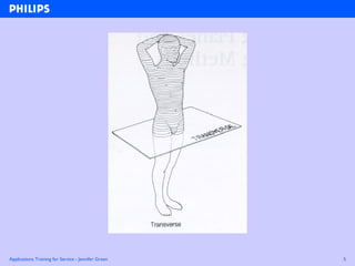
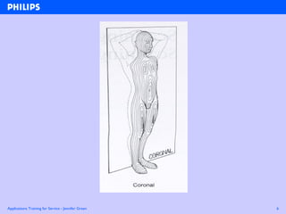
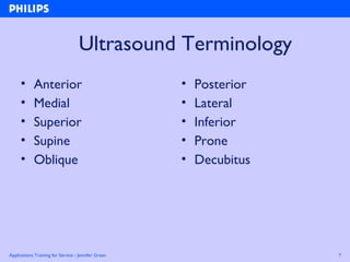
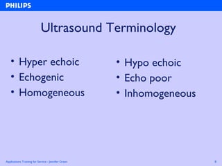
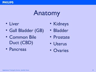
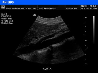
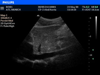
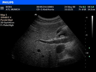
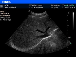
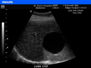
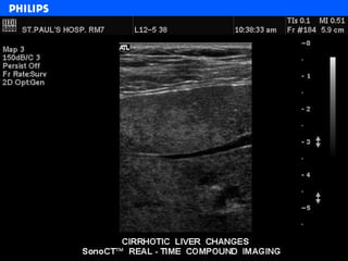
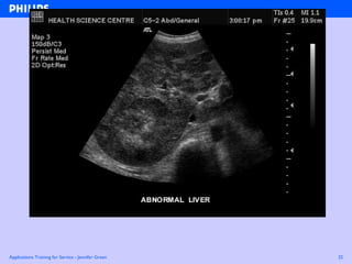
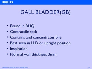
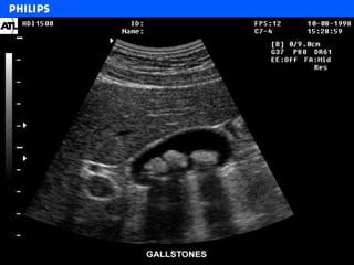
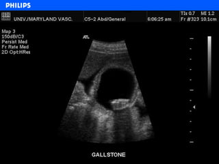
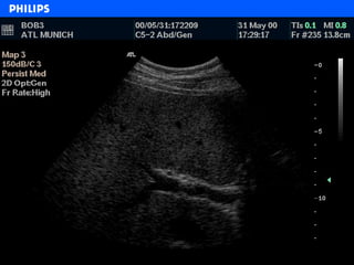
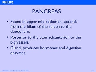
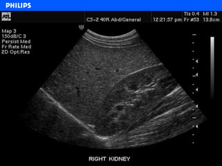
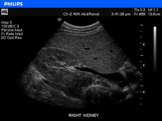
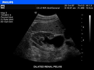
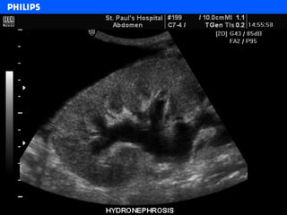
Abdominal Anatomy
Overview
Jennifer Green
General Imaging Training Manager
Applications Training for Service - Jennifer Green 2
• Scanning positions
• Sonographic anatomy
• System setup
ABDOMEN
Applications Training for Service - Jennifer Green 3
Ultrasound Scan Planes
• Sagittal
• Transverse
• Coronal
Applications Training for Service - Jennifer Green 4
Applications Training for Service - Jennifer Green 5
Applications Training for Service - Jennifer Green 6
Applications Training for Service - Jennifer Green 7
Ultrasound Terminology
• Anterior
• Medial
• Superior
• Supine
• Oblique
• Posterior
• Lateral
• Inferior
• Prone
• Decubitus
Applications Training for Service - Jennifer Green 8
Ultrasound Terminology
• Hyper echoic
• Echogenic
• Homogeneous
• Hypo echoic
• Echo poor
• Inhomogeneous
Applications Training for Service - Jennifer Green 9
Anatomy
Applications Training for Service - Jennifer Green 10
Anatomy
• Liver
• Gall Bladder (GB)
• Common Bile
Duct (CBD)
• Pancreas
• Kidneys
• Bladder
• Prostate
• Uterus
• Ovaries
Applications Training for Service - Jennifer Green 11
GREAT VESSELS
• Aorta
• Celiac Axis
• SVC
• Renal Arteries
• Hepatic Artery
• IVC
• Renal veins
• Portal vein
• Hepatic Veins
Applications Training for Service - Jennifer Green 18
LIVER
• Found in right upper quadrant (RUQ)
• Largest internal organ
• Stores and filters blood
• Produces bile
• Synthesizes many substances
• Performs vast number of metabolic
functions
Applications Training for Service - Jennifer Green 19
LIVER
• During deep inspiration!!!,
• Patient position – supine
- left oblique
- left lateral decubitus (LLD)
• Sonographic windows:
– Mid line, inferior to xiphoid process
• Left lobe Sagittal and Transverse
– Inferior & superior to right costal margin
• Right lobe Sagittal and Transverse
Applications Training for Service - Jennifer Green 21
Applications Training for Service - Jennifer Green 22
Applications Training for Service - Jennifer Green 23
Applications Training for Service - Jennifer Green 24
Applications Training for Service - Jennifer Green 26
Applications Training for Service - Jennifer Green 29
Applications Training for Service - Jennifer Green 31
Applications Training for Service - Jennifer Green 32
Applications Training for Service - Jennifer Green 34
GALL BLADDER(GB)
• Found in RUQ
• Contractile sack
• Contains and concentrates bile
• Best seen in LLD or upright position
• Inspiration
• Normal wall thickness 3mm
Applications Training for Service - Jennifer Green 35
Applications Training for Service - Jennifer Green 36
Applications Training for Service - Jennifer Green 40
COMMON BILE DUCT (CBD)
• Conducts bile from the G.B. to the
duodenum
• Found in RUQ, inferior to the G.B.
• CBD runs anterior to the portal vein; distal
part is lateral to the common hepatic
artery
• Best seen in LLD
Applications Training for Service - Jennifer Green 41
Applications Training for Service - Jennifer Green 42
PANCREAS
• Found in upper mid abdomen; extends
from the hilum of the spleen to the
duodenum.
• Posterior to the stomach,anterior to the
big vessels.
• Gland, produces hormones and digestive
enzymes.
Applications Training for Service - Jennifer Green 43
PANCREAS
• Best seen in supine position in inspiration
or pushed up abdomen.
• Sonographic appearance:
– smooth borders; homogeneous, mid gray
echoes
– echogenicity  with age.
Applications Training for Service - Jennifer Green 44
Applications Training for Service - Jennifer Green 45
Applications Training for Service - Jennifer Green 46
KIDNEYS
• Paired organ; 9 –13cms long
• Principal parts: cortex, medulla, sinus...
• Filters metabolic wastes from the blood.
• Normal variants:
– Dromedary humps
– Double collecting system
– Horseshoe
– Ectopic kidney
Applications Training for Service - Jennifer Green 48
Applications Training for Service - Jennifer Green 50
Applications Training for Service - Jennifer Green 52
Applications Training for Service - Jennifer Green 53
Applications Training for Service - Jennifer Green 56
BLADDER
• It is a symmetrical, hollow, muscular organ.
• Located posterior to the symphysis pubis.
• Shape varies with distention.
• Normal thickness of the distended wall is
< 1cm.
• Provides window for Ob/Gyn, prostate.
Applications Training for Service - Jennifer Green 57
BLADDER
• Function:
– Urine delivered through ureters, is
stored in the bladder, and released
through urethra.
• Sonographic appearance:
– Wall is a smooth echogenic line
– cavity is not seen if collapsed, otherwise
is non echoic.
Applications Training for Service - Jennifer Green 58
Applications Training for Service - Jennifer Green 59
Applications Training for Service - Jennifer Green 60
PROSTATE
• Lies inferior to the urinary bladder,and anterior
to the rectum
• The size of the gland 3.5x4x2.5 cm
• The base is the broadest and it is in contact with
bladder floor
• The urethra and the ejaculatory ducts pass
through the prostate
• Best seen endo rectally (ER), size can be
estimated trans abdominally (TA)
Applications Training for Service - Jennifer Green 61
Applications Training for Service - Jennifer Green 62
Applications Training for Service - Jennifer Green 63
Applications Training for Service - Jennifer Green 65
Applications Training for Service - Jennifer Green 67
UTERUS
• Pear shaped pelvic organ lying in midline.
• Found in the small pelvis infero-posterior
to the bladder
• Usual size: 7-8x3-5x3-5 cm.
• Uterus is divided into: cervix, corpus,
fundus, cornua.
• Has three layers : endometrium,
myometrium, serous.
Applications Training for Service - Jennifer Green 68
UTERUS
• Endometrial cavity is continuous with vagina via
cervix.
• Uterus can be retroverted/flexed or
anteverted/flexed.
• Best seen endo vaginally (EV), need full bladder
for trans abdominal (TA)
• Sonographic appearance:
– myometrium is midgray with even texture
– endometrium is a thin echogenic line that varies in
intensity with menstrual phase and age.
Applications Training for Service - Jennifer Green 69
Applications Training for Service - Jennifer Green 70
Applications Training for Service - Jennifer Green 71
OVARIES
• Paired pelvic organ, position very variable
alongside the uterus.
• Search lateral/posterior to the uterus.
• Best seen EV.
• Normal post pubertal size approx: 4x3x2 cm
• Sonographic appearance:
– homogenous midgray echogenicity
– follicles are round, anechoic, on the periphery.
Applications Training for Service - Jennifer Green 72
Applications Training for Service - Jennifer Green 73
Applications Training for Service - Jennifer Green 74
Grey Scale
• U/S is a dynamic examination needing frequent
optimization of the imaging parameters for
consistent good quality of images.
• Sound pathway changes constantly due to
movement of both the patient and the scanner.
• Important parameters: power (MI)
Gain
TGC
Dynamic Range (dB)
Gray Maps.
Applications Training for Service - Jennifer Green 75
Power and Gain
• Balance is needed to maintain safety
(ALARA principle) and to obtain the good
image quality through wider dynamic
range.
•  power, than  gain
Applications Training for Service - Jennifer Green 76
Time Gain Compensation(TGC)
• The purpose of TGC is to compensate for
attenuation along the propagation path.
• The desired effect is to equalize near to far
field brightness.
Applications Training for Service - Jennifer Green 77
Dynamic Range(dB)
• Ratio of largest to smallest echo power.
• High dynamic range image has low contrast due to
numerous available shades of gray, giving high tissue
differentiation
– good for easy patients, superficial structures
• Images of lower dynamic range show increased
contrast. Reduced number of available shades of gray
will poorly differentiate tissues
– good for challenging patients.
Applications Training for Service - Jennifer Green 78
Gray Scale Maps
• Apply nonlinear assignment of the display
brightness range among stored range of the
system.
• Manipulation enables emphasis of certain
image elements over each other.
Abdo 0verview

Abdo 0verview

  • 1.
  • 2.
    Applications Training forService - Jennifer Green 2 • Scanning positions • Sonographic anatomy • System setup ABDOMEN
  • 3.
    Applications Training forService - Jennifer Green 3 Ultrasound Scan Planes • Sagittal • Transverse • Coronal
  • 4.
    Applications Training forService - Jennifer Green 4
  • 5.
    Applications Training forService - Jennifer Green 5
  • 6.
    Applications Training forService - Jennifer Green 6
  • 7.
    Applications Training forService - Jennifer Green 7 Ultrasound Terminology • Anterior • Medial • Superior • Supine • Oblique • Posterior • Lateral • Inferior • Prone • Decubitus
  • 8.
    Applications Training forService - Jennifer Green 8 Ultrasound Terminology • Hyper echoic • Echogenic • Homogeneous • Hypo echoic • Echo poor • Inhomogeneous
  • 9.
    Applications Training forService - Jennifer Green 9 Anatomy
  • 10.
    Applications Training forService - Jennifer Green 10 Anatomy • Liver • Gall Bladder (GB) • Common Bile Duct (CBD) • Pancreas • Kidneys • Bladder • Prostate • Uterus • Ovaries
  • 11.
    Applications Training forService - Jennifer Green 11 GREAT VESSELS • Aorta • Celiac Axis • SVC • Renal Arteries • Hepatic Artery • IVC • Renal veins • Portal vein • Hepatic Veins
  • 12.
    Applications Training forService - Jennifer Green 18 LIVER • Found in right upper quadrant (RUQ) • Largest internal organ • Stores and filters blood • Produces bile • Synthesizes many substances • Performs vast number of metabolic functions
  • 13.
    Applications Training forService - Jennifer Green 19 LIVER • During deep inspiration!!!, • Patient position – supine - left oblique - left lateral decubitus (LLD) • Sonographic windows: – Mid line, inferior to xiphoid process • Left lobe Sagittal and Transverse – Inferior & superior to right costal margin • Right lobe Sagittal and Transverse
  • 14.
    Applications Training forService - Jennifer Green 21
  • 15.
    Applications Training forService - Jennifer Green 22
  • 16.
    Applications Training forService - Jennifer Green 23
  • 17.
    Applications Training forService - Jennifer Green 24
  • 18.
    Applications Training forService - Jennifer Green 26
  • 19.
    Applications Training forService - Jennifer Green 29
  • 20.
    Applications Training forService - Jennifer Green 31
  • 21.
    Applications Training forService - Jennifer Green 32
  • 22.
    Applications Training forService - Jennifer Green 34 GALL BLADDER(GB) • Found in RUQ • Contractile sack • Contains and concentrates bile • Best seen in LLD or upright position • Inspiration • Normal wall thickness 3mm
  • 23.
    Applications Training forService - Jennifer Green 35
  • 24.
    Applications Training forService - Jennifer Green 36
  • 25.
    Applications Training forService - Jennifer Green 40 COMMON BILE DUCT (CBD) • Conducts bile from the G.B. to the duodenum • Found in RUQ, inferior to the G.B. • CBD runs anterior to the portal vein; distal part is lateral to the common hepatic artery • Best seen in LLD
  • 26.
    Applications Training forService - Jennifer Green 41
  • 27.
    Applications Training forService - Jennifer Green 42 PANCREAS • Found in upper mid abdomen; extends from the hilum of the spleen to the duodenum. • Posterior to the stomach,anterior to the big vessels. • Gland, produces hormones and digestive enzymes.
  • 28.
    Applications Training forService - Jennifer Green 43 PANCREAS • Best seen in supine position in inspiration or pushed up abdomen. • Sonographic appearance: – smooth borders; homogeneous, mid gray echoes – echogenicity  with age.
  • 29.
    Applications Training forService - Jennifer Green 44
  • 30.
    Applications Training forService - Jennifer Green 45
  • 31.
    Applications Training forService - Jennifer Green 46 KIDNEYS • Paired organ; 9 –13cms long • Principal parts: cortex, medulla, sinus... • Filters metabolic wastes from the blood. • Normal variants: – Dromedary humps – Double collecting system – Horseshoe – Ectopic kidney
  • 32.
    Applications Training forService - Jennifer Green 48
  • 33.
    Applications Training forService - Jennifer Green 50
  • 34.
    Applications Training forService - Jennifer Green 52
  • 35.
    Applications Training forService - Jennifer Green 53
  • 36.
    Applications Training forService - Jennifer Green 56 BLADDER • It is a symmetrical, hollow, muscular organ. • Located posterior to the symphysis pubis. • Shape varies with distention. • Normal thickness of the distended wall is < 1cm. • Provides window for Ob/Gyn, prostate.
  • 37.
    Applications Training forService - Jennifer Green 57 BLADDER • Function: – Urine delivered through ureters, is stored in the bladder, and released through urethra. • Sonographic appearance: – Wall is a smooth echogenic line – cavity is not seen if collapsed, otherwise is non echoic.
  • 38.
    Applications Training forService - Jennifer Green 58
  • 39.
    Applications Training forService - Jennifer Green 59
  • 40.
    Applications Training forService - Jennifer Green 60 PROSTATE • Lies inferior to the urinary bladder,and anterior to the rectum • The size of the gland 3.5x4x2.5 cm • The base is the broadest and it is in contact with bladder floor • The urethra and the ejaculatory ducts pass through the prostate • Best seen endo rectally (ER), size can be estimated trans abdominally (TA)
  • 41.
    Applications Training forService - Jennifer Green 61
  • 42.
    Applications Training forService - Jennifer Green 62
  • 43.
    Applications Training forService - Jennifer Green 63
  • 44.
    Applications Training forService - Jennifer Green 65
  • 45.
    Applications Training forService - Jennifer Green 67 UTERUS • Pear shaped pelvic organ lying in midline. • Found in the small pelvis infero-posterior to the bladder • Usual size: 7-8x3-5x3-5 cm. • Uterus is divided into: cervix, corpus, fundus, cornua. • Has three layers : endometrium, myometrium, serous.
  • 46.
    Applications Training forService - Jennifer Green 68 UTERUS • Endometrial cavity is continuous with vagina via cervix. • Uterus can be retroverted/flexed or anteverted/flexed. • Best seen endo vaginally (EV), need full bladder for trans abdominal (TA) • Sonographic appearance: – myometrium is midgray with even texture – endometrium is a thin echogenic line that varies in intensity with menstrual phase and age.
  • 47.
    Applications Training forService - Jennifer Green 69
  • 48.
    Applications Training forService - Jennifer Green 70
  • 49.
    Applications Training forService - Jennifer Green 71 OVARIES • Paired pelvic organ, position very variable alongside the uterus. • Search lateral/posterior to the uterus. • Best seen EV. • Normal post pubertal size approx: 4x3x2 cm • Sonographic appearance: – homogenous midgray echogenicity – follicles are round, anechoic, on the periphery.
  • 50.
    Applications Training forService - Jennifer Green 72
  • 51.
    Applications Training forService - Jennifer Green 73
  • 52.
    Applications Training forService - Jennifer Green 74 Grey Scale • U/S is a dynamic examination needing frequent optimization of the imaging parameters for consistent good quality of images. • Sound pathway changes constantly due to movement of both the patient and the scanner. • Important parameters: power (MI) Gain TGC Dynamic Range (dB) Gray Maps.
  • 53.
    Applications Training forService - Jennifer Green 75 Power and Gain • Balance is needed to maintain safety (ALARA principle) and to obtain the good image quality through wider dynamic range. •  power, than  gain
  • 54.
    Applications Training forService - Jennifer Green 76 Time Gain Compensation(TGC) • The purpose of TGC is to compensate for attenuation along the propagation path. • The desired effect is to equalize near to far field brightness.
  • 55.
    Applications Training forService - Jennifer Green 77 Dynamic Range(dB) • Ratio of largest to smallest echo power. • High dynamic range image has low contrast due to numerous available shades of gray, giving high tissue differentiation – good for easy patients, superficial structures • Images of lower dynamic range show increased contrast. Reduced number of available shades of gray will poorly differentiate tissues – good for challenging patients.
  • 56.
    Applications Training forService - Jennifer Green 78 Gray Scale Maps • Apply nonlinear assignment of the display brightness range among stored range of the system. • Manipulation enables emphasis of certain image elements over each other.

Editor's Notes

  • #28 The portal vein is formed from the splenic vein,sup&amp;inf mesenteric veins and the gastric and cystic veins.It carries blood from the abdominal portion of the digestive system and spleen via the liver and then into the IVC. This way blood with a high concentration of nutrients goes to the liver first to be modified.