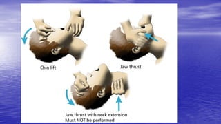
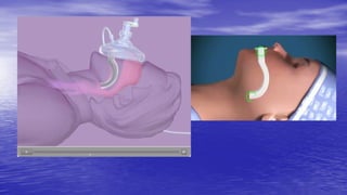
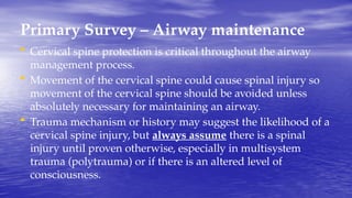
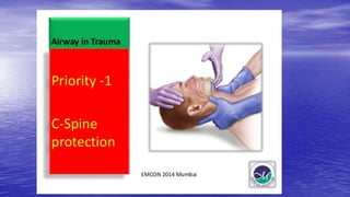
Trauma
Dr Yotham Phiri MMed(Surg)
Chainama College of Health Sciences
Traumatology
• from Greek trauma, meaning injury or wound,
• the study of wounds and injuries caused by accidents or
violence to a person, and the surgical therapy and repair
of the damage.
• It is often considered a subset of surgery, a surgical
specialty of trauma surgery
• Most often a sub-specialty to orthopaedic surgery.
• Traumatology may also be known as accident surgery.
Branches of traumatology
Branches of traumatology include
• Medical traumatology
• Psychological traumatology
Medical Traumatology
Medical traumatology
• defined as the study of specializing in the treatment of
wounds and injuries caused by violence or general
accidents.
• This type of traumatology focuses on the surgical
procedures and future physical therapy a patients need to
repair the damage and recover properly
Psychological Traumatology
Psychological traumatology
• a type of damage to one’s mind due to a distressing event.
• This type of trauma can also be the result of
overwhelming amounts of stress in one’s life.
• usually involves some type of physical trauma that poses
as a threat to one’s sense of security and survival.
• often leaves people feeling overwhelmed, anxious, and
threatened (Emotional and Psychological Trauma).
Types of trauma
• Types of trauma include car accidents, gunshot wounds,
concussions, PTSD from incidents, etc.
• Medical traumas are repaired with surgeries, however
they can still cause psychological trauma and other stress
factors.
• For example, a teenager in a car accident who broke their
wrist needed extensive surgery to save their arm. It is
possible for that child to get anxiety when driving in a car
after the accident.
Post Traumatic Stress Disorder
Post Traumatic Stress Disorder (PTSD)
• can be diagnosed after a person experiences one or more
intense and traumatic events and react with fear with
complaints from three categorical symptoms lasting one
month or longer.
• These categories are - re-experiencing the traumatic event,
avoiding anything associated with the trauma, and
increased symptoms of increased psychological arousal
Advanced Trauma Life Support (ATLS)
• A training program for health care medical providers in the
management of acute trauma cases, developed by the
American College of Surgeons.
• Similar programs exist for immediate care providers such
as paramedics.
• The program has been adopted worldwide in over 60
countries.
Advanced Trauma Life Support (ATLS)
• Zambia Trauma Course by the Surgical Society of Zambia
(SSZ)
• Its goal is to teach a simplified and standardized
approach to trauma patients.
• ATLS is now widely accepted as the standard of care for
initial assessment and treatment in trauma centres.
• The premise of the ATLS program is to treat the greatest
threat to life first.
ATLS - History
• ATLS has its origins in the United States in 1976,
when James K. Styner, an orthopedic surgeon
piloting a light aircraft, crashed his plane into a
field in Nebraska.
• His wife was killed instantly and three of his four
children sustained critical injuries.
• He carried out the initial triage of his children at
the crash site.
ATLS - History
• Dr. Styner had to flag down a car to transport him to the
nearest hospital; upon arrival, he found it closed.
• Even once the hospital was opened and a doctor called in,
he found that the emergency care provided at the small
regional hospital where they were treated was inadequate
and inappropriate.
ATLS – History (Cont…)
• Upon returning home, Dr. Styner declared: “When I can provide better care
in the field with limited resources than what my children and I received at the
primary care facility, there is something wrong with the system and the
system has to be changed”
• Upon returning to work, he set about developing a system for saving lives in
medical trauma situations.
• Styner and his colleague, with assistance from advanced cardiac life support
personnel and the Lincoln Medical Education Foundation, produced the
initial ATLS course which was held in 1978.
• In 1980, the American College of Surgeons Committee on Trauma adopted
ATLS and began US and international dissemination of the course.
ATLS
• Primary Survey - ABCDE
• Secondary Survey
• Tertiary Survey
Primary Survey
• The first and key part of the assessment of patients
presenting with trauma is called the primary survey.
• During this time, life-threatening injuries are identified
and simultaneously resuscitation is begun.
• A simple mnemonic, ABCDE, is used as a memory aid for
the order in which problems should be addressed
Primary Survey
• A - Airway maintenance with cervical spine protection
• B - Breathing and ventilation
• C - Circulation with haemorrhage control
• D - Disability/Neurologic assessment
• E - Exposure and environmental control
Secondary Survey
• When the primary survey is completed, resuscitation efforts are
well established, and the vital signs are normalizing, the
secondary survey can begin.
• The secondary survey is a head-to-toe evaluation of the trauma
patient, including a complete history and physical examination,
including the reassessment of all vital signs.
• Each region of the body must be fully examined. X-rays indicated
by examination are obtained.
• If at any time during the secondary survey the patient deteriorates,
another primary survey is carried out as a potential life threat may
be present.
Tertiary Survey
• A careful and complete examination followed by
• serial assessments help recognize missed injuries and
related problems,
• allowing a definitive care management.
Triage
• Sorting of patients according to their need for treatment
and the resources available
• Starts at the scene and continues at the receiving hospital.
• Priority given to patients most likely to deteriorate
clinically
• takes into account of vital signs, pre-hospital clinical course,
mechanism of injury, age and other medical conditions.
Teamwork
• In trauma centres, teamwork should ensure critically
injured patients are evaluated as diagnostic procedures
are performed simultaneously, thus reducing the time to
treatment.
• A team approach is demanding of personnel and
resources and, in smaller institutions, non-hospital
settings or with mass casualties, available personnel and
resources can rapidly be overwhelmed:
Triage
Triage: is done according to the ‘ABCDE’ principles
• Airway maintenance with cervical spine protection
• Breathing and ventilation
• Circulation with haemorrhage control
• Disability: neurological status
• Exposure/environmental control
Triage
• Multiple casualties: the number of patients and severity
of injury do not exceed the capacity of the treatment centre
- life-threatening injuries and multiple system injuries are
treated first.
• Mass casualties: the number of patients and severity of
injury exceeds capacity of the treatment centre - patients
are selected for treatment according to best chance of
survival with least expenditure of resources (time,
personnel, equipment, supplies).
Initial assessment
This comprises:
• Resuscitation and primary survey.
• Secondary survey.
• Definitive treatment or transfer for definitive care.
• Many mistakes are caused by not examining the patient carefully.
• The peripheral injuries are unlikely to kill the patient even if you
miss them but you can easily overlook serious central ones
especially injures to the chest and abdomen.
• You will not miss a bone sticking out of his trouser leg but you can
easily miss blood in his thoracic cavity or a slowly developing
haemoperitoneum.
• More lives are lost from failing to care for the airway than from
any other cause!!!
Primary Survey
A = Airway maintenance cervical spine protection
• Are there signs of airway obstruction, foreign bodies,
facial, mandibular or laryngeal fractures?
• Management may involve
– secretion control
– intubation or
– surgical airway e.g. cricothyroidotomy, emergency
tracheostomy).
Primary Survey – Airway maintenance
• Establish a clear airway (chin lift or jaw thrust) but protect
the cervical spine at all times.
• If the patient can talk, the airway is likely to be safe;
however, remain vigilant and recheck.
• An oropharyngeal airway in an unconscious patient with
no gag reflex.
• Definitive airway should be established if the patient is
unable to maintain integrity of airway;
Primary Survey – Airway maintenance
• Cervical spine protection is critical throughout the airway
management process.
• Movement of the cervical spine could cause spinal injury so
movement of the cervical spine should be avoided unless
absolutely necessary for maintaining an airway.
• Trauma mechanism or history may suggest the likelihood of a
cervical spine injury, but always assume there is a spinal
injury until proven otherwise, especially in multisystem
trauma (polytrauma) or if there is an altered level of
consciousness.
Primary Survey – Breathing
B = Breathing and ventilation
• Provide high-flow oxygen through a rebreather mask if
not intubated and ventilated.
• Evaluate breathing: lungs, chest wall, diaphragm. Chest
examination with adequate exposure: watch chest
movement, auscultate, percuss to detect lesions acutely
impairing ventilation
Lesions acutely impairing ventilation:
• Tension pneumothorax - requires needle thoracostomy
followed by drainage.
• Flail chest - management involves ventilation.
• Haemothorax - will usually require intercostal drain
insertion.
• Pneumothorax - may require intercostal drain insertion.
Tension pneumothorax
• The accumulation of air under pressure in the pleural space.
• An imminent danger is that the lung will collapse under the
pressure.
• In tension pneumothorax, air enters the pleural cavity and is
trapped there during expiration so the air pressure within the
thorax mounts higher than atmospheric pressure, compresses the
lung, may displace the mediastinum and its structures (including
the lung) toward the opposite side, and cause cardiopulmonary
impairment. Also called pressure pneumothorax,
Flail chest
• Occurs when a segment of the rib cage breaks due to trauma and
becomes detached from the rest of the chest wall.
• It occurs when multiple adjacent ribs are broken in multiple
places, separating a segment, so a part of the chest wall moves
independently.
• The flail segment moves in the opposite direction to the rest of the
chest wall:, it goes in while the rest of the chest is moving out, and
vice versa.
• This so-called “paradoxical breathing“ is painful and increases
the work involved in breathing.
Primary Survey - Circulation
C = Circulation with haemorrhage control
Blood loss is the main preventable cause of death after
trauma. To assess blood loss rapidly observe:
• Level of consciousness.
• Skin colour.
• Pulse.
• Bleeding - this should be assessed and controlled
Primary Survey - Circulation
Bleeding - this should be assessed and controlled:
• IV access with two large cannulae in an upper limb. Access by cut down or
central venous catheterisation may be done according to skills available.
• At cannula insertion, blood should be taken for crossmatch and baseline
investigations.
• IV fluids will need to be given rapidly. Ringer's lactate is the preferred initial
crystalloid solution.
• Direct manual pressure should be used to stem visible bleeding (not
tourniquets, except for traumatic amputation, as these cause distal
ischaemia).
• Occult bleeding into the abdominal cavity and around long-bone or pelvic
fractures is problematic but should be suspected in a patient not responding
to fluid resuscitation.
Primary Survey - Disability
D = Disability: neurological status
After A, B and C above, rapid neurological assessment is made to
establish:
• Level of consciousness, using GCS.
• Pupils: size, symmetry and reaction.
• Any lateralising signs.
• Level of any spinal cord injury (limb movements, spontaneous
respiratory effort).
• Oxygenation, ventilation, perfusion, drugs, alcohol and
hypoglycaemia may all also affect the level of consciousness.
Primary Survey - Exposure
E = Exposure/environmental control
• Undress the patient, but prevent hypothermia.
• Clothes may need to be cut off
• after examination, attend to prevention of heat loss with
warming devices, warmed blankets, etc.
• Also check blood glucose levels.
Additional Monitoring
• ECG monitoring: this can guide resuscitation by diagnosing dysrhythmias,
ischaemia, cardiac injury
• Urinary catheters:
• Nasogastric catheters are inserted to reduce aspiration risk. Suction should
be applied.
• Other monitoring: monitoring of resuscitation by measuring various
important parameters such as:
– Pulse rate,blood pressure, ventilatory rate, arterial blood gases, body
temperature and urinary output.
– Pulse oximetry measures oxygenation of haemoglobin colorimetrically
(sensor on finger, ear lobe, etc.).
• Remember: blood pressure is a poor measure of perfusion.
Secondary Survey
• This begins after the “ABCDE” of the primary survey,
once resuscitation is underway and the patient is
responding with normalisation of vital signs.
• The secondary survey is essentially a head-to-toe
examination
• It requires repeat physical examinations and may require
further X-ray and laboratory tests.
Secondary Survey
It comprises History( AMPLE) and Physical Examination
History (AMPLE)
• A = Allergies.
• M = Medication currently used.
• P = Past illnesses/Pregnancy.
• L = Last meal.
• E = Events/Environment related to injury
Secondary Survey – Physical Examination
• This will repeat some examinations already undertaken in the
primary survey and will be further informed by the progress of
the resuscitation.
• Examination aims to identify serious injuries, occult bleeding, etc.
• A review of neurological status including GCS score is also
undertaken.
• Back and spinal injuries are commonly missed and pelvic
fractures cause large blood loss which is often underestimated.
P/E - Head-to-toe examination
• Head - Head and face fractures, CSF leaks from nose and
ears
• Eyes
• Mouth
• Chest
• Abdomen
• Pelvis
• Limbs
Pupils
• Record the size and reaction to light (fixed and dilated pupils
suggest severe brain damage)
• Always ask the conscious patient if he can see with both eyes.
Look for eye injury in the unconscious patient.
• Examine the conjunctiva and tongue for pallor and cyanosis. You
are not looking for signs of anaemia but rather the pale white
tongue of massive haemorrhage or a discoloured purplish tongue
suggestive of hypoxia.
Mouth
• Can a patient open and close his mouth?
• Do the teeth occlude properly? Failure to do either of
these suggests a fracture or dislocation of the mandible. Is
the mandible stable?
• Make sure there are no loose teeth.
Chest - The important points to consider are
• Is there pain on breathing?
• Watch the breathing pattern; is there a paradoxical movement or inequality between
the sides of the chest?
• Assess the depth of breathing; does the patient use accessory muscles for breathing?
• Are there bruises or penetrating wounds?
• Palpate for fractures and surgical emphysema.
• Is the trachea centrally positioned or shifted?
• Percuss the chest; are both sides equal? , Is there any hyperesonance suggestive of a
pneumothorax or dullness suggestive of haemothorax?
• Auscultate for reduced air entry and added sounds. Coarse crepitations may suggest
aspiration of blood or vomit.
Abdomen
The following may suggest intra-abdominal injury;
• A. Abdominal or shoulder tip pain
• B. Reduced movement with respirations
• C. Distension
• D. Bruising
• E. Rigidity
• F. Tenderness
• G. Absent bowel sounds
If there is major haemorrhage within the abdomen, signs of hypovolaemic shock
will be present. The abdominal organs prone to trauma are spleen and liver.
These organs can rupture and cause massive hemorrhage.
Pelvis
• Compress the iliac bones and spring the pubis to
determine whether there is a fracture.
• Look for blood at external meatus of the urethra or if
catheterized check for haematuria (blood in urine), which
would suggest a urethral injury or bladder injury.
• Is the bladder full, empty, or ruptured?
Limbs
• Look for swelling, bruising, rotation or deformity
• Feel distal pulses e.g. the dorsalis pedis and radials
• Test sensation
• Palpate all bones unless there is severe pain or obvious injury
• Move all joints unless painful
• Always examine the distal pulses when there is a fracture or soft tissue injury
to a limb.
• Record all injuries and wounds carefully in the patient notes
• A trauma form with a two figures of a body, one front and one back view will
help to record injuries and it is quicker to draw the site of a laceration than to
describe it.
Additional investigations
A range of further diagnostic tests and procedures may be
required after the secondary survey. These include
• ultrasound investigations
• contrast X-rays
• angiography
• Bronchoscopy
• CT scans
Definitive care
• Choosing where definitive care should continue most
appropriately will depend on results of the primary and
secondary surveys (what's the final diagnosis?)
• Refer to appropriate closest facilities available
Thank You

9 trauma.pptx basic presentation based on trauma

  • 1.
    Trauma Dr Yotham PhiriMMed(Surg) Chainama College of Health Sciences
  • 2.
    Traumatology • from Greektrauma, meaning injury or wound, • the study of wounds and injuries caused by accidents or violence to a person, and the surgical therapy and repair of the damage. • It is often considered a subset of surgery, a surgical specialty of trauma surgery • Most often a sub-specialty to orthopaedic surgery. • Traumatology may also be known as accident surgery.
  • 3.
    Branches of traumatology Branchesof traumatology include • Medical traumatology • Psychological traumatology
  • 4.
    Medical Traumatology Medical traumatology •defined as the study of specializing in the treatment of wounds and injuries caused by violence or general accidents. • This type of traumatology focuses on the surgical procedures and future physical therapy a patients need to repair the damage and recover properly
  • 5.
    Psychological Traumatology Psychological traumatology •a type of damage to one’s mind due to a distressing event. • This type of trauma can also be the result of overwhelming amounts of stress in one’s life. • usually involves some type of physical trauma that poses as a threat to one’s sense of security and survival. • often leaves people feeling overwhelmed, anxious, and threatened (Emotional and Psychological Trauma).
  • 6.
    Types of trauma •Types of trauma include car accidents, gunshot wounds, concussions, PTSD from incidents, etc. • Medical traumas are repaired with surgeries, however they can still cause psychological trauma and other stress factors. • For example, a teenager in a car accident who broke their wrist needed extensive surgery to save their arm. It is possible for that child to get anxiety when driving in a car after the accident.
  • 7.
    Post Traumatic StressDisorder Post Traumatic Stress Disorder (PTSD) • can be diagnosed after a person experiences one or more intense and traumatic events and react with fear with complaints from three categorical symptoms lasting one month or longer. • These categories are - re-experiencing the traumatic event, avoiding anything associated with the trauma, and increased symptoms of increased psychological arousal
  • 8.
    Advanced Trauma LifeSupport (ATLS) • A training program for health care medical providers in the management of acute trauma cases, developed by the American College of Surgeons. • Similar programs exist for immediate care providers such as paramedics. • The program has been adopted worldwide in over 60 countries.
  • 9.
    Advanced Trauma LifeSupport (ATLS) • Zambia Trauma Course by the Surgical Society of Zambia (SSZ) • Its goal is to teach a simplified and standardized approach to trauma patients. • ATLS is now widely accepted as the standard of care for initial assessment and treatment in trauma centres. • The premise of the ATLS program is to treat the greatest threat to life first.
  • 10.
    ATLS - History •ATLS has its origins in the United States in 1976, when James K. Styner, an orthopedic surgeon piloting a light aircraft, crashed his plane into a field in Nebraska. • His wife was killed instantly and three of his four children sustained critical injuries. • He carried out the initial triage of his children at the crash site.
  • 11.
    ATLS - History •Dr. Styner had to flag down a car to transport him to the nearest hospital; upon arrival, he found it closed. • Even once the hospital was opened and a doctor called in, he found that the emergency care provided at the small regional hospital where they were treated was inadequate and inappropriate.
  • 12.
    ATLS – History(Cont…) • Upon returning home, Dr. Styner declared: “When I can provide better care in the field with limited resources than what my children and I received at the primary care facility, there is something wrong with the system and the system has to be changed” • Upon returning to work, he set about developing a system for saving lives in medical trauma situations. • Styner and his colleague, with assistance from advanced cardiac life support personnel and the Lincoln Medical Education Foundation, produced the initial ATLS course which was held in 1978. • In 1980, the American College of Surgeons Committee on Trauma adopted ATLS and began US and international dissemination of the course.
  • 13.
    ATLS • Primary Survey- ABCDE • Secondary Survey • Tertiary Survey
  • 14.
    Primary Survey • Thefirst and key part of the assessment of patients presenting with trauma is called the primary survey. • During this time, life-threatening injuries are identified and simultaneously resuscitation is begun. • A simple mnemonic, ABCDE, is used as a memory aid for the order in which problems should be addressed
  • 15.
    Primary Survey • A- Airway maintenance with cervical spine protection • B - Breathing and ventilation • C - Circulation with haemorrhage control • D - Disability/Neurologic assessment • E - Exposure and environmental control
  • 16.
    Secondary Survey • Whenthe primary survey is completed, resuscitation efforts are well established, and the vital signs are normalizing, the secondary survey can begin. • The secondary survey is a head-to-toe evaluation of the trauma patient, including a complete history and physical examination, including the reassessment of all vital signs. • Each region of the body must be fully examined. X-rays indicated by examination are obtained. • If at any time during the secondary survey the patient deteriorates, another primary survey is carried out as a potential life threat may be present.
  • 17.
    Tertiary Survey • Acareful and complete examination followed by • serial assessments help recognize missed injuries and related problems, • allowing a definitive care management.
  • 18.
    Triage • Sorting ofpatients according to their need for treatment and the resources available • Starts at the scene and continues at the receiving hospital. • Priority given to patients most likely to deteriorate clinically • takes into account of vital signs, pre-hospital clinical course, mechanism of injury, age and other medical conditions.
  • 19.
    Teamwork • In traumacentres, teamwork should ensure critically injured patients are evaluated as diagnostic procedures are performed simultaneously, thus reducing the time to treatment. • A team approach is demanding of personnel and resources and, in smaller institutions, non-hospital settings or with mass casualties, available personnel and resources can rapidly be overwhelmed:
  • 21.
    Triage Triage: is doneaccording to the ‘ABCDE’ principles • Airway maintenance with cervical spine protection • Breathing and ventilation • Circulation with haemorrhage control • Disability: neurological status • Exposure/environmental control
  • 22.
    Triage • Multiple casualties:the number of patients and severity of injury do not exceed the capacity of the treatment centre - life-threatening injuries and multiple system injuries are treated first. • Mass casualties: the number of patients and severity of injury exceeds capacity of the treatment centre - patients are selected for treatment according to best chance of survival with least expenditure of resources (time, personnel, equipment, supplies).
  • 24.
    Initial assessment This comprises: •Resuscitation and primary survey. • Secondary survey. • Definitive treatment or transfer for definitive care.
  • 25.
    • Many mistakesare caused by not examining the patient carefully. • The peripheral injuries are unlikely to kill the patient even if you miss them but you can easily overlook serious central ones especially injures to the chest and abdomen. • You will not miss a bone sticking out of his trouser leg but you can easily miss blood in his thoracic cavity or a slowly developing haemoperitoneum. • More lives are lost from failing to care for the airway than from any other cause!!!
  • 26.
    Primary Survey A =Airway maintenance cervical spine protection • Are there signs of airway obstruction, foreign bodies, facial, mandibular or laryngeal fractures? • Management may involve – secretion control – intubation or – surgical airway e.g. cricothyroidotomy, emergency tracheostomy).
  • 30.
    Primary Survey –Airway maintenance • Establish a clear airway (chin lift or jaw thrust) but protect the cervical spine at all times. • If the patient can talk, the airway is likely to be safe; however, remain vigilant and recheck. • An oropharyngeal airway in an unconscious patient with no gag reflex. • Definitive airway should be established if the patient is unable to maintain integrity of airway;
  • 34.
    Primary Survey –Airway maintenance • Cervical spine protection is critical throughout the airway management process. • Movement of the cervical spine could cause spinal injury so movement of the cervical spine should be avoided unless absolutely necessary for maintaining an airway. • Trauma mechanism or history may suggest the likelihood of a cervical spine injury, but always assume there is a spinal injury until proven otherwise, especially in multisystem trauma (polytrauma) or if there is an altered level of consciousness.
  • 36.
    Primary Survey –Breathing B = Breathing and ventilation • Provide high-flow oxygen through a rebreather mask if not intubated and ventilated. • Evaluate breathing: lungs, chest wall, diaphragm. Chest examination with adequate exposure: watch chest movement, auscultate, percuss to detect lesions acutely impairing ventilation
  • 37.
    Lesions acutely impairingventilation: • Tension pneumothorax - requires needle thoracostomy followed by drainage. • Flail chest - management involves ventilation. • Haemothorax - will usually require intercostal drain insertion. • Pneumothorax - may require intercostal drain insertion.
  • 38.
    Tension pneumothorax • Theaccumulation of air under pressure in the pleural space. • An imminent danger is that the lung will collapse under the pressure. • In tension pneumothorax, air enters the pleural cavity and is trapped there during expiration so the air pressure within the thorax mounts higher than atmospheric pressure, compresses the lung, may displace the mediastinum and its structures (including the lung) toward the opposite side, and cause cardiopulmonary impairment. Also called pressure pneumothorax,
  • 42.
    Flail chest • Occurswhen a segment of the rib cage breaks due to trauma and becomes detached from the rest of the chest wall. • It occurs when multiple adjacent ribs are broken in multiple places, separating a segment, so a part of the chest wall moves independently. • The flail segment moves in the opposite direction to the rest of the chest wall:, it goes in while the rest of the chest is moving out, and vice versa. • This so-called “paradoxical breathing“ is painful and increases the work involved in breathing.
  • 45.
    Primary Survey -Circulation C = Circulation with haemorrhage control Blood loss is the main preventable cause of death after trauma. To assess blood loss rapidly observe: • Level of consciousness. • Skin colour. • Pulse. • Bleeding - this should be assessed and controlled
  • 46.
    Primary Survey -Circulation Bleeding - this should be assessed and controlled: • IV access with two large cannulae in an upper limb. Access by cut down or central venous catheterisation may be done according to skills available. • At cannula insertion, blood should be taken for crossmatch and baseline investigations. • IV fluids will need to be given rapidly. Ringer's lactate is the preferred initial crystalloid solution. • Direct manual pressure should be used to stem visible bleeding (not tourniquets, except for traumatic amputation, as these cause distal ischaemia). • Occult bleeding into the abdominal cavity and around long-bone or pelvic fractures is problematic but should be suspected in a patient not responding to fluid resuscitation.
  • 48.
    Primary Survey -Disability D = Disability: neurological status After A, B and C above, rapid neurological assessment is made to establish: • Level of consciousness, using GCS. • Pupils: size, symmetry and reaction. • Any lateralising signs. • Level of any spinal cord injury (limb movements, spontaneous respiratory effort). • Oxygenation, ventilation, perfusion, drugs, alcohol and hypoglycaemia may all also affect the level of consciousness.
  • 51.
    Primary Survey -Exposure E = Exposure/environmental control • Undress the patient, but prevent hypothermia. • Clothes may need to be cut off • after examination, attend to prevention of heat loss with warming devices, warmed blankets, etc. • Also check blood glucose levels.
  • 52.
    Additional Monitoring • ECGmonitoring: this can guide resuscitation by diagnosing dysrhythmias, ischaemia, cardiac injury • Urinary catheters: • Nasogastric catheters are inserted to reduce aspiration risk. Suction should be applied. • Other monitoring: monitoring of resuscitation by measuring various important parameters such as: – Pulse rate,blood pressure, ventilatory rate, arterial blood gases, body temperature and urinary output. – Pulse oximetry measures oxygenation of haemoglobin colorimetrically (sensor on finger, ear lobe, etc.). • Remember: blood pressure is a poor measure of perfusion.
  • 56.
    Secondary Survey • Thisbegins after the “ABCDE” of the primary survey, once resuscitation is underway and the patient is responding with normalisation of vital signs. • The secondary survey is essentially a head-to-toe examination • It requires repeat physical examinations and may require further X-ray and laboratory tests.
  • 57.
    Secondary Survey It comprisesHistory( AMPLE) and Physical Examination History (AMPLE) • A = Allergies. • M = Medication currently used. • P = Past illnesses/Pregnancy. • L = Last meal. • E = Events/Environment related to injury
  • 58.
    Secondary Survey –Physical Examination • This will repeat some examinations already undertaken in the primary survey and will be further informed by the progress of the resuscitation. • Examination aims to identify serious injuries, occult bleeding, etc. • A review of neurological status including GCS score is also undertaken. • Back and spinal injuries are commonly missed and pelvic fractures cause large blood loss which is often underestimated.
  • 59.
    P/E - Head-to-toeexamination • Head - Head and face fractures, CSF leaks from nose and ears • Eyes • Mouth • Chest • Abdomen • Pelvis • Limbs
  • 60.
    Pupils • Record thesize and reaction to light (fixed and dilated pupils suggest severe brain damage) • Always ask the conscious patient if he can see with both eyes. Look for eye injury in the unconscious patient. • Examine the conjunctiva and tongue for pallor and cyanosis. You are not looking for signs of anaemia but rather the pale white tongue of massive haemorrhage or a discoloured purplish tongue suggestive of hypoxia.
  • 61.
    Mouth • Can apatient open and close his mouth? • Do the teeth occlude properly? Failure to do either of these suggests a fracture or dislocation of the mandible. Is the mandible stable? • Make sure there are no loose teeth.
  • 62.
    Chest - Theimportant points to consider are • Is there pain on breathing? • Watch the breathing pattern; is there a paradoxical movement or inequality between the sides of the chest? • Assess the depth of breathing; does the patient use accessory muscles for breathing? • Are there bruises or penetrating wounds? • Palpate for fractures and surgical emphysema. • Is the trachea centrally positioned or shifted? • Percuss the chest; are both sides equal? , Is there any hyperesonance suggestive of a pneumothorax or dullness suggestive of haemothorax? • Auscultate for reduced air entry and added sounds. Coarse crepitations may suggest aspiration of blood or vomit.
  • 63.
    Abdomen The following maysuggest intra-abdominal injury; • A. Abdominal or shoulder tip pain • B. Reduced movement with respirations • C. Distension • D. Bruising • E. Rigidity • F. Tenderness • G. Absent bowel sounds If there is major haemorrhage within the abdomen, signs of hypovolaemic shock will be present. The abdominal organs prone to trauma are spleen and liver. These organs can rupture and cause massive hemorrhage.
  • 64.
    Pelvis • Compress theiliac bones and spring the pubis to determine whether there is a fracture. • Look for blood at external meatus of the urethra or if catheterized check for haematuria (blood in urine), which would suggest a urethral injury or bladder injury. • Is the bladder full, empty, or ruptured?
  • 65.
    Limbs • Look forswelling, bruising, rotation or deformity • Feel distal pulses e.g. the dorsalis pedis and radials • Test sensation • Palpate all bones unless there is severe pain or obvious injury • Move all joints unless painful • Always examine the distal pulses when there is a fracture or soft tissue injury to a limb. • Record all injuries and wounds carefully in the patient notes • A trauma form with a two figures of a body, one front and one back view will help to record injuries and it is quicker to draw the site of a laceration than to describe it.
  • 66.
    Additional investigations A rangeof further diagnostic tests and procedures may be required after the secondary survey. These include • ultrasound investigations • contrast X-rays • angiography • Bronchoscopy • CT scans
  • 67.
    Definitive care • Choosingwhere definitive care should continue most appropriately will depend on results of the primary and secondary surveys (what's the final diagnosis?) • Refer to appropriate closest facilities available
  • 68.