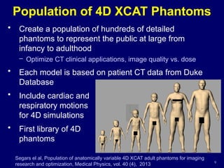
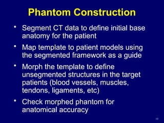
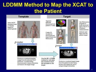
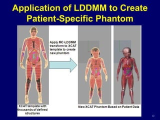
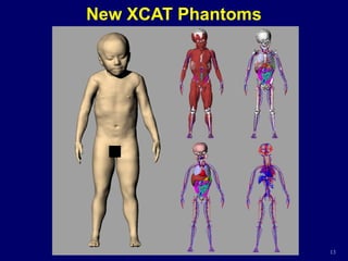
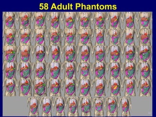
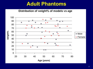
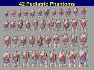
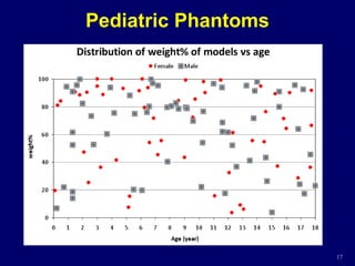
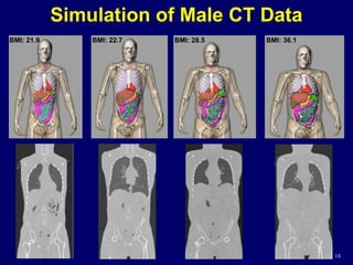
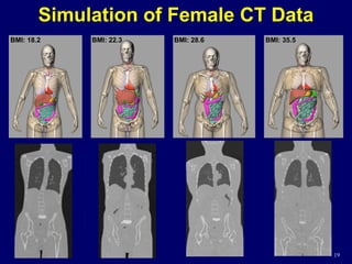
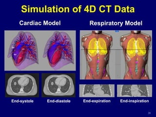
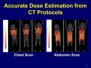
Population of 4D
Computational Phantoms for
CT Imaging Research and
Dosimetry
CARL E RAVIN
A D V A N C E D
I M A G I N G
LABORATORIES
W. Paul Segars, PhD
Carl E. Ravin Advanced Imaging
Labs
Duke University
Myocardial SPECT
Digital phantom
Emission
Computed
Tomography
(ECT)
X-ray CT
Transmission
Computed
Tomography
(TCT)
Medical Imaging Simulation
Computer model of
SPECT scanner
Images reconstructed
from projections
Computer model
of X-ray CT
scanner
Images reconstructed
from projections
Medical Imaging Simulation
• Advantages:
– Exact anatomy of the computer phantom is known
providing a gold standard to evaluate and improve
devices and techniques
– Computer phantoms can be altered easily to
model different anatomies and medical situations
providing a large population of subjects from
which to perform research
– No need to worry about overexposing the
phantom to radiation or being sued by the
phantom (IRB approval not needed)
4D eXtended CArdiac-Torso (XCAT) Phantoms
4
Segars el al, 4D XCAT phantom for multimodality imaging research, Medical Physics,
vol. 37 (9), 2010
5
Detailed Brain Model based on MRI
4D XCAT Phantom Anatomy
6
4D XCAT Phantom Anatomy
Detail in the hands and feet
Adding nervous and
lymphatic systems
Cardiac and Respiratory Models
Cardiac Model Based on 4D Tagged MRI and CT
Respiratory Model Based on 4D CT 7
Imaging Simulations using the
Computerized XCAT Phantoms
8
Population of 4D XCAT Phantoms
• Create a population of hundreds of detailed
phantoms to represent the public at large from
infancy to adulthood
– Optimize CT clinical applications, image quality vs. dose
• Each model is based on patient CT data from Duke
Database
• Include cardiac and
respiratory motions
for 4D simulations
• First library of 4D
phantoms
9
Segars el al, Population of anatomically variable 4D XCAT adult phantoms for imaging
research and optimization, Medical Physics, vol. 40 (4), 2013
Phantom Construction
• Segment CT data to define initial base
anatomy for the patient
• Map template to patient models using
the segmented framework as a guide
• Morph the template to define
unsegmented structures in the target
patients (blood vessels, muscles,
tendons, ligaments, etc)
• Check morphed phantom for
anatomical accuracy
10
11
LDDMM Method to Map the XCAT to
the Patient
12
Application of LDDMM to Create
Patient-Specific Phantom
13
New XCAT Phantoms
58 Adult Phantoms
14
Adult Phantoms
15
42 Pediatric Phantoms
16
Pediatric Phantoms
17
18
Simulation of Male CT Data
BMI: 21.9 BMI: 22.7 BMI: 28.5 BMI: 36.1
18
19
Simulation of Female CT Data
BMI: 18.2 BMI: 22.3 BMI: 28.6 BMI: 35.5
19
Simulation of 4D CT Data
End-diastole
End-systole End-expiration End-inspiration
Cardiac Model Respiratory Model
20
Accurate Dose Estimation from
CT Protocols
21
Chest Scan Abdomen Scan
Ultimate Goal
22
• Create hundreds of models
representing both genders with varying
ages, heights, and weights
encompassing the full range from
pediatric to adult patients
• Optimize CT clinical applications in
terms of image quality versus radiation
dose
• Distribute the phantoms for research
Conclusions
• The phantoms developed in this work
will have a widespread use in CT
imaging research to quantitatively
evaluate and improve imaging devices
and techniques and to investigate the
effects of anatomy and motion
• They can also be used to investigate
patient and population-based dose
correlations in CT and to enable
prospective estimation of CT dose and
radiation risk
23

4D computed tomography imaging in radiotherapy

  • 1.
    Population of 4D ComputationalPhantoms for CT Imaging Research and Dosimetry CARL E RAVIN A D V A N C E D I M A G I N G LABORATORIES W. Paul Segars, PhD Carl E. Ravin Advanced Imaging Labs Duke University
  • 2.
    Myocardial SPECT Digital phantom Emission Computed Tomography (ECT) X-rayCT Transmission Computed Tomography (TCT) Medical Imaging Simulation Computer model of SPECT scanner Images reconstructed from projections Computer model of X-ray CT scanner Images reconstructed from projections
  • 3.
    Medical Imaging Simulation •Advantages: – Exact anatomy of the computer phantom is known providing a gold standard to evaluate and improve devices and techniques – Computer phantoms can be altered easily to model different anatomies and medical situations providing a large population of subjects from which to perform research – No need to worry about overexposing the phantom to radiation or being sued by the phantom (IRB approval not needed)
  • 4.
    4D eXtended CArdiac-Torso(XCAT) Phantoms 4 Segars el al, 4D XCAT phantom for multimodality imaging research, Medical Physics, vol. 37 (9), 2010
  • 5.
    5 Detailed Brain Modelbased on MRI 4D XCAT Phantom Anatomy
  • 6.
    6 4D XCAT PhantomAnatomy Detail in the hands and feet Adding nervous and lymphatic systems
  • 7.
    Cardiac and RespiratoryModels Cardiac Model Based on 4D Tagged MRI and CT Respiratory Model Based on 4D CT 7
  • 8.
    Imaging Simulations usingthe Computerized XCAT Phantoms 8
  • 9.
    Population of 4DXCAT Phantoms • Create a population of hundreds of detailed phantoms to represent the public at large from infancy to adulthood – Optimize CT clinical applications, image quality vs. dose • Each model is based on patient CT data from Duke Database • Include cardiac and respiratory motions for 4D simulations • First library of 4D phantoms 9 Segars el al, Population of anatomically variable 4D XCAT adult phantoms for imaging research and optimization, Medical Physics, vol. 40 (4), 2013
  • 10.
    Phantom Construction • SegmentCT data to define initial base anatomy for the patient • Map template to patient models using the segmented framework as a guide • Morph the template to define unsegmented structures in the target patients (blood vessels, muscles, tendons, ligaments, etc) • Check morphed phantom for anatomical accuracy 10
  • 11.
    11 LDDMM Method toMap the XCAT to the Patient
  • 12.
    12 Application of LDDMMto Create Patient-Specific Phantom
  • 13.
  • 14.
  • 15.
  • 16.
  • 17.
  • 18.
    18 Simulation of MaleCT Data BMI: 21.9 BMI: 22.7 BMI: 28.5 BMI: 36.1 18
  • 19.
    19 Simulation of FemaleCT Data BMI: 18.2 BMI: 22.3 BMI: 28.6 BMI: 35.5 19
  • 20.
    Simulation of 4DCT Data End-diastole End-systole End-expiration End-inspiration Cardiac Model Respiratory Model 20
  • 21.
    Accurate Dose Estimationfrom CT Protocols 21 Chest Scan Abdomen Scan
  • 22.
    Ultimate Goal 22 • Createhundreds of models representing both genders with varying ages, heights, and weights encompassing the full range from pediatric to adult patients • Optimize CT clinical applications in terms of image quality versus radiation dose • Distribute the phantoms for research
  • 23.
    Conclusions • The phantomsdeveloped in this work will have a widespread use in CT imaging research to quantitatively evaluate and improve imaging devices and techniques and to investigate the effects of anatomy and motion • They can also be used to investigate patient and population-based dose correlations in CT and to enable prospective estimation of CT dose and radiation risk 23

Editor's Notes

  • #2 Computational phantoms are used as part of medical imaging simulation studies. These basically involve performing a clinical experiment entirely on your computer.
  • #4 These are the 4D XCAT series of phantoms. XCAT stands for extended cardiac-torso. Originally these were just chest models then they got extended. Organs in the phantoms are formed using NURBS surfaces which can accurately model anatomy and are flexible so they can be manipulated. Anatomy originally based on Visible Human data, but using the flexibility of NURBS they underwent an extreme makeover to get them to match 50th percentile measurements and to pretty up the faces.
  • #7 The XCAT phantom includes realistic models for the cardiac and respiratory motions. The beating heart model was based on tagged MRI and cardiac gated CT data. The respiratory motion is based on respiratory mechanics. The extents of its motion were based on respiratory-gated CT data. Both motions are parameterized to simulate different normal and abnormal conditions.
  • #8 Combined with freely available models of the imaging process, the phantoms are capable of simulating realistic imaging data, including all the artifacts of current scanners. The phantoms give you a method to perform a clinical experiment entirely on the computer.
  • #9 In addition to the original phantoms based on the Visible Human data, we are now creating a series of new 4D models based on patient CT data. Modeling patient variability is essential to mimic clinical trials. You can’t be limited to one or two anatomies. Each new phantom we create includes the same amount of detail as the original XCAT models. They also include the cardiac and respiratory motions. This work will result in the first library of 4D computational phantoms.
  • #10 These are the steps used to create the new adult phantoms. We first segment the CT data to create an initial model for the patient. We then …
  • #11 A template (I0) and target image (I1) are required in order to calculate the LDDMM transform. For our application, the templates are our existing XCAT adult phantoms. The template and target images for each patient case were created by voxelizing the template XCAT phantom and the initial patient model. Each structure is assigned a unique integer ID in the target image to drive the LDDMM transform. The template image was set to contain the same structures and intensities as the target so the images have a 1:1 correspondence. Given the template and target images, the LDDMM method calculates the high level transform to map the whole-body template (XCAT) to the target (patient framework).
  • #12 Once the transform was determined, it was applied to the template XCAT to create the patient-specific XCAT phantom containing all anatomical structures. So now the mystery is taken out of the miracle.
  • #18 One can see rapidly the advantage of having a patient-specific set of phantoms, which is that multiple body shapes and sizes can be used for imaging studies.
  • #19 One can see rapidly the advantage of having a patient-specific set of phantoms, which is that multiple body shapes and sizes can be used for imaging studies.
  • #20 With 4D modeling, our phantoms have pushed the envelope in what we can do with simulations.
  • #23 Mention Rachel Tian’s talk to be delivered Wednesday - “Patient-Specific Radiation Dose and Risk Estimation for Abdominopelvic CT…” Ultimately, we will expand this database to include 400 phantoms. Each will be 4D with parameterized heart and lung motion.