
Recommended
More Related Content
Similar to neuroaxialanaesthesia-160620135003.pptx
Similar to neuroaxialanaesthesia-160620135003.pptx (20)
Recently uploaded
Recently uploaded (20)
neuroaxialanaesthesia-160620135003.pptx
- 2. Neuraxial Anesthesia • Neuraxial anesthesia is a type of regional anesthesia that involves injection ofanesthetic medication in the fatty tissue that surround the nerve roots as they exist the spine (also known as an epidural and caudal) or into the cerebrospinal fluid which surrounds the spinal cord (also known as aspinal).
- 3. HISTORY • 1885 - J.Leonard Corning – first spinal anesthetic was administered accidentally Theneedle wasmade of gold • 1898 -August Bier - first planned spinalanesthesia for surgery • In 1921, Spanishmilitary surgeon Fidel Pagés(1886– 1923) developed the modern technique of lumbar epidural anesthesia
- 4. Advantages of Regional Anesthesia overGA Safe, reliable technique in patients at risk of apnoea, bradycardia, desaturation, cardiac or respiratory complications afterGA Good alternative for day caresurgeries Minimal risk of postoperative respiratorydepression Limited stress response tosurgery Cost effective
- 5. Spinal anesthesia Spinal anesthesia involves the use of small amounts of local anesthetic injected into the subarachnoid space to produce a reversible loss of sensation and motor function. The anesthesia provider places the needle below L2in the adult patient to avoid trauma to the spinal cord. Spinalanesthesia provides excellentoperating conditions for – Hernia (Inguinal or epigastric). – Haemorrhoidectomy , fistula ,fissure. – cystectoscopy – Transurethral resection of the prostate and transurethral resection of the bladder tumors. – Abdominal and vaginal hysterectomies – Caesareansections. – Lower limb surgery(orthopedic,plastic,vascular)
- 6. Vertebral Anatomy • Eachvertebra consists of apedicle, transverse process, superior and inferior articular processes, and aspinous process. • Eachvertebra is connected to the nextby intervertebral disks. • There are 2 superior and inferior articular processes (synovial joints) on each vertebra that allows for articulation. • Pedicles contain anotch superiorly and inferiorly to allow the spinal nerve root to exit the vertebral column.
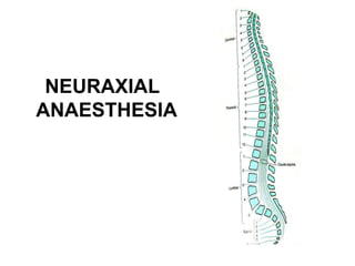
- 7. VERTEBRA 33Vertebrae ◦ 7 Cervical ◦ 12Thoracic ◦ 5 Lumbar ◦ 5 Sacral ◦ 4Coccygeal
- 8. Vertebral Anatomy- TopView T ransverse Process V ertebral Body SpinalCanal Spinous Process Lamina
- 9. Ligaments that support thevertebral column Ventral side: Anterior and posterior longitudinal ligaments Dorsalside: Important since these are the structures your needle will pass through!
- 10. Spinal Cord • Spinal Cord – Adult • Begins: Foramen Magnum • Ends:L1 – Newborn • Begins: Foramen Magnum • Ends:L3 – Terminal End:Conus Medullaris – Filum Terminale:Anchors in sacralregion – CaudaEquina: Nerve group of lower dural sac
- 11. Termination of SpinalCord In adults usually ends at L1. Infants L3 There are anatomical variations. For most adults it is generally safeto place aspinal needle below L2unless there is aknown anatomic variation.
- 12. Sagittal Section Through LumberVertebrae Supraspinous Ligament (Outer most layer) Intraspinous Ligament (Middle layer) Ligamentum Flavum (Inner most layer)
- 13. CSF • Clear fluid that fills the subarachnoidspace • Total volume in adults is 100-150ml • Volume found in the subarachnoid spaceis25-35 ml • Continually produced at arate of450 ml per 24 hour period replacing itself 3-4times • Reabsorbed into the blood stream byarachnoid villi andgranulations • Specific gravity is between 1.003-1.009 (this will play acrucial role in the baracity of local anesthetic that onechooses)
- 14. Membranes that surround the spinalcord • Piamater- highly vascular,covers the spinal cord and brain • Arachnoid mater- non vascular and attached to the dura mater. Principal barrier to the migration of medications in and out of theCSF • Dura mater (“tough mother”)- extension ofthe cranial dura mater, extends from the foramen magnum to S2(ending at the filum terminale)
- 15. Adapted with permission from “Unintended subdural injection: a complication of epidural anesthesia- a case report”, AANA Journal, vol. 74, no. 3, 2006.
- 16. CONTRAINDICATIONS Absolute Patient Refusal Infection At TheSite Of Injection Coagulopathy And Other BleedingDisorders SevereHypovolemia Increased Intracranial Pressure SevereAortic Stenosis SevereMitral Stenosis
- 17. CONTRAINDICATIONS Relative Sepsis Uncoperative Patient Preexisting Neurological Deficits SevereSpinal Deformity Controversial Prior SurgeryAt TheSite Of Injection Complicated Surgery ProlongedOperation Major BloodLoss
- 18. Spinal Technique • Preparation & Monitoring – EKG – NBP – PulseOximeter • Patient Positioning – Lateral decubitous – Sitting – Prone (hypobaric technique)
- 24. • Midline Approach – Skin – Subcutaneoustissue – Supraspinous ligament – Interspinous ligament – Ligamentum flavum – Epidural space – Dura mater – Arachnoid mater • Paramedian or LateralApproach – Sameasmidline excluding supraspinous & interspinous ligaments Anatomic Approach
- 26. Complications of SpinalAnaesthesia • Hypotension • Bradycardia • Total SpinalAnesthesia • Neurological Complecations – CaudaEquina Syndrome • PostDural Puncture Headache • Infection • Backache
- 27. Spinal Pharmacology Factors Effecting Distribution Site of injection Shape of spinal column Patient height Angulation of needle Volume of CSF Characteristics of local anesthetic Density Specific gravity Baracity Dose Volume Patient position (during & after)
- 28. Spinal Pharmacology Anesthesia level is determined by patient position Uptake of local anesthetic occurs by diffusion Elimination determines duration of block Lipid solubility decreases vascular absorption Vasoconstriction can decrease rate of elimination
- 29. Cardiovascular Effects Blockade of Sympathetic Preganglionic Neurons Send signals to both arteries and veins Predominant action is venodilation Reduces: Venous return Stroke volume Cardiac output Blood pressure T1-T4 Blockade Causes unopposed vagal stimulation Bradycardia Associated with decrease venous return & cardioaccelerator fibers blockade Decreased venous return to right atrium causes decreased stretch receptor response
- 30. Hypotension Treatment Best way to treat is physiologic not pharmacologic Primary Treatment Increase the cardiac preload Large IV fluid bolus within 30 minutes prior to spinal placement, minimum 1 liter of crystalloids Secondary Treatment Pharmacologic Ephedrine is more effective than Phenylephrine
- 31. Respiratory System Healthy Patients Appropriate spinal blockade has little effect on ventilation High Spinal Decrease functional residual capacity (FRC) Paralysis of abdominal muscles Intercostal muscle paralysis interferes with coughing and clearing secretions Apnea is due to hypoperfusion of respiratory center
- 33. Introduction: • Epidural anesthesia is acentral neuraxialblock technique with manyapplications. • Theepidural spacewasfirst described by Corning in 1901, and Fidel Pagesfirst used epidural anaesthesiain humans in 1921. • In 1945 Tuohyintroduced the needle which is stillmost commonly used for epiduralanesthesia. • it can be used asan anesthetic, asan analgesic adjuvant to general anesthesia, and forpostoperative analgesia in procedures involving the lower limbs, perineum, pelvis, abdomen andthorax.
- 34. Indications: • Epidural anaesthesia canbe used assole anaesthetic for procedures involving the lower limbs, pelvis, perineum and lower abdomen. • It is possible to perform upper abdominal and thoracic procedures under epidural anaesthesia alone, but the height of block required, with its attendant side effects, make it difficult to avoid significant patient discomfort and risk. • Theadvantage of epidural over spinal anaesthesia isthe ability to maintain continuous anaesthesia after placement of an epidural catheter, thus making it suitable for procedures of longduration.
- 37. Technique of Epidural Anesthesia Loss of resistance technique to identify the epidural space. – 0.5% Bupivacaine (mainly) or lidocaine (2.0%) is usually used to produce epidural anaesthesia. – Local anaesthetic solutions are deposited in the epidural space between the dura mater and the periosteum lining the vertebral canal. – The epidural space contains adipose tissue, lymphatics and blood vessels. – The injected local anaesthetic solution produces analgesia by blocking conduction at the spinal nerve roots. 32
- 38. 33
- 39. Indication and Contraindication: • The same of spinal anaesthesia. • Additional indication is the post operative Pain management using the epidural catheter technique. • Complications: the same of spinal anaesthesia, except the postdural puncture headache. 35
- 40. Detail Technique of Epidural Anaesthesia • Usinglocal anaesthetic raise asubcutaneous wheal at the midpoint between two adjacentvertebrae. • Inflitrate deeper in the midline and paraspinously toanaesthetise the posterior structures. • Insert epidural needle to the skin at this point,and advance through the supraspinous ligament, with the needle pointing in aslightly cephalad (upward) direction. • Then advance the needle into the interspinous ligament, whichis encountered at adepth of 2-3cm Until distinct sensation of increased resistance is felt astheneedle passesinto the ligamentum flavum.
- 41. With 5-10ml of air in the syringe, attach it to the hub of the needle once it hasentered the interspinous ligament. Theplunger is gently pressed,and if there is resistance ("bounce"), the needle is very carefully advanced, with the dorsum of both hands resting against the back to providestability. After 2-3mm, the plunger is again gently pressed,and thisprocedure is repeated asthe needle is carefully advancedthrough thetissues. Thedistinctive decreasein resistance when the needle entersthe ligamentum flavum is felt, and theprocessis continued in 2mm increments. There is usually a distinctive "click" when the needle enters the epidural space, and provided great care is taken, and the needle only advanced in 2mm increments, the needle should stop before it reaches the dura. At this point air can be injected into the epidural spacevery easily.The syringe is removed and the catheterthreaded
- 42. • Removethe syringe and thread the catheter gently via theneedle into the epiduralspace. • Thecatheter hasmarkings showing the distance from its tip, and should be advancedto 15-18cm at the hub of the needle, to ensure that asufficient length of catheter hasentered the epiduralspace. • Removethe needle carefully, ensuring that the catheter isnot drawn back with it. • The markings on the needle will show the depth of the needle from the skin to the epidural space, and this distance will help determine the depth to which the catheter should be inserted at the skin. • For example, if the needle entered the epidural space at a depth of 5cm, the catheter should be withdrawn so that the 10cm mark is at the skin, thus leaving approximately 5cm of the catheter inside the epidural space,which is an appropriate length.
- 45. 34
- 46. Choice of drugs: • Thechoice of drugs administered epidurally depends onthe indication for theepidural: • Surgical anaesthesia:Requires dense sensory block and usually moderate to dense motor block. T oachieve this, concentrated local anaesthetic preparations are required. Themost commonly usedlocal anaesthetics in this setting are 2% lignocaine 10-20ml (with or without adrenaline 1:200 000) or 0.5% bupivacaine 10-20ml. The latter hasalonger duration of action, but aslower onset time, compared with lignocaine. • For analgesia during labour: 0.1-0.25% bupivacaine 5-10ml ismore popular, asit produces lessmotor block. • Postoperative analgesia: weaker concentrations of bupivacaine, e.g. 0.1-0.166%with or without added lowdose opioids.
- 47. Differences between Spinal and Epidural Anesthesia Spinal anaesthesia Epidural Anaesthesia Level: below L1/L2, where the spinal cord ends Level: at any level of the vertebral column. Injection: subarachnoid space i.e, puncture of the dura mater Injection: epidural space (between Ligamentum flavum and dura mater) i.e ,without puncture of the dura mater Identification of the subarachnoid space: When CSF appears Identification of the epidural space: Using the Loss of Resistance technique. Doses: 2.5- 3.5 ml Bupivacaine 0.5% heavy Doses: 15- 20 ml Bupivacaine 0.5% Onset of action: Rapid (2-5 min) Onset of action: Slow (15-20 min) Density of block: More dense Density of block: Less dense Hypotension: Rapid Hypotension: Slow Headache: Is a probable complication Headache: Is not a probable complication. 41
- 48. Complications and SideEffects • Hypotension • Inadvertent high epiduralblock • Local anaesthetictoxicity • T otalspinal block
- 49. CAUDALANESTHESIA
- 50. Introduction: • Caudalanesthesia hasbeen used for many yearsandis the easiest and safest approach to the epidural space. When correctly performed there is little danger of either the spinal cord ordura being damaged. • It is used to provide peri and post operative analgesia in adults andchildren. • It may be the sole anesthetic for some procedures, or it may be combined with generalanesthesia.
- 51. Indications: 1. Anesthesia and analgesia below theumbilicus 2. Obstetric analgesia :For the 2nd stageor instrumental deliveries. Careshould be taken as the foetal head lies close to the site of injection and there is real risk of injectinglocal anesthetic into the foetus. 3. Chronic pain problems relating to lowerlimbs and lower abdominalpains.
- 52. Contraindications: 1. Infection near the site of theneedle insertion. 2. Coagulopathy or anti coagulation. 3. Congenital abnormalities of the lowerspine or meninges, becauseof the unclear or impalpable anatomy.
- 53. Anatomy: • Thecaudal epidural space is the lowest portion ofthe epidural space and is entered through the sacral hiatus. • Thesacrum is atriangular bone that consists of the five fused sacralvertebrae (S1-S5). • It articulates with the fifth lumber vertebra and the coccyx.
- 54. • Thesacral hiatus is adefect in the lower part of the posterior wall of the sacrum formed by the failure of the laminae of S5and/or S4to meet and fuse in themidline. • Thesacral canal is a continuation of the lumbar spinal canal which terminates at the sacralhiatus.
- 55. Choice of drugs &dosage: • Drugs that are commonly usedinclude : Lignocaine 1%and Bupivacaine 0.25%.
- 56. Technique: Thepatient is prepared as for generalanaesthesia: (1)He/she should be fasted (2)All appropriate equipment for resuscitation mustbe available. (3)An intravenous cannula should always be inserted in an upper limb, in caseof accidental intravenous injection, or profound sympathetic blockade from ahigh epiduralblock. (4)The procedure must be carried out with a strict aseptic technique. The skin should be thoroughly prepared and sterile glovesworn.
- 57. (5) Thereare three mainapproaches: • Theprone, the semi-prone, and the lateral. Thechoice depends on the preference of the anesthetist and the degree of sedation of the patient. Thecaudal spaceismade more prominent by asking the patient to internally rotate their ankles. Thesemi-prone position is preferred for the anesthetised or heavily sedated patient asthe airway is easier to control in this position, while still allowing reasonably easyaccessto the sacralhiatus. Thelateral position is often used in children, asthe landmarks are easier to find than in adults. Careshould be taken to avoid over flexing the hips (asforlumber epidurals) asthis can makethe landmarks more difficult to palpate
- 58. 6) Thelandmarks are palpated. Thesacral hiatus and the posterior superior iliac spines form an equilateral triangle pointing inferiorly. • Thesacral hiatus canbe located by first palpating the coccyx,and thensliding the palpating finger in a cephalad direction until a depression in the skin is felt.
- 60. 7) Oncethe sacral hiatus is identified the areaabove is carefully cleaned with antiseptic solution, anda 22 gaugecannula or needle is directed at about 90 degree to skin and inserted till a"click" is felt asthe sacro- coccygeal ligament is pierced.
- 61. Careshould be taken not to insert the needle too far asthe dura lies at orbelow the S2level in the child. Theneedle should be aspirated looking for either CSFor blood. Theinjection should never be more than 10ml/30 seconds • Further tests to confirm the correct position include: Introduction of asmall amount of air will not produce subcutaneous emphysema, and will be heard asa"woosh" sound if astethoscope is place further up the lumbarspine. There should be no local pain during injection.
- 62. (9)Asmall amount of local anaesthetic should beinjected asatest dose (2-4mls). It should not produce either a lump in the subcutaneous tissues, or afeeling of resistance to the injection, nor any systemic effects such asarrhythmias or hypotension. If the test dose does not produce any side effects then the rest of the drug is injected, the needle removed and the patient positioned for surgery. In the post-operative period, motor function must be checkedand the patient should not be allowed to try and walk until complete return of motor function is assured. The patient should not be discharged from hospital until he/she haspassedurine, asurinary retention is a recognized complication.
- 63. Complications: Intravascular or intraosseous injection:Thismay lead to grandmal seizuresand/or cardio-respiratory arrest. Dural puncture: Extreme care must be taken to avoid this asatotal spinal block will occur if the dose for acaudal block is injected into the subarachnoid space. If this occurs then the patient will become rapidly apnoeic and profoundly hypotensive. Perforation of the rectum: Contamination of the needleis extremely dangerous if it is then inserted into the epidural space. Sepsis:This should be avery rare occurrence if strict aseptic procedures are followed. Urinary retention. Haematoma
Editor's Notes
- Join the highest points on the iliac crest ,crosses at midline at the level of l4-5 interspace (tuffiers line) Most common site for giving spinal
- Cauda equine syndrome: injury to cauda equine l/to low backache,radiating to leg, bowel bladder dysfunction