
Recommended
More Related Content
Similar to COMPARATIVE PROTEIN ANALYSIS
Similar to COMPARATIVE PROTEIN ANALYSIS (20)
Recently uploaded
Recently uploaded (20)
COMPARATIVE PROTEIN ANALYSIS
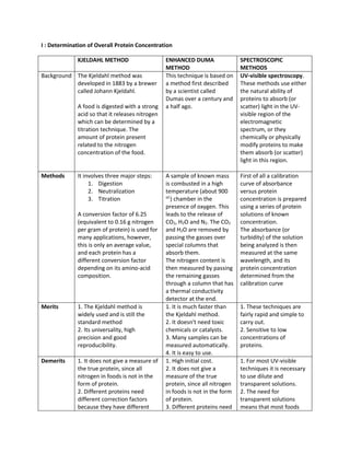
- 1. I : Determination of Overall Protein Concentration KJELDAHL METHOD ENHANCED DUMA METHOD SPECTROSCOPIC METHODS Background The Kjeldahl method was developed in 1883 by a brewer called Johann Kjeldahl. A food is digested with a strong acid so that it releases nitrogen which can be determined by a titration technique. The amount of protein present related to the nitrogen concentration of the food. This technique is based on a method first described by a scientist called Dumas over a century and a half ago. UV-visible spectroscopy. These methods use either the natural ability of proteins to absorb (or scatter) light in the UV- visible region of the electromagnetic spectrum, or they chemically or physically modify proteins to make them absorb (or scatter) light in this region. Methods It involves three major steps: 1. Digestion 2. Neutralization 3. Titration A conversion factor of 6.25 (equivalent to 0.16 g nitrogen per gram of protein) is used for many applications, however, this is only an average value, and each protein has a different conversion factor depending on its amino-acid composition. A sample of known mass is combusted in a high temperature (about 900 oC ) chamber in the presence of oxygen. This leads to the release of CO2, H2O and N2. The CO2 and H2O are removed by passing the gasses over special columns that absorb them. The nitrogen content is then measured by passing the remaining gasses through a column that has a thermal conductivity detector at the end. First of all a calibration curve of absorbance versus protein concentration is prepared using a series of protein solutions of known concentration. The absorbance (or turbidity) of the solution being analyzed is then measured at the same wavelength, and its protein concentration determined from the calibration curve Merits 1. The Kjeldahl method is widely used and is still the standard method 2. Its universality, high precision and good reproducibility. 1. It is much faster than the Kjeldahl method. 2. It doesn't need toxic chemicals or catalysts. 3. Many samples can be measured automatically. 4. It is easy to use. 1. These techniques are fairly rapid and simple to carry out. 2. Sensitive to low concentrations of proteins. Demerits 1. It does not give a measure of the true protein, since all nitrogen in foods is not in the form of protein. 2. Different proteins need different correction factors because they have different 1. High initial cost. 2. It does not give a measure of the true protein, since all nitrogen in foods is not in the form of protein. 3. Different proteins need 1. For most UV-visible techniques it is necessary to use dilute and transparent solutions. 2. The need for transparent solutions means that most foods
- 2. amino acid sequences. 3. The use of concentrated sulfuric acid at high temperatures poses a considerable hazard, as does the use of some of the possible catalysts. 4. The technique is time consuming to carry-out. different correction factors because they have different amino acid sequences. 4. The small sample size makes it difficult to obtain a representative sample. must undergo significant amounts of sample preparation before they can be analyzed. 3. It can be time consuming and laborious. II: UV VISIBLE methods for determining the protein content of Samples Direct measurement at 280nm Biuret method Lowry method Dye binding method Merit: simple to carry out, it is nondestructive, and no special reagents are required. Demerits: nucleic acids also absorb strongly at 280 nm and can interfere with the measurement of the protein if they are present in sufficient concentrations Principle: A violet- purplish color is produced when cupric ions (Cu2+) interact with peptide bonds under alkaline conditions. Merits: there is no interference from materials that adsorb at lower wavelengths, Demerits: it has a relatively low sensitivity compared to other UV- visible methods. Principle: The Lowry method combines the biuret reagent with another reagent (the Folin-Ciocalteau phenol reagent) which reacts with tyrosine and tryptophan residues in proteins. This gives a bluish color which can be read somewhere between 500 - 750 nm depending on the sensitivity required. Merits: This method is more sensitive to low concentrations of proteins than the biuret method. Demerits: It can be time consuming and laborious Principle: A known excess of a negatively charged (anionic) dye is added to a protein solution whose pH is adjusted so that the proteins are positively charged (i.e. < the isoelectric point). The proteins form an insoluble complex with the dye but the unbound dye remains soluble. The amount of unbound dye remaining in solution is determined by measuring its absorbance. Merits: These techniques are fairly rapid and simple to carry out. 2. Sensitive to low concentrations of proteins. Demerits It can be time consuming and laborious