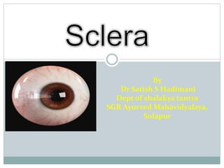
Anatomy, Layers and Structures of the Sclera
•Download as PPTX, PDF•
6 likes•591 views

Detailed explanation of anatomy of sclera

Recommended
More Related Content
What's hot
What's hot (20)
Similar to Anatomy, Layers and Structures of the Sclera
Similar to Anatomy, Layers and Structures of the Sclera (20)
Recently uploaded
Recently uploaded (20)
Anatomy, Layers and Structures of the Sclera
- 1. By Dr Satish S Hadimani Dept of shalakya tantra SGR Ayurved Mahavidyalaya, Solapur
- 3. Sclera Introduction Gross anatomy Layers Blood supply, drainage and nerve supply
- 4. Introduction Its dense connective tissue that accounts for five sixths of the outer coat of the eyeball Sklera mannix- hard membrane 1. Protects intraocular components from trauma, light, and mechanical displacement 2. Withstands the considerable expansive force generated by the intraocular pressure maintaining the shape of the globe 3. Provides attachment sites for the extraocular muscles.
- 6. Insertion Of Rectus Muscle
- 7. Tenons capsule • Fascial sheath of the eyeball • And it’s connection with sclera and optic nerve
- 8. Ant scleral foramen & Scleral spur
- 9. Post scleral foramen & lamina cribrosa
- 10. Apertures
- 12. Lamina cribrosa: It is a sieve-like sclera from which the fibres of the optic nerve pass. Scleral apertures(emissaria): Sclera has three sets of apertures : Posterior aperture : situated around the optic nerve. Middle apertures: situated 4-7mm posterior to the equator. Anterior aperture: situated 3-4mm away from the limbus
- 13. Microscopic structure Histologically, sclera consist of following three layers Episcleral tissue Sclera proper Lamina fusca
- 14. Episclera Superficial aspect of sclera bundles of collagen circumferentially arranged rich blood supply anteriorly thickest anterior to the rectus muscle insertions and becomes progressively thinner toward the back of the eye.
- 15. Scleral stroma bundles of collagen fibers with fibroblasts, melanocytes, elastic fibers, proteoglycans, and glycoproteins variability in collagen fiber diameter, interlacing in bundles of collagen, and relative deficiency in water-binding substances accounts for the scleral dull-white color.
- 16. Lamina fusca Brown colour due to melanocytes grooves for the passage of ciliary vessels and nerves (emissary canals) attached to the choroid by fine collagen fibers
- 17. Blood supply Episclera-anterior ciliary arteries posterior ciliary arteries Scleral stroma-relatively avasculature structure
- 19. Venous drainage Episcleral collecting veins Vortex veins Anterior ciliary veins
- 20. Nerve supply Rich in nerve supply Anterior sclera- long posterior ciliary nerves Posterior sclera- short posterior ciliary nerves Pain- inflammation, stretching due to oedema and movement of eye
- 21. Thank you