The concept of peripheral nerve repair
•Download as PPTX, PDF•
8 likes•2,210 views

illustrations and material from internet

Recommended
More Related Content
What's hot
What's hot (20)
Similar to The concept of peripheral nerve repair
Similar to The concept of peripheral nerve repair (20)
More from Rekha Pathak
More from Rekha Pathak (20)
Recently uploaded
Recently uploaded (20)
The concept of peripheral nerve repair
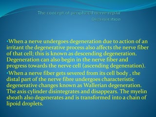
- 1. •When a nerve undergoes degeneration due to action of an irritant the degenerative process also affects the nerve fiber of that cell; this is known as descending degeneration. Degeneration can also begin in the nerve fiber and progress towards the nerve cell (ascending degeneration). •When a nerve fiber gets severed from its cell body , the distal part of the nerve fibre undergoes characteristic degenerative changes known as Wallerian degeneration. The axis cylinder disintegrates and disappears. The myelin sheath also degenerates and is transformed into a chain of lipoid droplets.
- 2. The cells of the sheath of Schwann proliferate and get converted into phagocytes which remove the remnants of axis cylinder and the lipoid droplets. Similar changes occur in the proximal part up to the first node of Ranvier.
- 3. Regeneration In a degenerated nerve fiber there are also attempts at repair. Nerve fiber in the central nervous system cannot regenerate but the peripheral nerves regenerate fairly rapidly. Schwannn cells play a leading role in the healing of nerves. If the sheath of schwann is intact, the Schwann cells proliferate and arrange themselves in both proximal and distal ends in the form of a tube.
- 4. Along this tube new axis cylinders grow and unite the two severed ends. They fail to heal the gap if it is more than one inch. In such casesthe gap is filled in by granulation tissue which originates from the three connective
- 5. tissue coverings of the nerve and its bundles. The Schwann cells proliferate at both ends. In case of amputation the axon fibrils coil up to and form a nodule called amputation neuroma which is covered by fibrous tissue.
- 6. When a peripheral nerve is cut degenerative chances occur in the neurons. These changes are called Nissl’s Degeneration in which the cells become enlarged and the nucleus is pushed to a side . Chromatolysis of Nissl’s substance occurs after breaking up. When regeneration of the nerve fiber starts the neuron tens to return to normal. Nissl granule reappear n nucleus takes up to a central position and the cell become smaller. Repair in a nerve fibre is a prolonged process requiring 10- 12 months for complete healing. Repair of the nerve. Repair of the nerve fibers of central nervous system lacking a sheath of Schwann does not occur.
- 7. The Peripheral Nerve System is responsible for the relaying of messages between the brain and spinal cord (Central Nervous System) to the muscles, joints, organs and skin. Many millions of people unfortunately suffer from varying degrees of nerve injuries and loss in limb use.
- 8. These injuries may be are most commonly caused by trauma. When a nerve is compressed or injured, a variety of damage types can usually be found – each has its own time course for recovery. A simple compression will recover very rapidly, where as if the nerve sheath is destroyed, then weeks may be required for a new insulating lining to be rebuilt.
- 9. If the axon itself is disrupted, then a replacement must be grown from the cut point all the way to the skin or muscle which happens at a rate of 1-4 millimetres per day. If the conduit itself is destroyed, then a regrowing nerve may never find its way past the point of injury meaning permanent loss. If a muscle loses its nerve supply, then the muscle nerve receptors disappear within 12-18 months.
- 10. PRIMARY NERVE REPAIR INDICATIONS Any nerve which has been completely transected, If the wound is clean, attempt of the repair should be taken up immediately. If it is grossly contaminated, control infection first.
- 11. Equipments Finest monofilament sutures, needles, 8/0 polypropylene/ prolene sutures on 3 mm atraumatic needles and needle holder should be used. Use ophthalmic forceps and needle holders, and operating spectacles
- 12. Don’t use silk, catgut, human hair, or dexon because these are irritant. Coarse sutures may cause so much fibrosis that the nerve will never function again.Find the cut ends of the nerve. Put his limb in the position which will help to bring them together.
- 13. Trim back both the cut ends of the nerve with a new sterile razor blade as in A, Usually about 2 mm is enough. Match the cut ends in their correct anatomical positions, without rotation. There are usually very fine blood vessels on one side of a nerve which will enable you to distinguish its two sides. Study the cross section of its fasciculi carefully, and get the two cut ends to match. The exposed nerve must be freed from the surrounding tissues for some distance proximal and distal to the site of injury.
- 14. Haemastasis is vital during peripheral nerve surgery. Try to put all sutures into the outer sheath of the nerve. Sutures deep inside it will interfere with its function seriously. Pass two stay sutures through the outer sheath of the nerve on either side. Tie them and leave the ends long (B). Carefully hold the two ends of the nerve together, and ask an assistant to hold the ends in artery forceps. Put one or two sutures into the front of the nerve (C). Pass one of your stay sutures behind the anastomosis (D), and cross the other one in front of it, so that you rotate the nerve as you pull them and expose its back (E).
- 15. Note: (1) Try not to put more than 8 sutures into the nerve, or there will be unnecessary fibrosis. (2) Don’t let any nerve fibres stick out of the suture line. Manage the wound . If you are leaving the wound open for delayed closure, try to cover the sutured nerve with muscle or skin, and don’t leave it naked in the wound.
- 16. If necessary, make relieving incisions, so that you can move skin over to cover the nerve, or cover it with a transposition flap,. or, least satisfactorily, cover it with a split skin graft. Splint the patient’s limb in the position which best relieves tension on the nerve. If you fail to do this, the sutures may pull out.
- 18. Nerve repair