Treating Basal Cell Carcinoma
•
1 like•729 views

Dr Patrick Treacy shares some of his most challenging cases. This month he talks about treating basal cell cancer

Recommended
More Related Content
What's hot
What's hot (20)
Viewers also liked
Similar to Treating Basal Cell Carcinoma
Similar to Treating Basal Cell Carcinoma (20)
More from Dr. Patrick J. Treacy
More from Dr. Patrick J. Treacy (20)
Recently uploaded
Recently uploaded (20)
Treating Basal Cell Carcinoma
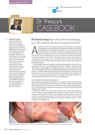
- 1. 48 S K I N/ D E RMATOLOGY Aesthetic Medicine • November 2014 SPONSORED BY Dr Patrick Treacy shares some of his most challenging cases. This month he talks about treating basal cell cancer Dr Treacy’s CASEBOOK DR PATRICK TREACY is chairman of the Irish Association of Cosmetic Doctors and Irish regional representative of the British College of Aesthetic Medicine (BCAM). He is European medical advisor to Network Lipolysis and Consulting Rooms and holds higher qualifications in dermatology, laser technology and skin resurfacing. In 2012 and 2013 he won awards for ‘Best Innovative Techniques’ for his contributions to facial aesthetics and hair transplants. Dr Treacy also sits on the editorial boards of three international journals and features regularly on international television and radio programmes. He was a faculty member at IMCAS Paris 2013, AMWC Monaco 2013, EAMWC Moscow 2013 and a keynote speaker for the American Academy of Anti-Ageing Medicine in Mexico City this year. >> A 43-year-old Irish male patient was referred to Ailesbury with a slowly enlarging lesion on the right side of his neck that wouldn’t heal and bled when traumatised. The patient described it as an acne lump that had appeared about a year before and he felt in the beginning that he could express some pus or fluid from it from time to time. More recently he had become concerned because a course of antibiotics from his GP had failed to make any impact on the lesion. The patient gave a history of occupational sun exposure as he worked in the construction industry. There was no relevant medical history and the patient did not have any clinical evidence of autoimmune or allergic diseases. On examination the lesion appeared eroded and ulcerated and bled easily when traumatised. There was some crusting at the anterior margins. The presence of rolled borders, pearly edges and telangiectases gave a clinical suspicion of basal cell carcinoma (BCC) and a decision was made to proceed to removal rather than do a biopsy. The author feels that any doctor should consider BCC in any patient with a history of a sore or skin anomaly that does not heal within four to six weeks and occurs on sun-exposed skin, especially if it is dimpled in the middle. These tumors may take many months or years to reach even 1cm in diameter. TREATMENT (EXCISIONAL SURGERY) After numbing the area with local anesthesia, an 11 scalpel to remove the entire growth along with a surrounding border of normal skin as a safety margin. The skin around the surgical site is then closed with a number of stitches, and the excised tissue is sent to the laboratory for microscopic examination to verify that all the malignant cells have been removed. People who sunburn are more likely to develop skin cancer than those who do not; however, sunlight damages the skin with or without sunburn. History of any prior treatment to the index tumor should be elicited, as well as history of any prior non-melanoma skin cancer. In patients with recurrent tumors, deeper invasion should be expected. Recurrence following radiation therapy is often biologically more aggressive. CASE FILES www.aestheticmed.co.uk ABOVE AND OPPOSITE PAGE: Resection of basal cell carcinoma from neck area of patient with deep sutures and primary closure
- 2. SKIN/DERMATOLOGY Characteristic features of BCC tumors include the following: Slow growing (0.5 cm in 1-2 y) Erosion or ulceration, often central Telangiectases over the surface Rolled (raised) border Waxy papules with central depression Pearly appearance Bleeding, especially when traumatized Crusting Translucency BCC seldom causes regional or distant metastasis, with the exception of the basosquamous type. To evaluate for lymph node metastasis, particular attention should be taken to examine the parotid posterior auricular, suboccipital, and upper cervical groups of lymph nodes. HISTOPATHOLOGICAL TYPES There are several different histopathological types of BCC exist, each with distinct clinical presentation.1 Nodular - Cystic, pigmented, keratotic Infiltrative Micronodular Morpheaform Superficial Nodular basal cell carcinoma Nodular basal cell carcinoma is the most common type of basal cell carcinoma and usually presents as a round, pearly, flesh- colored papule with telangiectases. More than 60% of BCCs belong to this subtype. As it enlarges, it frequently ulcerates centrally, leaving a raised, pearly border with telangiectases, which aids in making the diagnosis. The tumor may present as a cyst or pigmented with brown-black macules making it like a melanoma. Keratotic BCC is a variant of nodular BCC and is usually clinically indistinguishable from nodular BCC histologically. Infiltrative basal cell carcinoma With this variant of BCC, tumor infiltrates the dermis in thin strands between collagen fibers, making tumor margins less clinically apparent. Mohs micrographic surgery is the treatment of choice for infiltrative basal cell carcinoma. Because of its growth pattern, electrodessication and curettage has a significantly higher recurrence rate when used to treat infiltrative BCC compared to the treatment of nodular BCC; other treatment methods should be sought. Micronodular basal cell carcinoma This aggressive BCC subtype has the typical BCC distribution. It is not prone to ulceration, it may appear yellow-white when stretched, and it is firm to the touch. It may have a seemingly well-defined border. Morpheaform (sclerosing) basal cell carcinoma Morpheaform basal cell carcinoma is an uncommon variant in which tumor cells induce a proliferation of fibroblasts within the dermis and an increased collagen deposition (sclerosis) that clinically resembles a scar. This form accounts for 10% of lesions. Such lesions appear as flat or slightly depressed, fibrotic, and firm. The morpheaform (sclerosing) type of basal cell carcinoma is often the most difficult type to diagnose, as it bears little resemblance to the typical nodular BCC. Ulceration, bleeding, and crusting are uncommon and these tumors are commonly mistaken for scar tissue. 49 Aesthetic Medicine • November 2014 SPONSORED BY CASE FILES www.aestheticmed.co.uk
- 3. 50 Aesthetic Medicine • November 2014 CASE FILES SPONSORED BY www.aestheticmed.co.uk Superficial basal cell carcinoma Superficial basal cell carcinomas are seen mostly on the upper trunk or shoulders. This type of BCC grows slowly, has minimal tendency to be invasive, and appears clinically as an erythematous, well-circumscribed patch or plaque, often with a whitish scale. Occasionally, minute eschars may appear within the patch or plaque. The tumor often appears multi-centric, with areas of clinically normal skin intervening among clinically involved areas. Gorlin syndrome or basal cell nevus syndrome Basal cell carcinoma (BCC) is also a feature of basal cell nevus syndrome (Gorlin syndrome)2 an autosomal dominant inherited condition. The gene responsible for this syndrome is located on arm 9q, and chromosome abnormalities develop in some patients. Multiple BCCs begin to appear after puberty on the face, trunk, and extremities. In many cases, the tumors are highly invasive and may involve areas around the eyes and nose.3 CONCLUSION With an incidence of 70 to over 800 new cases per 100,000 persons per year, basal cell carcinoma (BCC) is the commonest skin cancer, accounting for about 80% of all cases of non-melanoma skin cancer. BCCs are slow-growing, locally invasive, epidermal skin tumours which mainly affect white skinned people and very rarely metastasize. Of all BCCs, 85% occur in the head and neck region4 and the incidence of this carcinoma (BCC) is increasing worldwide. As a result therefore, the cosmetic outcome is a particularly important issue the demand for novel and effective treatment modalities for BCC is high.5 BCC is commonly treated with surgical excision, curettage, carbon dioxide (CO2) laser ablation or cryotherapy, depending on the tumor depth and the histological subtype of the BCC6. The effectiveness of excisional surgery does not match that of Mohs, but produces cure rates around 90%. Photodynamic therapy (PDT) has been widely used for the treatment of superficial BCC in preference to excision due to its minimal invasiveness and satisfactory cosmetic results.7,8 However, the efficacy of this treatment modality is limited in the treatment of deeper lesions and the more aggressive subtypes of BCC9. Retreatment and recurrences of the disease are frequent if the tumor depth is >2 mm.10 In order to improve the outcomes of BCC treatment, it is possible to combine CO2 laser ablation with topical methyl aminolevulinate (MAL) PDT and modified cryotherapy for the treatment of variable BCC. Conventional cryotherapy following PDT additionally kills the remaining cancer cells by interrupting vital metabolic cycles, destabilizing cell membranes, creating an adverse hyperosmolar environment and forming water crystals.9 The gold standard of treatment is Mohs surgical excision with histological control of excision margins, which has a five-year recurrence rate of less than 3% on the face. For superficial BCC, approved medications such as imiquimod (total remission rate, 82-90%) and topical 5-fluorouracil (80%) are available, as is photodynamic therapy (71-87%). Other ablative methods (laser, cryosurgery) are applicable in some cases. Radiotherapy is an alternative treatment for invasive, inoperable BCC, with five-year tumor control rates of 89-96%. Recently, drugs that inhibit an intracellular signaling pathway have become available for the treatment of locally advanced or metastatic BCC.11. AM REFERENCES 1. (Medscape) Robert S Bader, MD Dermatologist, Section of Dermatology, Department of Medicine, Broward Health - North Coauthor(s) Luigi Santacroce, MD Assistant Professor, Medical School, State University at Bari, Italy Laura Diomede University of Bari School of Medicine, Italy Andrew Scott Kennedy, MD Physician-in-Chief, Radiation Oncology 2. Gorlin RJ. Nevoid basal cell carcinoma (Gorlin) syndrome: unanswered issues. J Lab Clin Med. Dec 1999;134(6):551-2 3. Bernardini FP. Management of malignant and benign eyelid lesions. Curr Opin Ophthalmol. Oct 2006;17(5):480-4. 4. Miller SJ. Biology of basal cell carcinoma (Part I) J Am Acad Dermatol. 1991;24:1–13. 5. Barry J, Oon SF, Watson R, Barnes L. The management of basal cell carcinomas. Ir Med J. Jun 2006;99(6):179-81 6. Arits AH, Schlangen MH, Nelemans PJ, Kelleners-Smeets NW. Trends in the incidence of basal cell carcinoma by histopathological subtype. J Eur Acad Dermatol Venereol. 2011;25:565–569 7. Szeimies RM, Ibbotson S, Murrell DF, et al. A clinical study comparing methyl aminolevulinate photodynamic therapy and surgery in small superficial basal cell carcinoma (8–20 mm), with a 12-month follow-up. J Eur Acad Dermatol Venereol. 2008;22:1302–1311. 8. Fantini F, Greco A, Del Giovane C, et al. Photodynamic therapy for basal cell carcinoma: clinical and pathological determinants of response. J Eur Acad Dermatol Venereol. 2011;25:896–901. 9. Dandurand M, Petit T, Martel P, Guillot B. Management of basal cell carcinoma in adults Clinical practice guidelines. Eur J Dermatol. Jul-Aug 2006;16(4):394-401 10. J, Oon SF, Watson R, Barnes L. The management of basal cell carcinomas. Ir Med J. Jun 2006;99(6):179-81 11. Messeguer F, Serra-Guillen C, Echeverria B, et al. A pilot study of clinical efficacy of imiquimod and cryotherapy for the treatment of basal cell carcinoma with incomplete response to imiquimod. J Eur Acad Dermatol Venereol. 2012;26:879–881. 12. Dtsch Arztebl Int. 2014 May 30;111(22):389-95. doi: 10.3238/arztebl.2014.0389. Basal cell carcinoma-treatments for the commonest skin cancer. Berking C1, Hauschild A, Kölbl O, Mast G, Gutzmer R. S K I N/ D E RMATOLOGY HISTOLOGY CLINICAL DETAILS: Rule out basal cell carcinoma. MACROSCOPY: Labelled NC. R pos triangle neck: 5.2cm x 3.2cm of cream skin. All embedded. 1/1. MICROSCOPY: Skin, R pos triangle, Basal cell carcinoma, nodular and invasion of 1mm. PATHOLOGISTS: Prof.Kieran Sheahan MCRN 02276