
Recommended
More Related Content
Similar to OCT ango in ARMD and telangictasia.pptx
Similar to OCT ango in ARMD and telangictasia.pptx (20)
Recently uploaded
Recently uploaded (20)
OCT ango in ARMD and telangictasia.pptx
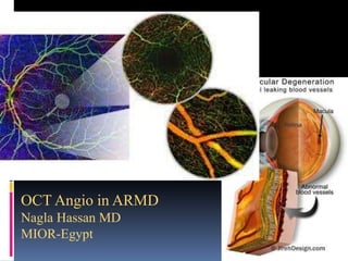
- 1. OCT Angio in ARMD Nagla Hassan MD MIOR-Egypt
- 2. ARMD ARMD is thought to be a group of genetically, environmentally-determined retinal degenerative , age related diseases, of the RPE cells ,which are essential for photoreceptor metabolism and removal of waste products. Dysfunctional cells accumulate undigested waste, which is clinically evident as focal yellow subretinal clumps, termed drusen. Drusen are the herald of ARMD, and large or numerous drusen are poor prognostic factors for future visual loss. The disruption of photoreceptor metabolism eventually causes areas of retinal atrophy or neovascularization.
- 4. Risk factors forAMD 4 Age1 -Genetic factors -Smoking -Hypertension and cardiovascular disease -Race -High cholesterol -Low intake of antioxidants
- 11. Dry type ARMD Areas of impaired choriocapillaris flow typically extended beyond the borders of the geographic atrophy GA. Eyes with dry AMD were shown to have a generalized decrease in choriocapillaris density, which was sometimes associated with drusen
- 13. The soft drusen shown are associated with areas of decreased signal in the choriocapillaris, which could indicate flow impairment
- 14. Soft drusen demonstrating an area of decreased signal in the choriocapillaris underlying the drusen.
- 16. OCTA in Neovascular AMD Calcifications: Type 1 40% under the RPE Type 2 9 % subretinal Type 3 34% intraretinal (RAP) Type 4 17% mixed
- 17. Choriocapillaris and the outer retina are shown two nets of abnormal vessels are shown surrounded by relatively homogenous choriocapillaris. The abnormal vessels exist both below and above Bruch’s membrane. Sulzbacher F, Pollreisz et al.
- 18. Abnormal vessels are shown surrounded by relatively homogenous choriocapillaris Kuehlewein L, Bansal M, et al.
- 19. OCT angiogram segmented so the choriocapillaris are shown. A circular net of abnormal vessels are shown surrounded by relatively homogenous choriocapillaris.The abnormal vessels exist both below and above Bruch’s membrane (in the outer retina).
- 20. LUMBROSO B, RISPOLI M ET AL
- 28. OCTA showed a membrane was identified as sea fan
- 29. OCTA showed a membarne idinfied as medussa
- 30. OCTA shaowed membranes lacking such distinct membrane morphology-ill-defined
- 31. The membrane was identified as long filamentous if the membrane had a dead-tree
- 34. Sulzbacher F, Pollreisz et al.
- 35. Sarks SH. Br J Ophthalmol.
- 36. Role of VEGF-Ain wetAMD progression VEGF-A Migrating endothelial cells form new blood vessels in formerly avascular space Hypoxia Proliferation Migration Proteolysis Vascular endothelial cell Other angiogenic growth factors Basement membrane
- 37. VEGF inhibitors Anti-VEGF therapies are established as a key molecule in neovascular AMD therapies 37
- 39. Before and after treatment
- 40. Before and after injection
- 42. Dead tree
- 43. DEAD TREE
- 44. PED without any neovessles Huang D, Swanson EA, Lin CP, Schuman JS, Stinson WG, et al.
- 46. Stages Stage I: Intraretinal Neovascularization (IRN) Vascular proliferation originates from the deep capillary plexus of the retina in the paramacular area and is confined within the retina, as a retinal-retinal anastomosis. Intraretinal haemorrhage and edema are common. Stage II: Subretinal Neovascularization (SRN) Neovascularization invades sub retinal space (above/superficial to the retinal pigment epithelium). Neurosensory and serous pigment epithelial detachment can be found, together with increasing edema of the retina and haemorrhages in the pre-retinal and intraretinal spaces. Stage III: Choroidal Neovascularization (CNV) Choroidal neovascularization (subretinal pigment epithelium) is present. It can be associated with vascularized pigment epithelial detachment. A retinal-choroidal anastomosis is formed.
- 48. Shows a large anomalous vessel in the deep retinal plexus The hyper-reflective foci observed on structural SD-OCT (middle) within this particular location have abnormal flow
- 49. OCTA en face images show the RAP lesion in the superficial, deep, and avascular segments of the retina, respectively
- 50. Type 3 RAP High flow transverses the inner and outer retina and extends below the RPE which corresponding to hyperreflective lesion in the B scan
- 52. RAP with no extension to sub RPE before and after injection
- 53. Myopic CNVM
- 57. Stage 2 macular Telangiectasia with dilated feeder and draining vessels
- 59. Stage 2 macular Telangiectasia with dilated feeder and draining vessels
- 60. Chronic CSR CNVM
- 61. The disease may be complicated by type 1 (CNV) However ,this complication may be difficult to diagnose because chronic CSC and type 1 CNV share many signs on both FA and OCT and CNVM is more common in certain eyes with chronic CSCR Old age undergone laser photocoagulation Diffuse RPE irregularities &undulating RPED Hindawi -Journal of Ophthalmology 2018. Intraretinal fluid or diffuse SRF
- 62. Types of CNVM in Chronic SCCR by OCTA Sea Fan Poorly circumscribed