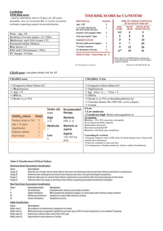
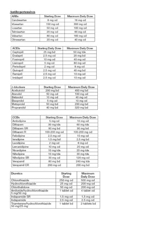
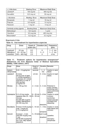
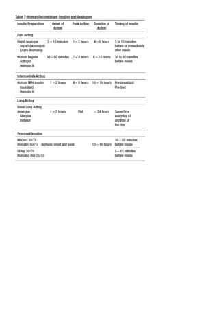
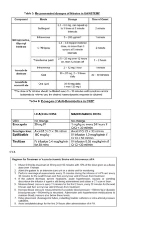
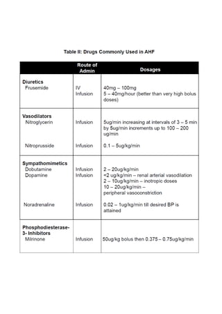
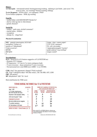
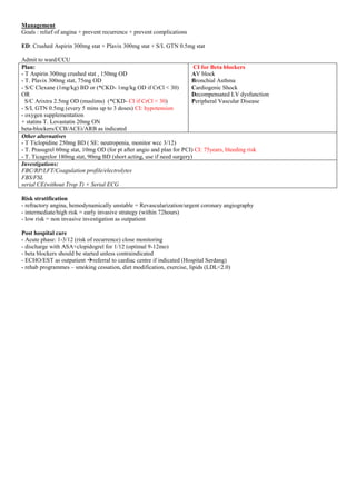
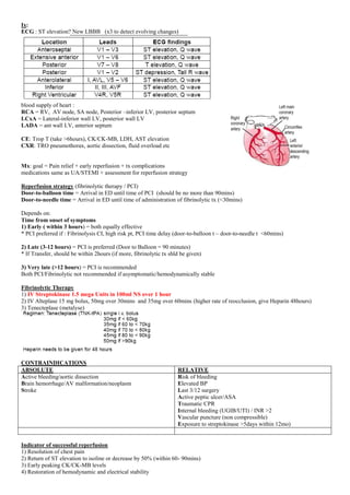
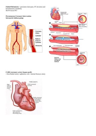
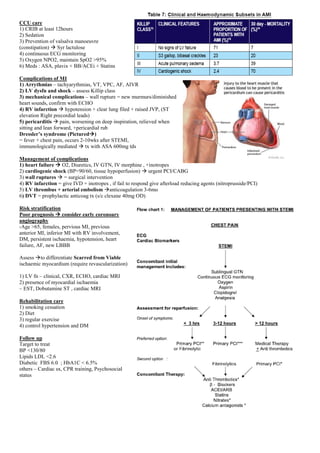
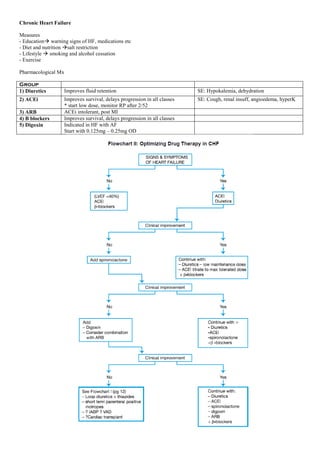
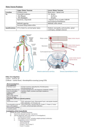
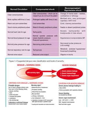
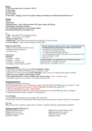
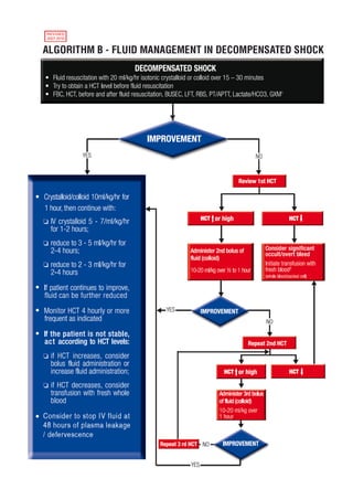
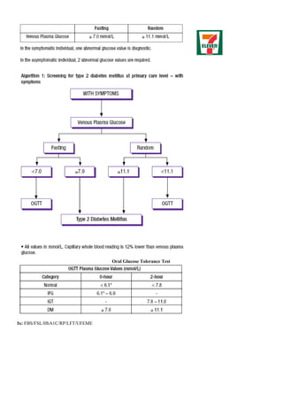
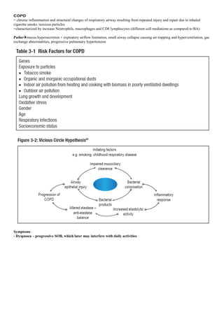
This document provides guidelines for house officers on managing patients with acute coronary syndrome. It outlines the classifications of ACS, including unstable angina, NSTEMI, and STEMI. For each type, it describes the typical history, physical exam findings, investigations, and management approach. Priority is given to relieving chest pain, preventing complications, and pursuing early reperfusion through fibrinolytic therapy or percutaneous coronary intervention depending on timing from symptom onset and patient risk factors. Complications of myocardial infarction are also summarized. The overall aim is to equip house officers with the essential knowledge for initial evaluation and treatment of ACS patients.

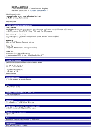


![CKD is an irreversible loss of renal function for at least three months and poses a major public health problem.
Who should be screened?
Patients with diabetes mellitus and/or hypertension should be screened at least yearly for chronic kidney disease o Age >65 years
old
o Family history of stage 5 CKD or hereditary kidney disease
o Structural renal tract disease, renal calculi or prostatic hypertrophy
o Opportunistic (incidental) detection of haematuria or proteinuria
o Chronic use of non-steroidal anti-inflammatory drugs (NSAIDs) or other nephrotoxic drugs
o Cardiovascular disease (CVD)
o Multisystem diseases with potential kidney involvement such as systemic lupus erythematosus
Screening method
1)Proteinuria
Factors Increases protein excretion Decreases protein excretion
• Strenuous exercise
• Poorly controlled DM
• Heart failure
• UTI
• Acute febrile illness
• Uncontrolled hypertension
• Haematuria
• Menstruation
• Pregnancy
2)Hematuria
3)Renal function (RP)
Equations for estimation of renal function (suggest to use online calculators/ apps)
i. MDRD eGFR =
175 x serum Cr -1.154
x age-0.203
x constant [constant = 1.212 [if black] or 0.742 [if female] ]
* where GFR is expressed as ml/min/1.73m2 of body surface area and sCr is expressed in mg/dl
ii. CKD-epi eGFR (Chronic Kidney Disease Epidemiology Collaboration )
- complexed formula calculation, suggest to use online app
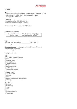
iii. Cockcroft-Gault Creatinine Clearance
CrCl (ml/min) = (140 - age (yrs)) x body weight (kg)
sCr (μmol/l) x Constant [constant = 1.23 in male or 1.04 in female]
4)Renal tract US (US KUB)
identifies obstructive uropathy, renal size and symmetry, renal scarring and polycystic disease.
Indications for renal ultrasound in patients with CKD:
• a rapid deterioration of renal function (eGFR >5 ml/min/1.73m2 within one year or 10 ml/min/1.73m2 within five years)
• visible or persistent non-visible haematuria
• symptoms or history of urinary tract obstruction
• a family history of polycystic kidney disease and age over 20 years
• stage 4 or 5 CKD
• when a renal biopsy is required
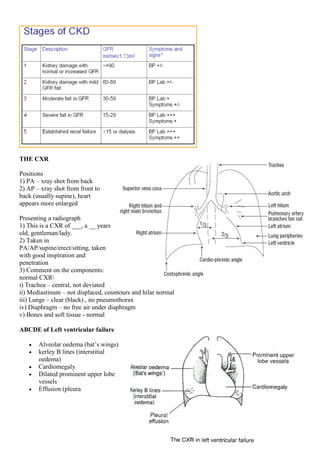
CKD classification:
Stages of CKD Stage GFR (ml/min/1.73m2) Description
1 ≥90 Normal or increased GFR, with other evidence of
kidney damage
2 60 – 89 Slight decrease in GFR, with other evidence of kidney
damage
3A 45 - 59 Moderate decrease in GFR, with or without other evidence
of kidney damage
3B 30 - 44
4 15 – 29 Severe decrease in GFR, with or without other evidence of
kidney damage
5 <15 Established renal failure](https://image.slidesharecdn.com/ampangmedical-guide1-220215225019/85/Ampang-medical-guide-59-320.jpg)
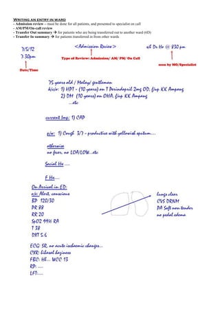
![A patient with chronic kidney disease (CKD) and any of the following criteria should be referred to a nephrologist/physician:
o heavy proteinuria (urine protein ≥1 g/day or urine protein: creatinine ratio (uPCR) ≥0.1 g/mmol) unless known to be due to
diabetes and optimally treated
o haematuria with proteinuria (urine protein ≥0.5 g/day or uPCR ≥0.05 g/mmol)
o rapidly declining renal function (loss of glomerular filtration rate/GFR >5 ml/min/1.73m2 in one year or >10 ml/min/1.73m2
within five years)
o resistant hypertension (failure to achieve target blood pressure despite three antihypertensive agents including a diuretic)
o suspected renal artery stenosis
o suspected glomerular disease
o suspected genetic causes of CKD
o pregnant or when pregnancy is planned
o estimated GFR <30 ml/min or serum creatinine >200 μmol/L
o unclear cause of CKD.
Uremic symptoms:
Neural and muscular
Fatigue
Peripheral neuropathy
Decreased mental acuity
Seizures
Anorexia
Nausea
Decreased taste and smell
Cramps]
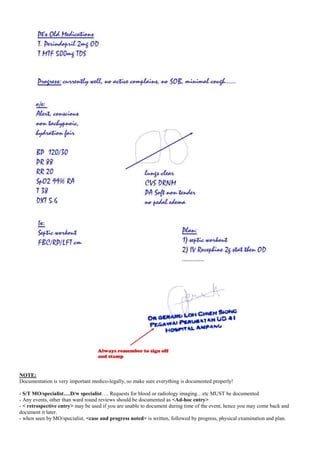
Sleep disturbance
Coma
Endocrine and metabolic
Amenorrhea
Sexual dysfunction
Reduced body temperature
Altered levels of amino acids
Bone disease by
hyperphosphatemia,
hyperparathyroidism, and
vitamin D deficiency
Reduced basal metabolic rate
Insulin resistance
Increased muscle protein
catabolism[3]
Other
Itching
Hiccups
granulocyte and lymphocyte dysfunction[3]
Platelet dysfunction](https://image.slidesharecdn.com/ampangmedical-guide1-220215225019/85/Ampang-medical-guide-60-320.jpg)