SlideShare a Scribd company logo
1 of 144
Prehospital: Emergency Care
Eleventh Edition
Chapter 15
Shock and Resuscitation
Copyright © 2018, 2014, 2010 Pearson Education, Inc. All Rights Reserved
Learning Readiness
Copyright © 2018, 2014, 2010 Pearson Education, Inc. All Rights Reserved
• EMS Education Standards, text p. 435.
• Chapter Objectives, text p. 435.
• Key Terms, text p. 436.
• Purpose of lecture presentation versus textbook reading
assignments.
Setting the Stage
Copyright © 2018, 2014, 2010 Pearson Education, Inc. All Rights Reserved
• Overview of Lesson Topics
– Shock
– Resuscitation in Cardiac Arrest
– Automated External Defibrillation and
Cardiopulmonary Resuscitation
– Recognizing and Treating Cardiac Arrest
– Special Considerations for the AED
Case Study Introduction (1 of 2)
Copyright © 2018, 2014, 2010 Pearson Education, Inc. All Rights Reserved
EMTs Tess Price and Steph Bowman are responding to a
report of chest pain, along with the crew from Engine 9.
Their general impression is of a man in his 50s who is
sitting in a recliner, hand on chest, and in apparent
distress. He is pale and extremely sweaty.
Case Study Introduction (2 of 2)
Copyright © 2018, 2014, 2010 Pearson Education, Inc. All Rights Reserved
The EMTs confirm a chief complaint of chest pain, and
continue their assessment as they begin treatment. Tess is
immediately concerned with the weak, rapid radial pulse
and labored respirations.
Case Study (1 of 6)
Copyright © 2018, 2014, 2010 Pearson Education, Inc. All Rights Reserved
• What is the significance of the weak, rapid pulse and
labored respirations?
• What could explain the the weak, rapid pulse and labored
respirations?
• What should the EMTs do to manage this patient?
Introduction
Copyright © 2018, 2014, 2010 Pearson Education, Inc. All Rights Reserved
• Shock is a state of inadequate perfusion of the cells that
can lead to death.
• The body’s attempts to restore homeostasis result in
many signs and symptoms of shock.
• Resuscitation is the emergency care provided to restore
vital body functions.
Shock (1 of 38)
Copyright © 2018, 2014, 2010 Pearson Education, Inc. All Rights Reserved
• Defined as inadequate tissue perfusion or hypoperfusion
• Inadequate amounts of oxygen and glucose are available
to cells to meet metabolic needs
• Inadequate waste product removal
Shock (2 of 38)
Copyright © 2018, 2014, 2010 Pearson Education, Inc. All Rights Reserved
• Shift from aerobic to anaerobic metabolism results in
decreased energy production and waste product
accumulation.
• Cellular sodium/potassium pump fails, leading to cell
death.
• Cell death results in organ failure.
Shock (3 of 38)
Copyright © 2018, 2014, 2010 Pearson Education, Inc. All Rights Reserved
• Treatment of shock is aimed at restoring perfusion to
provide the cells with glucose and oxygen.
Shock (4 of 38)
Copyright © 2018, 2014, 2010 Pearson Education, Inc. All Rights Reserved
• Oxygen delivery to the cells is critical, and requires:
– Breathing in an adequate amount of oxygen
– Diffusion of oxygen from alveoli to pulmonary
capillaries
– Oxygen transport to the cellular level
– Release of oxygen at the cellular level
Shock (5 of 38)
Copyright © 2018, 2014, 2010 Pearson Education, Inc. All Rights Reserved
• Etiologies of Shock
– Poor tissue perfusion is caused by one or more of
these problems:
▪ Inadequate volume
▪ Inadequate pump function
▪ Inadequate vessel tone
Shock (6 of 38)
Copyright © 2018, 2014, 2010 Pearson Education, Inc. All Rights Reserved
• Etiologies of Shock
– Inadequate Volume
▪ Decreased blood volume decreases preload
▪ Decreased preload causes stroke volume and
cardiac output to fall
▪ Decreased cardiac output causes a drop in systolic
blood pressure
▪ Decreased systolic blood pressure results in
inadequate tissue perfusion
Shock (7 of 38)
Copyright © 2018, 2014, 2010 Pearson Education, Inc. All Rights Reserved
• Etiologies of Shock
– Inadequate Volume
▪ May result from loss of whole blood or plasma
volume
– Bleeding
– Vomiting, diarrhea
– Excessive urination
– Increased capillary leakage
Shock (8 of 38)
Copyright © 2018, 2014, 2010 Pearson Education, Inc. All Rights Reserved
• Etiologies of Shock
– Inadequate Volume
▪ Patient requires an increase in blood volume
▪ If red blood cells have been lost, there is a
decrease in oxygen-carrying capacity, as well as a
decrease in pressure and perfusion
Shock (9 of 38)
Copyright © 2018, 2014, 2010 Pearson Education, Inc. All Rights Reserved
• Etiologies of Shock
– Inadequate Pump Function
▪ If the heart fails as a pump, regardless of the blood
volume, the delivery of oxygen and glucose to cells
decreases
Shock (10 of 38)
Copyright © 2018, 2014, 2010 Pearson Education, Inc. All Rights Reserved
• Etiologies of Shock
– Inadequate Pump Function
▪ May occur from myocardial infarction or heart
failure, or mechanical obstruction to blood flow
– Pericardial tamponade
– Tension pneumothorax
▪ Giving fluids may worsen the condition
Shock (11 of 38)
Copyright © 2018, 2014, 2010 Pearson Education, Inc. All Rights Reserved
• Etiologies of Shock
– Inadequate Vessel Tone
▪ Blood pressure is a function of cardiac output and
Systemic Vascular Resistance (SVR)
▪ If SVR decreases from vasodilation, blood
pressure decreases
Etiology of Shock: Vasodilation
Copyright © 2018, 2014, 2010 Pearson Education, Inc. All Rights Reserved
Shock (12 of 38)
Copyright © 2018, 2014, 2010 Pearson Education, Inc. All Rights Reserved
• Etiologies of Shock
– Inadequate Vessel Tone
▪ Massive vasodilation can occur from loss of
sympathetic nervous system function or chemicals
released within the body.
▪ Treatment includes vasoconstriction and volume
restoration.
Shock (13 of 38)
Copyright © 2018, 2014, 2010 Pearson Education, Inc. All Rights Reserved
• Etiologies of Shock
– Inadequate Vessel Tone
▪ Treatment of shock depends on underlying cause
▪ Consider requesting ALS, but weigh the benefit
against any potential delay in reaching the hospital
Categories and Types of Shock
Copyright © 2018, 2014, 2010 Pearson Education, Inc. All Rights Reserved
Shock (14 of 38)
Copyright © 2018, 2014, 2010 Pearson Education, Inc. All Rights Reserved
• Categories of Shock
– Hypovolemic shock is caused by low blood volume
▪ Hemorrhage is the most common cause of
hypovolemic shock
▪ Also caused by burns and dehydration
(a) Hemorrhagic Hypovolemia: Loss of Whole
Blood; (b) Nonhemorrhagic Hypovolemia: Loss of
Plasma
Copyright © 2018, 2014, 2010 Pearson Education, Inc. All Rights Reserved
Shock (15 of 38)
Copyright © 2018, 2014, 2010 Pearson Education, Inc. All Rights Reserved
• Categories of Shock
– Distributive Shock
▪ Caused by vasodilation leading to a relative
reduction in intravascular volume
▪ Can also involve fluid loss from increased capillary
permeability
Shock (16 of 38)
Copyright © 2018, 2014, 2010 Pearson Education, Inc. All Rights Reserved
• Categories of Shock
– Cardiogenic Shock
▪ Caused by inability of heart to contract effectively
▪ Generally occurs with loss of 40 percent or greater
of left ventricular volume
▪ Stroke volume and cardiac output are reduced
Heart Attack as a Cause of Cardiogenic
Shock
Damaged heart muscle results in reduced force of contractions,
reduced stroke volume, and reduced cardiac output
Copyright © 2018, 2014, 2010 Pearson Education, Inc. All Rights Reserved
Shock (17 of 38)
Copyright © 2018, 2014, 2010 Pearson Education, Inc. All Rights Reserved
• Categories of Shock
– Obstructive Shock
▪ Results from a condition that obstructs forward
blood flow
▪ Causes include pulmonary embolism, tension
pneumothorax, and pericardial tamponade
▪ Treat by relieving the obstruction
Causes of Obstructive Shock
(a) pulmonary embolism; (b) tension pneumothorax; (c)
pericardial tamponade
Copyright © 2018, 2014, 2010 Pearson Education, Inc. All Rights Reserved
Shock (18 of 38)
Copyright © 2018, 2014, 2010 Pearson Education, Inc. All Rights Reserved
• Categories of Shock
– Metabolic or Respiratory Shock
▪ Some sources list metabolic or respiratory shock
as a fifth category
▪ Dysfunction in the ability of oxygen to diffuse into
the blood, be carried by hemoglobin, off-load at the
cell, or be used effectively by the cell for
metabolism
Shock (19 of 38)
Copyright © 2018, 2014, 2010 Pearson Education, Inc. All Rights Reserved
• Specific Types of Shock
– Hemorrhagic Hypovolemic Shock
▪ Loss of whole blood from medical or traumatic
causes
▪ Results in decreased perfusion pressure and
decreased oxygen-carrying capacity
▪ Treat by stopping the bleeding and replacing blood
or blood components
Shock (20 of 38)
Copyright © 2018, 2014, 2010 Pearson Education, Inc. All Rights Reserved
• Specific Types of Shock
– Nonhemorrhagic Hypovolemic Shock
▪ Loss of fluid without loss of red blood cells
▪ Can result from vomiting, diarrhea, sweating
▪ Administration of IV fluids can be helpful
Shock (21 of 38)
Copyright © 2018, 2014, 2010 Pearson Education, Inc. All Rights Reserved
• Specific Types of Shock
– Burn Shock
▪ Is a specific form of nonhemorrhagic hypovolemic
shock resulting from a burn injury
▪ Plasma and plasma proteins leak from damaged
capillaries
▪ Loss of fluid and plasma oncotic pressure leads to
edema
Shock (22 of 38)
Copyright © 2018, 2014, 2010 Pearson Education, Inc. All Rights Reserved
• Specific Types of Shock
– Anaphylactic Shock
▪ Chemical substances released in anaphylactic
reaction cause massive systemic vasodilation and
increased capillary permeability
▪ Epinephrine is the medication of choice
Shock (23 of 38)
Copyright © 2018, 2014, 2010 Pearson Education, Inc. All Rights Reserved
• Specific Types of Shock
– Septic Shock
▪ Septic shock is another type of distributive shock
▪ Bacteria and toxins in the blood lead to
vasodilation and increased capillary permeability
▪ Patient can benefit from ALS treatment
Table 15-1 EMS Screening Tools to Identify
Sepsis
Copyright © 2018, 2014, 2010 Pearson Education, Inc. All Rights Reserved
Robson
Screening Tool
Sepsis is suspected if two of the following findings are present with a
suspected active infection:
• Temperature >38.3°C (100.9°F ) or <36°C (96.8°F )
• Heart rate >90 bpm
• Respiratory rate >20 breaths/minute
• Acute altered mental status
• BGL <120 milligram/dL
BAS 90-30-90
Scale
Sepsis is suspected if one or more of the following findings are present in a
patient with a suspected active infection:
• Systolic blood pressure <90 millimeterHg
• Respiratory rate >30 breaths/minute
• SpO2 <90%
qSOFA Score Sepsis is suspected if two or more of the following findings are present
in a patient with a suspected active infection:
•Respiratory rate 22 breaths/minute or greater
•Altered mental status (GCS <13)
Systolic BP 100 millimeterHg or less
Shock (24 of 38)
Copyright © 2018, 2014, 2010 Pearson Education, Inc. All Rights Reserved
• Specific Types of Shock
– Neurogenic Shock
▪ Spinal cord injury can cause loss of sympathetic
nerve fiber functions responsible for maintaining
blood vessel tone
▪ Loss of systemic vascular resistance
Shock (25 of 38)
Copyright © 2018, 2014, 2010 Pearson Education, Inc. All Rights Reserved
• Specific Types of Shock
– Cardiogenic Shock
▪ Causes include acute myocardial infarction,
congestive heart failure, abnormal cardiac rhythm,
and overdose on certain drugs
▪ Patients can benefit from ALS interventions
Click on the Term That Best Describes the Type of
Shock That Occurs When There is Massive Systemic
Vasodilation
Copyright © 2018, 2014, 2010 Pearson Education, Inc. All Rights Reserved
A. Obstructive
B. Metabolic
C. Distributive
D. Cardiogenic
Shock (26 of 38)
Copyright © 2018, 2014, 2010 Pearson Education, Inc. All Rights Reserved
• The Body’s Response to Shock
– The body attempts to compensate to return perfusion
to normal
– Many signs and symptoms of shock are related to
compensatory mechanisms
– Sympathetic nervous system stimulation and the
release of hormones are the two major compensatory
mechanisms
Shock (27 of 38)
Copyright © 2018, 2014, 2010 Pearson Education, Inc. All Rights Reserved
• The Body’s Response to Shock
– Direct Nerve Stimulation
▪ The effects of sympathetic stimulation are
immediate
– Increase in heart rate
– Increase in force of ventricular contraction
– Vasoconstriction
– Stimulation of the release of epinephrine and
norepinephrine from the adrenal gland
Shock (28 of 38)
Copyright © 2018, 2014, 2010 Pearson Education, Inc. All Rights Reserved
• The Body’s Response to Shock
– Release of Hormones
▪ The effects of hormones are more sustained
– Epinephrine has alpha and beta effects that
cause vasoconstriction and increased cardiac
output
– Norepinephrine has alpha effects that cause
vasoconstriction
– Other hormones are also released
Table 15-2 Effects of Alpha and Beta
Stimulation
Copyright © 2018, 2014, 2010 Pearson Education, Inc. All Rights Reserved
Receptor Stimulatory Effect Sign or Symptom
Alpha 1 Contraction of the muscles controlling the iris Dilated pupils
Contraction of vascular smooth muscle causing
vasoconstriction
Pale, cool skin, narrow pulse
pressure
Stimulation of sweat glands Localized sweating, clammy skin
Beta 1 Increased heart rate Tachycardia
Increased speed of impulse through conduction
system
Tachycardia
Increased force of contraction Pounding heart
Beta 2 Bronchial smooth muscle dilation Decreased resistance in airway
Skeletal muscle contractility Tremors
Table 15-3 Effects of Hormones Released in
Shock (1 of 2)
Copyright © 2018, 2014, 2010 Pearson Education, Inc. All Rights Reserved
Hormone Effect on Body Sign or Symptom
Epinephrine Increased heart rate (beta 1) Tachycardia
Increased contractility (beta 1) Pounding heart
Vasoconstriction (alpha 1) Pale, cool skin
Sweat gland stimulation (alpha 1) Clammy skin
Decreased insulin secretion (alpha 2) Increased blood glucose level
Conversion of stored glucose in liver to blood
glucose
Conversion of noncarbohydrates into sugar
Iris muscle contraction (alpha 1) Pupillary dilation
Norepinephrine Vasoconstriction (alpha 1) Pale, cool skin
Sweat gland stimulation (alpha 1) Clammy skin
Increased heart rate (beta 1) Tachycardia
Table 15-3 Effects of Hormones Released in
Shock (2 of 2)
Copyright © 2018, 2014, 2010 Pearson Education, Inc. All Rights Reserved
Hormone Effect on Body Sign or Symptom
Antidiuretic Hormone
(Vasopressin)
Increased sodium reabsorption in the
kidneys
Decreased urine output
Vasoconstriction Increased blood pressure
Angiotensin II Vasoconstriction Pale, cool skin
Sodium reabsorption in the kidney Decreased urine output
Aldosterone Sodium reabsorption in the kidney Decreased urine output
Glucagon Conversion of stored glucose in liver to
blood glucose
Increased blood glucose level
Conversion of noncarbohydrates into
sugar
Increased heart rate and contractility Tachycardia
Shock (29 of 38)
Copyright © 2018, 2014, 2010 Pearson Education, Inc. All Rights Reserved
• Stages of Shock
– The stages of shock are:
▪ Compensatory
▪ Decompensatory
The Cycle of Hemorrhagic Shock
Copyright © 2018, 2014, 2010 Pearson Education, Inc. All Rights Reserved
Shock (30 of 38)
Copyright © 2018, 2014, 2010 Pearson Education, Inc. All Rights Reserved
• Stages of Shock
– Compensatory Shock
▪ The body is able to maintain near-normal blood
pressure and perfusion of vital organ
▪ Blood is shunted away from non-vital areas, such
as the skin and gastrointestinal tract
▪ Pulse pressure may be narrowed
Shock (31 of 38)
Copyright © 2018, 2014, 2010 Pearson Education, Inc. All Rights Reserved
• Stages of Shock
– Decompensatory Shock
▪ Compensatory mechanisms are overwhelmed
▪ The body can no longer maintain a blood pressure
and perfusion of the vital organs
▪ Anaerobic metabolism is occurring
▪ Vital organs are not perfused
Shock (32 of 38)
Copyright © 2018, 2014, 2010 Pearson Education, Inc. All Rights Reserved
• Stages of Shock
– Multiple Organ Dysfunction Syndrome (MODS)
▪ The stage in which multiple organs begin to fail
throughout the body from extreme and prolonged
hypoxia, altered metabolism, and elevated carbon
dioxide and acid levels
▪ Sometimes referred to as irreversible shock
Shock (33 of 38)
Copyright © 2018, 2014, 2010 Pearson Education, Inc. All Rights Reserved
• Shock Assessment
– Signs may be subtle or profound, but rapid
recognition of shock is key to treatment
– Consider history findings, physical assessment
findings, signs of perfusion disturbance, and vital
signs
– Do not rely on one finding, sign, or symptom
Shock (34 of 38)
Copyright © 2018, 2014, 2010 Pearson Education, Inc. All Rights Reserved
• Shock Assessment
– History
▪ Chief complaint
▪ Sample history
▪ Beta blockers and calcium channel blockers can
alter the response to shock
Shock (35 of 38)
Copyright © 2018, 2014, 2010 Pearson Education, Inc. All Rights Reserved
• Shock Assessment
– Physical exam
▪ Vital signs can appear normal in compensatory
shock
▪ Look for signs of poor perfusion
Table 15-5 Physical Assessment Indicators
of Hypovolemic Shock
Copyright © 2018, 2014, 2010 Pearson Education, Inc. All Rights Reserved
Vital Signs Signs of Poor Perfusion
Decreasing blood pressure Anxiety, anxiousness that Progresses to a decreased
mental status
Narrowing pulse pressure Pale, cool, clammy skin
Tachycardia Delayed capillary refill
Tachypnea Weak or absent peripheral pulses
Pale, cool, and clammy skin Decreased urine output
Unobtainable or poor SpO2
reading
Table 15-6 Physical Assessment Indicators
of Cardiogenic Shock
Copyright © 2018, 2014, 2010 Pearson Education, Inc. All Rights Reserved
Vital Signs Signs of Poor Perfusion
Decreasing blood pressure Anxiety, anxiousness that progresses to a
decreased mental status
Narrowing pulse pressure Pale, cool, clammy skin; cyanotic or mottled
skin
Tachycardia or bradycardia; may
be irregular
Jugular venous distention and peripheral
edema (right-sided heart failure)
Tachypnea Weak or absent peripheral pulses
Pale, cool, and clammy skin;
cyanotic or mottled skin
Decreased urine output
Decreased SpO2 reading Other sign: Crackles or rales upon
auscultation (left-sided heart failure)
Table 15-7 Physical Assessment Indicators
of Distributive Shock
Copyright © 2018, 2014, 2010 Pearson Education, Inc. All Rights Reserved
Vital Signs Signs of Poor Perfusion
Decreasing blood pressure Anxiety, anxiousness that progresses to a decreased
mental status
Tachycardia (anaphylactic and septic shock)
Relative bradycardia or normal heart rate (shock associated with
a spinal cord injury)
Mottled, cyanosis (late—sepsis, anaphylactic and
neurogenic)
Tachypnea with respiratory distress and wheezing (anaphylactic
shock)
Weak or absent peripheral pulses
Tachypnea (septic)
Normal respiratory rate (neurogenic)
Normal to flushed (early sepsis)
Warm, flushed skin (neurogenic)
Warm, flushed skin with hives, possible cyanosis (anaphylactic)
Mottled, cyanosis (late: sepsis, anaphylactic, and neurogenic)
Severely decreased SpO2 reading (anaphylactic) Other signs:
Fever (sepsis)
Loss of motor/sensory function (neurogenic due to
spinal cord injury)
Edema (anaphylactic)
Table 15-8 Physical Assessment Indicators
of Obstructive Shock
Copyright © 2018, 2014, 2010 Pearson Education, Inc. All Rights Reserved
Vital Signs Signs of Poor Perfusion
Decreasing blood pressure
Pulsus paradoxus (tension pneumothorax
and pericardial tamponade)
Anxiety, anxiousness that progresses to a decreased
mental status
Narrowing pulse pressure Pale, cool, clammy skin; cyanotic or mottled skin
Tachycardia Jugular venous distention (pericardial tamponade and
tension pneumothorax)
Tachypnea Weak or absent peripheral pulses
Pale, cool, and clammy skin; cyanotic or
mottled skin
Decreased urine output
Decreased SpO2 reading Severely
decreased SpO2 reading
(tension pneumothorax and massive
pulmonary embolism)
Other sign: Severely decreased to absent breath sounds
of one hemithorax (tension pneumothorax)
Shock (36 of 38)
Copyright © 2018, 2014, 2010 Pearson Education, Inc. All Rights Reserved
• Age Considerations in Shock
– Age may influence the development, presentation,
management, and recovery from shock.
– Children can compensate well, but deteriorate
quickly.
– Geriatric patients do not compensate well.
Shock (37 of 38)
Copyright © 2018, 2014, 2010 Pearson Education, Inc. All Rights Reserved
• General Goals of Prehospital Management of Shock
– Secure and maintain a patent airway.
– Establish and maintain adequate ventilation.
– Establish and maintain adequate oxygenation.
– Do not hyperventilate the shock patient.
– Stop bleeding as quickly as possible.
Shock (38 of 38)
Copyright © 2018, 2014, 2010 Pearson Education, Inc. All Rights Reserved
• General Goals of Prehospital Management of Shock
– Splint fractures, don’t delay transport.
– Do not remove an impaled object.
– Maintain the body temperature.
– Keep the patient supine.
– Apply PASG, according to protocol.
– Rapid transport.
– Consider ALS intercept.
Case Study (2 of 6)
Copyright © 2018, 2014, 2010 Pearson Education, Inc. All Rights Reserved
Finding that the patient is confused, the EMTs complete a
rapid secondary assessment and obtain a history from the
patient's wife, as the engine crew helps them prepare the
patient for transport.
Case Study (3 of 6)
Tess finds that the patient’s respirations are 28 and
labored, and that he has crackles in his lungs. His heart
rate is 116, with a blood pressure of
92 percent.
Copyright © 2018, 2014, 2010 Pearson Education, Inc. All Rights Reserved
100
,
72
and an SpO2 of
Case Study (4 of 6)
Copyright © 2018, 2014, 2010 Pearson Education, Inc. All Rights Reserved
• Is this patient in shock? Explain your answer?
• What interventions should the patient be receiving?
• What are the potential consequences of failing to
intervene appropriately or in a timely manner?
Resuscitation in Cardiac Arrest (1 of 12)
Copyright © 2018, 2014, 2010 Pearson Education, Inc. All Rights Reserved
• Resuscitation means bringing a patient back from a
potential or apparent death.
• Resuscitation focuses on management of the airway,
ventilation, and oxygenation, and restoring adequate
circulation.
Resuscitation in Cardiac Arrest (2 of 12)
Copyright © 2018, 2014, 2010 Pearson Education, Inc. All Rights Reserved
• Cardiac arrest occurs when the ventricles of the heart are
not contracting or cardiac output is ineffective and no
pulses can be felt.
– Brain cells begin to die within four to six minutes
following cardiac arrest.
– Cardiac arrest patients are described as having
suffered sudden death when the patient dies within
one hour of the onset of the signs and symptoms.
Resuscitation in Cardiac Arrest (3 of 12)
Copyright © 2018, 2014, 2010 Pearson Education, Inc. All Rights Reserved
• A common cause of cardiac arrest is ventricular
fibrillation.
• Ventricular fibrillation can be treated with defibrillation.
Resuscitation in Cardiac Arrest (4 of 12)
Copyright © 2018, 2014, 2010 Pearson Education, Inc. All Rights Reserved
• Pathophysiology of Cardiac Arrest
– The patient goes through three phases of cardiac
arrest that lead to biological death
▪ Electrical phase
▪ Circulatory phase
▪ Metabolic phase
Resuscitation in Cardiac Arrest (5 of 12)
Copyright © 2018, 2014, 2010 Pearson Education, Inc. All Rights Reserved
• Pathophysiology of Cardiac Arrest
– Electrical Phase
▪ First four minutes.
▪ The heart still has a supply of oxygen and glucose.
▪ Conditions are favorable for resuscitation.
▪ The heart is prepared to respond to defibrillation.
Resuscitation in Cardiac Arrest (6 of 12)
Copyright © 2018, 2014, 2010 Pearson Education, Inc. All Rights Reserved
• Pathophysiology of Cardiac Arrest
– Circulatory Phase
▪ Four through ten minutes after cardiac arrest.
▪ Oxygen stores are exhausted; myocardial cells
switch to anaerobic metabolism.
▪ CPR is needed to restore a supply of oxygen and
glucose to enhance the possibility of successful
defibrillation.
Resuscitation in Cardiac Arrest (7 of 12)
Copyright © 2018, 2014, 2010 Pearson Education, Inc. All Rights Reserved
• Pathophysiology of Cardiac Arrest
– Metabolic Phase
▪ Begins ten minutes after cardiac arrest.
▪ The heart muscle is acidic and ischemic, and
begins to die.
▪ Chances of resuscitation are unfavorable.
Resuscitation in Cardiac Arrest (8 of 12)
Copyright © 2018, 2014, 2010 Pearson Education, Inc. All Rights Reserved
• Terms Related to Out-of-Hospital Cardiac Arrest (OHCA)
Resuscitation
– Downtime
– Total downtime
– Return of spontaneous circulation (ROSC)
– Survival
– Witnessed cardiac arrest
– Unwitnessed cardiac arrest
Resuscitation in Cardiac Arrest (9 of 12)
Copyright © 2018, 2014, 2010 Pearson Education, Inc. All Rights Reserved
• Withholding a Resuscitation Attempt
– Do not resuscitate orders
– Physician orders for life-sustaining treatment
– Medical orders for life-sustaining treatment
– Injuries incompatible with life
Resuscitation in Cardiac Arrest (10 of 12)
Copyright © 2018, 2014, 2010 Pearson Education, Inc. All Rights Reserved
• The 2015 AHA Chain of Survival
– Successful resuscitation depends on a sequence of
events
▪ The Chain of Survival is slightly different for
pediatric patients.
Resuscitation in Cardiac Arrest (11 of 12)
Copyright © 2018, 2014, 2010 Pearson Education, Inc. All Rights Reserved
• The 2015 AHA Chain of Survival
– AHA 2015 OHCA Adult Chain of Survival
▪ Immediate recognition and activation.
▪ Immediate high-quality CPR.
▪ Rapid defibrillation.
▪ Basic and advanced emergency medical services.
▪ Advanced life support and post-arrest care.
Resuscitation in Cardiac Arrest (12 of 12)
Copyright © 2018, 2014, 2010 Pearson Education, Inc. All Rights Reserved
• The 2015 AHA Chain of Survival
– AHA 2015 OHCA Pediatric Chain of Survival
▪ Prevention of arrest.
▪ Early high-quality CPR.
▪ Rapid activation of EMS.
▪ Effective advanced life support and rapid transport.
▪ Integrated post-cardiac-arrest care.
Automated External Defibrillation and
CPR (1 of 9)
Copyright © 2018, 2014, 2010 Pearson Education, Inc. All Rights Reserved
• Rationale for early defibrillation
– The most frequent initial rhythm in sudden cardiac
arrest is ventricular fibrillation.
– The most effective treatment for ventricular fibrillation
is defibrillation.
– The probability of successful defibrillation decreases
over time.
Automated External Defibrillation and
CPR (2 of 9)
Copyright © 2018, 2014, 2010 Pearson Education, Inc. All Rights Reserved
• Rationale for early defibrillation
– Successful defibrillation depends on effective CPR;
interruptions in chest compressions for defibrillation
must be minimized.
– Without intervention, ventricular fibrillation
degenerates into asystole.
Automated External Defibrillation and
CPR (3 of 9)
Copyright © 2018, 2014, 2010 Pearson Education, Inc. All Rights Reserved
• “Push hard and push fast” to provide effective chest
compressions in CPR.
– Compressions at a rate of at least 100/minute.
– Compression-to-ventilation ratio of 30:2.
– Minimize interruptions for defibrillation.
– Avoid excess ventilation.
Automated External Defibrillation and
CPR (4 of 9)
Copyright © 2018, 2014, 2010 Pearson Education, Inc. All Rights Reserved
• Types of Defibrillators
– Manual defibrillators require extensive training to use.
– Automated defibrillators are simpler to use.
Automated External Defibrillation and
CPR (5 of 9)
Copyright © 2018, 2014, 2010 Pearson Education, Inc. All Rights Reserved
• Types of Defibrillators
– Advantages of AEDs
▪ Speed of operation
▪ Safer, more effective shock delivery
▪ More efficient monitoring
Automated External Defibrillation and
CPR (6 of 9)
Copyright © 2018, 2014, 2010 Pearson Education, Inc. All Rights Reserved
• Types of Defibrillators
– Types of AEDs
▪ Fully automated
▪ Semiautomated
– AEDs may use a monophasic or biphasic waveform
Physio-Control Lifepak® 1000 Defibrillator
(© Physio-Control, Inc.)
Copyright © 2018, 2014, 2010 Pearson Education, Inc. All Rights Reserved
Infant/Child Pads for Physiocontrol Lifepak® 1000
Copyright © 2018, 2014, 2010 Pearson Education, Inc. All Rights Reserved
Automated External Defibrillation and
CPR (7 of 9)
Copyright © 2018, 2014, 2010 Pearson Education, Inc. All Rights Reserved
• Analysis of Cardiac Rhythms
– Ventricular fibrillation
– Ventricular tachycardia
– Asystole
– Pulseless electrical activity
Ventricular Fibrillation is Associated with
Chaotic Electrical Discharge in the Ventricles
Copyright © 2018, 2014, 2010 Pearson Education, Inc. All Rights Reserved
Ventricular Tachycardia Originates in the
Conduction System of the Ventricle
Copyright © 2018, 2014, 2010 Pearson Education, Inc. All Rights Reserved
Asystole, or “Flatline,” is the Complete
Absence of Electrical Activity in the Heart
Copyright © 2018, 2014, 2010 Pearson Education, Inc. All Rights Reserved
Automated External Defibrillation and
CPR (8 of 9)
Copyright © 2018, 2014, 2010 Pearson Education, Inc. All Rights Reserved
• Analysis of Cardiac Rhythms
– No one should touch the patient during AED rhythm
analysis or shock delivery
▪ Movement interferes with rhythm analysis.
▪ The electrical energy can be transmitted to anyone
touching the patient.
Automated External Defibrillation and
CPR (9 of 9)
Copyright © 2018, 2014, 2010 Pearson Education, Inc. All Rights Reserved
• When and When Not to Use the AED
– AEDs can be used in patients of all ages.
– Manual defibrillation is preferred for those less than
one year of age.
– A dose attenuating system is preferred for use in
children.
– Use adult pads with children greater than eight years
of age.
AED Pads Applied to a Pediatric Patient
Copyright © 2018, 2014, 2010 Pearson Education, Inc. All Rights Reserved
AED Pads Applied to an Infant
Copyright © 2018, 2014, 2010 Pearson Education, Inc. All Rights Reserved
Recognizing and Treating Cardiac
Arrest (1 of 16)
Copyright © 2018, 2014, 2010 Pearson Education, Inc. All Rights Reserved
• Assessment-Based Approach: Cardiac Arrest
– Scene Size-Up and Primary Assessment
▪ If a patient appears unresponsive without signs of
life, quickly check for breathing and carotid pulse.
▪ Assess for no longer than ten seconds.
▪ If the patient is apneic or has agonal respirations,
begin CPR with a C-A-B approach.
Recognizing and Treating Cardiac
Arrest (2 of 16)
Copyright © 2018, 2014, 2010 Pearson Education, Inc. All Rights Reserved
• Assessment-Based Approach: Cardiac Arrest
– Patients less than one year of age
▪ If heart rate is >60 with inadequate ventilation,
assist or provide ventilations at 12–20 per minute.
▪ If heart rate is <60 or pulse is absent, begin chest
compressions.
Recognizing and Treating Cardiac
Arrest (3 of 16)
Copyright © 2018, 2014, 2010 Pearson Education, Inc. All Rights Reserved
• Assessment-Based Approach: Cardiac Arrest
– Patients less than one year of age
▪ 30 compressions: two ventilations for one EMT
performing CPR.
▪ 15 compressions: two ventilations for two EMTs
performing CPR.
Recognizing and Treating Cardiac
Arrest (4 of 16)
Copyright © 2018, 2014, 2010 Pearson Education, Inc. All Rights Reserved
• Assessment-Based Approach: Cardiac Arrest
– Patients one year of age to puberty
▪ 30 compressions: two ventilations for one EMT
performing CPR.
▪ 15 compressions: two ventilations for two EMTs
performing CPR.
Recognizing and Treating Cardiac
Arrest (5 of 16)
Copyright © 2018, 2014, 2010 Pearson Education, Inc. All Rights Reserved
• Assessment-Based Approach: Cardiac Arrest
– Adolescents with signs of puberty and adults
▪ 30 compressions: two ventilations
▪ Rate of at least 100/minute.
▪ Compression depth of at least two inches
▪ For an obviously pregnant patient, displace the
uterus laterally.
Recognizing and Treating Cardiac
Arrest (6 of 16)
Copyright © 2018, 2014, 2010 Pearson Education, Inc. All Rights Reserved
• Assessment-Based Approach: Cardiac Arrest
– Secondary Assessment
▪ Attempt to obtain a history from bystanders or
relatives, but do not interrupt chest compressions
or delay defibrillation.
Recognizing and Treating Cardiac
Arrest (7 of 16)
Copyright © 2018, 2014, 2010 Pearson Education, Inc. All Rights Reserved
• Assessment-Based Approach: Cardiac Arrest
– Emergency medical care
▪ Continue CPR and defibrillation, as indicated.
– Reassessment
▪ Once perfusion is restored, continue
reassessment.
▪ Patients may revert into cardiac arrest.
Recognizing and Treating Cardiac
Arrest (8 of 16)
Copyright © 2018, 2014, 2010 Pearson Education, Inc. All Rights Reserved
• Performing Defibrillation
– Using an AED
▪ Ideally, at least two EMTs are present.
▪ Take standard precautions.
▪ Perform a brief primary assessment.
▪ Perform CPR minimizing breaks.
▪ Prepare and apply the AED.
▪ Analyze the rhythm and defibrillate as
recommended by the AED.
EMT Skills 15-1
Copyright © 2018, 2014, 2010 Pearson Education, Inc. All Rights Reserved
Using a Semiautomated AED
In the Unresponsive Patient Suspected of Being in
Cardiac Arrest, Quickly Assess for Apnea or Agonal
Ventilations and a Pulse
Copyright © 2018, 2014, 2010 Pearson Education, Inc. All Rights Reserved
One EMT Should Immediately Initiate CPR
Beginning with Chest Compressions While the Other
EMT Prepares the AED
Copyright © 2018, 2014, 2010 Pearson Education, Inc. All Rights Reserved
Turn on the AED and Follow the Prompts
Copyright © 2018, 2014, 2010 Pearson Education, Inc. All Rights Reserved
Apply the Defibrillation Pads While Chest
Compressions are Being Performed - Minimize Any
Interruption in Chest Compressions
Copyright © 2018, 2014, 2010 Pearson Education, Inc. All Rights Reserved
Clear the Patient for Rhythm Analysis
Copyright © 2018, 2014, 2010 Pearson Education, Inc. All Rights Reserved
Deliver a Defibrillation If Advised
Copyright © 2018, 2014, 2010 Pearson Education, Inc. All Rights Reserved
Immediately Resume CPR Beginning with Chest
Compressions Following the Defibrillation
Copyright © 2018, 2014, 2010 Pearson Education, Inc. All Rights Reserved
After Two Minutes of CPR, Follow the AED Prompts
to Check Breathing and Pulse in a Non-Shockable
Rhythm
Copyright © 2018, 2014, 2010 Pearson Education, Inc. All Rights Reserved
Recognizing and Treating Cardiac
Arrest (9 of 16)
Copyright © 2018, 2014, 2010 Pearson Education, Inc. All Rights Reserved
• Performing Defibrillation
– Use of the AED by a Single EMT
▪ Simultaneously verify that the patient is
unresponsive, with no breathing and no pulse.
▪ Call for additional EMS and the AED.
▪ Immediately begin chest compressions and apply
the AED as soon as it is available.
▪ Minimize interruption of compressions.
Recognizing and Treating Cardiac
Arrest (10 of 16)
Copyright © 2018, 2014, 2010 Pearson Education, Inc. All Rights Reserved
• Cardiac Arrest in a Pregnant Patient
– A pregnant patient in cardiac arrest who is at 20
weeks of gestation or greater, it is necessary to place
the patient in a supine position and to manually
displace the uterus off the vena cava when doing
chest compressions.
Recognizing and Treating Cardiac
Arrest (11 of 16)
Copyright © 2018, 2014, 2010 Pearson Education, Inc. All Rights Reserved
• Transporting the Cardiac Arrest Patient
– After emergency care procedures and operating the
AED, if ALS is not responding to the scene, transport
if:
▪ The patient regains a pulse.
▪ Your protocol indicates transport after a
prescribed number of shocks or no shock
indicated messages.
Recognizing and Treating Cardiac
Arrest (12 of 16)
Copyright © 2018, 2014, 2010 Pearson Education, Inc. All Rights Reserved
• Transporting the Cardiac Arrest Patient
– Transporting a Patient with a Pulse
▪ Provide oxygen/ventilation as needed.
▪ Have suction ready.
▪ Transfer the patient to the ambulance.
▪ Consider getting ACLS to the patient.
▪ Keep AED attached.
▪ Perform secondary assessment.
▪ Reassess every five minutes.
Recognizing and Treating Cardiac
Arrest (13 of 16)
Copyright © 2018, 2014, 2010 Pearson Education, Inc. All Rights Reserved
• Transporting the Cardiac Arrest Patient
– Transporting a patient without a pulse
▪ Continue to CPR and defibrillation.
▪ Follow protocol.
▪ Use extreme caution when defibrillating in the
ambulance.
▪ Rendezvous with ALS as early as possible.
Recognizing and Treating Cardiac
Arrest (14 of 16)
Copyright © 2018, 2014, 2010 Pearson Education, Inc. All Rights Reserved
• Post-Resuscitation Care
– Indications that ROSC has occurred
▪ A pulse is felt after the AED indicates a no shock
advisory.
▪ Patient regains spontaneous breathing.
▪ The patient begins to move.
– Upon ROSC
▪ Assess the patient’s ventilation.
▪ Avoid potential oxygen toxicity issues.
Recognizing and Treating Cardiac
Arrest (15 of 16)
Copyright © 2018, 2014, 2010 Pearson Education, Inc. All Rights Reserved
• Providing for Advanced Cardiac Life Support
– The AHA’s 2015 Chain of Survival advocates for
advanced cardiac life support.
– Minimize the time from the delivery of CPR and
defibrillation to the arrival of ACLS.
Recognizing and Treating Cardiac Arrest (16
of 16)
Copyright © 2018, 2014, 2010 Pearson Education, Inc. All Rights Reserved
• Summary: Assessment and Care
– Review assessment findings that may be associated
with cardiac arrest and emergency care for cardiac
arrest.
Assessment Summary
Cardiac Arrest
The following are findings that indicate cardiac arest. These
findings are obtained during the primary assessment:
• Unresponsive
• Apneic (not breathing)
• Pulseless
Emergency Care Protocol: Cardiac Arrest (1 of 3)
Copyright © 2018, 2014, 2010 Pearson Education, Inc. All Rights Reserved
1. Take Standard Precautions.
2. Perform a brief primary assesment of the patient. It the patient is
unresponsive, is apneic or has agonal ventilation, has no pulse (checked
simulataneously with ventilation and for no less than 5 seconds but more
than 10 seconds), and no other signs of life, immediately initiate CPR
beginning with chest compressions.
3. Immediately intitate CPR beginning with chest compressions (CAB
intervention sequence). As soon as the AED is available, apply it while
chest compressions are being performed. When the AED is ready to start
the rhythm analysis, stop chest compressions and proceed with AED
protocol.
– If bystanders or first responders are already performing CPR when
you arrive, instruct them to continue while you prepare application of
the AED.
Emergency Care Protocol: Cardiac Arrest (2 of 3)
Copyright © 2018, 2014, 2010 Pearson Education, Inc. All Rights Reserved
4. Continue with chest compressions while the AED is readled for operation.
5. Turn on power to the AED.
6. Attach the adhesive monitoring-defibrillation pads to the chest while chest
compressions are being performed. Minimize any interruption to chest
compressions.
7. Begin analysis of the patient’s cardiac rhythm.
8. If the AED’s analysis indicates a shock, it provides a “deliver shock”
message. In that case, proceed with defibrillation by depressing the shock
or defibrillation button. If the AED’s analysis determines a nonshockable
rhythm, it gives a “no shock” message. In that case, immediately resume
CPR beginning with chest compressions.
9. After a shock has been delivered, immediately resume CPR beginning
with chest compressions. Perform CPR for approximately 2 minutes.
Emergency Care Protocol: Cardiac Arrest (3 of 3)
Copyright © 2018, 2014, 2010 Pearson Education, Inc. All Rights Reserved
10. After 2 minutes, the AED reanalyzes the rhythm. If a shock is indicated,
proceed with the defibrillation, then immediately resume CPR beginning
with chest compressions. If the AED gives a prompt to check breathing and
pulse, quickly asses the patient’s breathing and pulse. The AED may
indicate the patient now has a pulse or no longer has a shockable rhythm. If
the patient is unresponsive, apenic or has agonal ventilations, and has no
pulse, immediately resume CPR beginning with chest compressions.
Continue to repeat this sequence. If the patient has a pulse, continue with
ventilation. Continuously reassess the patient.
11. Follow your local protocol regarding when to transport the patient in cardiac
arrest.
Click on the Compression-to-Ventilation Ratio Used
When Two EMTs Are Performing CPR on a Five-
Year-Old Child
Copyright © 2018, 2014, 2010 Pearson Education, Inc. All Rights Reserved
A. 30:2
B. 15:2
C. 30:1
D. 15:1
Case Study (5 of 6)
Copyright © 2018, 2014, 2010 Pearson Education, Inc. All Rights Reserved
Tess is assisting the patient’s ventilations with a bag-valve-
mask and supplemental oxygen as they begin transport to
the emergency department. Two minutes into the transport,
the patient becomes unresponsive and has agonal
respirations. Steph quickly checks for a carotid pulse, but
cannot detect one.
Case Study (6 of 6)
Copyright © 2018, 2014, 2010 Pearson Education, Inc. All Rights Reserved
• What is the sequence of steps that Tess and Steph must
take to maximize the chances of successful
resuscitation?
• What safety concerns are there in carrying out those
steps?
Special Considerations for the AED (1 of 7)
Copyright © 2018, 2014, 2010 Pearson Education, Inc. All Rights Reserved
• Safety Considerations
– Clear everyone from patient before delivering a
shock.
– Do not defibrillate a patient who is wet.
– Use caution defibrillating on metal surfaces.
– Remove transdermal medication patches on the
chest.
– Remove excessive chest hair.
Special Considerations for the AED (2 of 7)
Copyright © 2018, 2014, 2010 Pearson Education, Inc. All Rights Reserved
• AED maintenance
– Scheduled maintenance is critical.
– Replace batteries on schedule.
– Allow machine to perform self-check at the beginning
of the shift.
– Have extra batteries available.
Special Considerations for the AED (3 of 7)
Copyright © 2018, 2014, 2010 Pearson Education, Inc. All Rights Reserved
• Training and Skills Maintenance
– Refresh and maintain skills through continuing
education.
– Incident review.
– Maintain knowledge of protocols.
Special Considerations for the AED (4 of 7)
Copyright © 2018, 2014, 2010 Pearson Education, Inc. All Rights Reserved
• Medical Direction and the AED
– AEDs are used under the authority of medical
direction.
– Medical directors play several roles in an AED
program, including ensuring EMTs’ skills and quality
improvement.
Special Considerations for the AED (5 of 7)
Copyright © 2018, 2014, 2010 Pearson Education, Inc. All Rights Reserved
• Cardiac pacemakers
– Do not place AED pads directly over the pacemaker.
An Implanted Pacemaker in an Adult Patient
(© Michal Heron)
Copyright © 2018, 2014, 2010 Pearson Education, Inc. All Rights Reserved
Pacemakers May Also Be Found in Children
(© Michal Heron)
Copyright © 2018, 2014, 2010 Pearson Education, Inc. All Rights Reserved
Special Considerations for the AED (6 of 7)
Copyright © 2018, 2014, 2010 Pearson Education, Inc. All Rights Reserved
• Automatic Implanted Cardioverter Defibrillators
– Detect rhythm disturbances and deliver shocks.
– Do not place AED pads over the ICD.
– If the ICD is operating, allow 30 to 60 seconds for it to
complete its cycle before attaching the AED.
Special Considerations for the AED (7 of 7)
Copyright © 2018, 2014, 2010 Pearson Education, Inc. All Rights Reserved
• Automated Chest Compression Devices
– Mechanical piston device
– Load-distributing band or vest
– Impedance threshold device
The Autopulse, a Load-Distributing-Band CPR
Device, Compresses the Thorax
Copyright © 2018, 2014, 2010 Pearson Education, Inc. All Rights Reserved
Case Study Conclusion (1 of 2)
Copyright © 2018, 2014, 2010 Pearson Education, Inc. All Rights Reserved
Signaling the EMT who is driving to pull to the side of the
road, Steph immediately begins chest compressions, as
Tess continues airway management. They perform CPR
with ratio of 30 compressions to two ventilations, as an EMT
from the engine crew attaches the AED.
Case Study Conclusion (2 of 2)
Copyright © 2018, 2014, 2010 Pearson Education, Inc. All Rights Reserved
The AED detects a shockable rhythm and, after clearing
the patient, the EMT delivers a shock. Tess immediately
resumes chest compressions as they resume transport.
Two minutes after defibrillation, Steph stops CPR to check
a pulse. The patient has a weak radial pulse. Tess
continues ventilations, as Steph gives an update to the
receiving hospital.
Summary (1 of 2)
Copyright © 2018, 2014, 2010 Pearson Education, Inc. All Rights Reserved
• Shock is a critical condition related to a decrease in
vascular volume, poor cardiac function, or vessel
disturbances.
• Shock results in a shift to anaerobic cell metabolism.
• Shock management focuses on airway, ventilation,
oxygenation, circulation, and rapid transport.
Summary (2 of 2)
Copyright © 2018, 2014, 2010 Pearson Education, Inc. All Rights Reserved
• The Chain of Survival from cardiac arrest includes:
– Immediate recognition and activation
– Early CPR
– Rapid defibrillation
– Effective ACLS
– Integrated post-cardiac arrest care
Correct! (1 of 2)
Copyright © 2018, 2014, 2010 Pearson Education, Inc. All Rights Reserved
When the blood vessels dilate, resulting in inadequate
systemic vascular resistance to maintain perfusion, the
type of shock is classified as distributive. Anaphylactic,
neurogenic, and septic shock are all forms of distributive
shock.
Click here to return to the program.
Incorrect (1 of 5)
Copyright © 2018, 2014, 2010 Pearson Education, Inc. All Rights Reserved
Obstructive shock results when forward flow of blood
through the circulatory system is prevented by an
obstruction. Causes include pulmonary embolism, tension
pneumothorax, and pericardial tamponade.
Click here to return to the quiz.
Incorrect (2 of 5)
Copyright © 2018, 2014, 2010 Pearson Education, Inc. All Rights Reserved
Metabolic shock occurs when something interferes with on-
loading of oxygen to hemoglobin, off-loading of oxygen
from hemoglobin, or use of oxygen by the cell. Causes
include cyanide poisoning and carbon monoxide poisoning.
Click here to return to the quiz.
Incorrect (3 of 5)
Copyright © 2018, 2014, 2010 Pearson Education, Inc. All Rights Reserved
Cardiogenic shock results when the heart fails as a pump
and cannot maintain adequate cardiac output and blood
pressure. Causes include myocardial infarction and heart
failure.
Click here to return to the quiz.
Correct! (2 of 2)
Copyright © 2018, 2014, 2010 Pearson Education, Inc. All Rights Reserved
The compression-to-ventilation ratio for a 1- to 8-year-old
child, with two rescuers performing CPR, is 15:2.
Click here to return to the program.
Incorrect (4 of 5)
Copyright © 2018, 2014, 2010 Pearson Education, Inc. All Rights Reserved
30:2 is the compression-to-ventilation ratio used when one
rescuer is performing CPR on a one- to eight-year-old
child.
Click here to return to the quiz.
Incorrect (5 of 5)
Copyright © 2018, 2014, 2010 Pearson Education, Inc. All Rights Reserved
Neither 15:1 nor 30:1 are compression-to-ventilation ratios
used in CPR.
Click here to return to the quiz.
Copyright
Copyright © 2018, 2014, 2010 Pearson Education, Inc. All Rights Reserved

More Related Content

What's hot

Pec11 chap 17 cardiovscular emergencies
Pec11 chap 17 cardiovscular emergenciesPec11 chap 17 cardiovscular emergencies
Pec11 chap 17 cardiovscular emergenciesMichael Bedford
 
Pec11 chap 33 eye, face, neck trauma
Pec11 chap 33 eye, face, neck traumaPec11 chap 33 eye, face, neck trauma
Pec11 chap 33 eye, face, neck traumaMichael Bedford
 
Pec11 chap 42 ambulance operations and air medical
Pec11 chap 42 ambulance operations and air medicalPec11 chap 42 ambulance operations and air medical
Pec11 chap 42 ambulance operations and air medicalMichael Bedford
 
Pec11 chap 01 ems systems
Pec11 chap 01   ems systemsPec11 chap 01   ems systems
Pec11 chap 01 ems systemsMichael Bedford
 
Pec11 chap 31 head trauma
Pec11 chap 31 head traumaPec11 chap 31 head trauma
Pec11 chap 31 head traumaMichael Bedford
 
Pec11 chap 36 multisystem trauma
Pec11 chap 36 multisystem traumaPec11 chap 36 multisystem trauma
Pec11 chap 36 multisystem traumaMichael Bedford
 
Pec11 chap 20 acute diabetic emergencies
Pec11 chap 20 acute diabetic emergenciesPec11 chap 20 acute diabetic emergencies
Pec11 chap 20 acute diabetic emergenciesMichael Bedford
 
Pec11 chap 30 musculoskeletal trauma
Pec11 chap 30 musculoskeletal traumaPec11 chap 30 musculoskeletal trauma
Pec11 chap 30 musculoskeletal traumaMichael Bedford
 
Pec11 chap 28 bleeding and soft tissue trauma
Pec11 chap 28 bleeding and soft tissue traumaPec11 chap 28 bleeding and soft tissue trauma
Pec11 chap 28 bleeding and soft tissue traumaMichael Bedford
 
Pec11 chap 03 medical-legal
Pec11 chap 03   medical-legalPec11 chap 03   medical-legal
Pec11 chap 03 medical-legalMichael Bedford
 
Pec11 chap 38 pediatrics
Pec11 chap 38 pediatricsPec11 chap 38 pediatrics
Pec11 chap 38 pediatricsMichael Bedford
 
DMACC EMT Chapter 29
DMACC EMT Chapter 29DMACC EMT Chapter 29
DMACC EMT Chapter 29MedicKern
 
Pec11 chap 21 anaphylactic reactions
Pec11 chap 21 anaphylactic reactionsPec11 chap 21 anaphylactic reactions
Pec11 chap 21 anaphylactic reactionsMichael Bedford
 
Pec11 chap 13 patient assessment
Pec11 chap 13 patient assessmentPec11 chap 13 patient assessment
Pec11 chap 13 patient assessmentMichael Bedford
 
Pec11 chap 34 chest trauma
Pec11 chap 34 chest traumaPec11 chap 34 chest trauma
Pec11 chap 34 chest traumaMichael Bedford
 
Pec11 chap 11 vital signs, monitoring devices, history taking
Pec11 chap 11 vital signs, monitoring devices, history takingPec11 chap 11 vital signs, monitoring devices, history taking
Pec11 chap 11 vital signs, monitoring devices, history takingMichael Bedford
 
Pec11 chap 18 ams, stroke, and headache
Pec11 chap 18 ams, stroke, and headachePec11 chap 18 ams, stroke, and headache
Pec11 chap 18 ams, stroke, and headacheMichael Bedford
 
DMACC EMT Chapter 23
DMACC EMT Chapter 23DMACC EMT Chapter 23
DMACC EMT Chapter 23MedicKern
 
Pec11 chap 22 toxicological emergencies
Pec11 chap 22   toxicological emergenciesPec11 chap 22   toxicological emergencies
Pec11 chap 22 toxicological emergenciesMichael Bedford
 
Pec11 chap 07 anatomy, physiology, and medical terminology
Pec11 chap 07   anatomy, physiology, and medical terminologyPec11 chap 07   anatomy, physiology, and medical terminology
Pec11 chap 07 anatomy, physiology, and medical terminologyMichael Bedford
 

What's hot (20)

Pec11 chap 17 cardiovscular emergencies
Pec11 chap 17 cardiovscular emergenciesPec11 chap 17 cardiovscular emergencies
Pec11 chap 17 cardiovscular emergencies
 
Pec11 chap 33 eye, face, neck trauma
Pec11 chap 33 eye, face, neck traumaPec11 chap 33 eye, face, neck trauma
Pec11 chap 33 eye, face, neck trauma
 
Pec11 chap 42 ambulance operations and air medical
Pec11 chap 42 ambulance operations and air medicalPec11 chap 42 ambulance operations and air medical
Pec11 chap 42 ambulance operations and air medical
 
Pec11 chap 01 ems systems
Pec11 chap 01   ems systemsPec11 chap 01   ems systems
Pec11 chap 01 ems systems
 
Pec11 chap 31 head trauma
Pec11 chap 31 head traumaPec11 chap 31 head trauma
Pec11 chap 31 head trauma
 
Pec11 chap 36 multisystem trauma
Pec11 chap 36 multisystem traumaPec11 chap 36 multisystem trauma
Pec11 chap 36 multisystem trauma
 
Pec11 chap 20 acute diabetic emergencies
Pec11 chap 20 acute diabetic emergenciesPec11 chap 20 acute diabetic emergencies
Pec11 chap 20 acute diabetic emergencies
 
Pec11 chap 30 musculoskeletal trauma
Pec11 chap 30 musculoskeletal traumaPec11 chap 30 musculoskeletal trauma
Pec11 chap 30 musculoskeletal trauma
 
Pec11 chap 28 bleeding and soft tissue trauma
Pec11 chap 28 bleeding and soft tissue traumaPec11 chap 28 bleeding and soft tissue trauma
Pec11 chap 28 bleeding and soft tissue trauma
 
Pec11 chap 03 medical-legal
Pec11 chap 03   medical-legalPec11 chap 03   medical-legal
Pec11 chap 03 medical-legal
 
Pec11 chap 38 pediatrics
Pec11 chap 38 pediatricsPec11 chap 38 pediatrics
Pec11 chap 38 pediatrics
 
DMACC EMT Chapter 29
DMACC EMT Chapter 29DMACC EMT Chapter 29
DMACC EMT Chapter 29
 
Pec11 chap 21 anaphylactic reactions
Pec11 chap 21 anaphylactic reactionsPec11 chap 21 anaphylactic reactions
Pec11 chap 21 anaphylactic reactions
 
Pec11 chap 13 patient assessment
Pec11 chap 13 patient assessmentPec11 chap 13 patient assessment
Pec11 chap 13 patient assessment
 
Pec11 chap 34 chest trauma
Pec11 chap 34 chest traumaPec11 chap 34 chest trauma
Pec11 chap 34 chest trauma
 
Pec11 chap 11 vital signs, monitoring devices, history taking
Pec11 chap 11 vital signs, monitoring devices, history takingPec11 chap 11 vital signs, monitoring devices, history taking
Pec11 chap 11 vital signs, monitoring devices, history taking
 
Pec11 chap 18 ams, stroke, and headache
Pec11 chap 18 ams, stroke, and headachePec11 chap 18 ams, stroke, and headache
Pec11 chap 18 ams, stroke, and headache
 
DMACC EMT Chapter 23
DMACC EMT Chapter 23DMACC EMT Chapter 23
DMACC EMT Chapter 23
 
Pec11 chap 22 toxicological emergencies
Pec11 chap 22   toxicological emergenciesPec11 chap 22   toxicological emergencies
Pec11 chap 22 toxicological emergencies
 
Pec11 chap 07 anatomy, physiology, and medical terminology
Pec11 chap 07   anatomy, physiology, and medical terminologyPec11 chap 07   anatomy, physiology, and medical terminology
Pec11 chap 07 anatomy, physiology, and medical terminology
 

Similar to Prehospital Emergency Care Chapter on Shock and Resuscitation

Pec11 chap 39 geriatrics
Pec11 chap 39 geriatricsPec11 chap 39 geriatrics
Pec11 chap 39 geriatricsMichael Bedford
 
DMACC EMT Chapter 17
DMACC EMT Chapter 17DMACC EMT Chapter 17
DMACC EMT Chapter 17MedicKern
 
Pec11 chap 16 respiratory emergencies
Pec11 chap 16 respiratory emergenciesPec11 chap 16 respiratory emergencies
Pec11 chap 16 respiratory emergenciesMichael Bedford
 
DMACC EMT Chapter 15
DMACC EMT Chapter 15DMACC EMT Chapter 15
DMACC EMT Chapter 15MedicKern
 
Pec11 chap 32 spinal cord injury
Pec11 chap 32 spinal cord injuryPec11 chap 32 spinal cord injury
Pec11 chap 32 spinal cord injuryMichael Bedford
 
DMACC EMT Chapter 39
DMACC EMT Chapter 39DMACC EMT Chapter 39
DMACC EMT Chapter 39MedicKern
 
Haemorrhage control in combat
Haemorrhage control in combatHaemorrhage control in combat
Haemorrhage control in combatHomoud Alenezi
 
Essay On Heart Failure
Essay On Heart FailureEssay On Heart Failure
Essay On Heart FailureHeidi Owens
 
DMACC EMT Chapter 11
DMACC EMT Chapter 11DMACC EMT Chapter 11
DMACC EMT Chapter 11MedicKern
 
DMACC EMT Chapter 18
DMACC EMT Chapter 18DMACC EMT Chapter 18
DMACC EMT Chapter 18MedicKern
 
Alexander ch17 lecture
Alexander ch17 lectureAlexander ch17 lecture
Alexander ch17 lecturecorynava00
 
Pec11 chap 35 abdominal trauma
Pec11 chap 35 abdominal traumaPec11 chap 35 abdominal trauma
Pec11 chap 35 abdominal traumaMichael Bedford
 
DMACC EMT Chapter 25
DMACC EMT Chapter 25DMACC EMT Chapter 25
DMACC EMT Chapter 25MedicKern
 
Reaction Paper On Ramipril
Reaction Paper On RamiprilReaction Paper On Ramipril
Reaction Paper On RamiprilOlga Bautista
 
Nurs212 Code Brain Attack
Nurs212 Code Brain AttackNurs212 Code Brain Attack
Nurs212 Code Brain Attackernursediane
 
Nurs212 Code Brain Attack
Nurs212 Code Brain AttackNurs212 Code Brain Attack
Nurs212 Code Brain Attackernursediane
 
Pec11 chap 24 environmental emergencies
Pec11 chap 24 environmental emergenciesPec11 chap 24 environmental emergencies
Pec11 chap 24 environmental emergenciesMichael Bedford
 

Similar to Prehospital Emergency Care Chapter on Shock and Resuscitation (20)

Pec11 chap 39 geriatrics
Pec11 chap 39 geriatricsPec11 chap 39 geriatrics
Pec11 chap 39 geriatrics
 
DMACC EMT Chapter 17
DMACC EMT Chapter 17DMACC EMT Chapter 17
DMACC EMT Chapter 17
 
Pec11 chap 16 respiratory emergencies
Pec11 chap 16 respiratory emergenciesPec11 chap 16 respiratory emergencies
Pec11 chap 16 respiratory emergencies
 
DMACC EMT Chapter 15
DMACC EMT Chapter 15DMACC EMT Chapter 15
DMACC EMT Chapter 15
 
Pec11 chap 32 spinal cord injury
Pec11 chap 32 spinal cord injuryPec11 chap 32 spinal cord injury
Pec11 chap 32 spinal cord injury
 
Cardiomyopathy
CardiomyopathyCardiomyopathy
Cardiomyopathy
 
DMACC EMT Chapter 39
DMACC EMT Chapter 39DMACC EMT Chapter 39
DMACC EMT Chapter 39
 
Ch15 lecture
Ch15 lectureCh15 lecture
Ch15 lecture
 
prac
pracprac
prac
 
Haemorrhage control in combat
Haemorrhage control in combatHaemorrhage control in combat
Haemorrhage control in combat
 
Essay On Heart Failure
Essay On Heart FailureEssay On Heart Failure
Essay On Heart Failure
 
DMACC EMT Chapter 11
DMACC EMT Chapter 11DMACC EMT Chapter 11
DMACC EMT Chapter 11
 
DMACC EMT Chapter 18
DMACC EMT Chapter 18DMACC EMT Chapter 18
DMACC EMT Chapter 18
 
Alexander ch17 lecture
Alexander ch17 lectureAlexander ch17 lecture
Alexander ch17 lecture
 
Pec11 chap 35 abdominal trauma
Pec11 chap 35 abdominal traumaPec11 chap 35 abdominal trauma
Pec11 chap 35 abdominal trauma
 
DMACC EMT Chapter 25
DMACC EMT Chapter 25DMACC EMT Chapter 25
DMACC EMT Chapter 25
 
Reaction Paper On Ramipril
Reaction Paper On RamiprilReaction Paper On Ramipril
Reaction Paper On Ramipril
 
Nurs212 Code Brain Attack
Nurs212 Code Brain AttackNurs212 Code Brain Attack
Nurs212 Code Brain Attack
 
Nurs212 Code Brain Attack
Nurs212 Code Brain AttackNurs212 Code Brain Attack
Nurs212 Code Brain Attack
 
Pec11 chap 24 environmental emergencies
Pec11 chap 24 environmental emergenciesPec11 chap 24 environmental emergencies
Pec11 chap 24 environmental emergencies
 

More from Michael Bedford

Principles of assessment for ems chapter 22
Principles of assessment for ems chapter 22Principles of assessment for ems chapter 22
Principles of assessment for ems chapter 22Michael Bedford
 
Principles of assessment for ems chapter 21
Principles of assessment for ems chapter 21Principles of assessment for ems chapter 21
Principles of assessment for ems chapter 21Michael Bedford
 
Principles of assessment for ems chapter 20
Principles of assessment for ems chapter 20Principles of assessment for ems chapter 20
Principles of assessment for ems chapter 20Michael Bedford
 
Principles of assessment for ems chapter 19
Principles of assessment for ems chapter 19Principles of assessment for ems chapter 19
Principles of assessment for ems chapter 19Michael Bedford
 
Principles of assessment for ems chapter 17
Principles of assessment for ems chapter 17Principles of assessment for ems chapter 17
Principles of assessment for ems chapter 17Michael Bedford
 
Principles of assessment for ems chapter 16
Principles of assessment for ems chapter 16Principles of assessment for ems chapter 16
Principles of assessment for ems chapter 16Michael Bedford
 
Principles of assessment for ems chapter 15
Principles of assessment for ems chapter 15Principles of assessment for ems chapter 15
Principles of assessment for ems chapter 15Michael Bedford
 
Principles of assessment for ems chapter 14
Principles of assessment for ems chapter 14Principles of assessment for ems chapter 14
Principles of assessment for ems chapter 14Michael Bedford
 
Principles of assessment for ems chapter 13
Principles of assessment for ems chapter 13Principles of assessment for ems chapter 13
Principles of assessment for ems chapter 13Michael Bedford
 
Principles of assessment for ems chapter 12
Principles of assessment for ems chapter 12Principles of assessment for ems chapter 12
Principles of assessment for ems chapter 12Michael Bedford
 
Principles of assessment for ems chapter 11
Principles of assessment for ems chapter 11Principles of assessment for ems chapter 11
Principles of assessment for ems chapter 11Michael Bedford
 
Principles of assessment for ems chapter 10
Principles of assessment for ems chapter 10Principles of assessment for ems chapter 10
Principles of assessment for ems chapter 10Michael Bedford
 
Principles of assessment for ems chapter 09
Principles of assessment for ems chapter 09Principles of assessment for ems chapter 09
Principles of assessment for ems chapter 09Michael Bedford
 
Principles of assessment for ems chapter 08
Principles of assessment for ems chapter 08Principles of assessment for ems chapter 08
Principles of assessment for ems chapter 08Michael Bedford
 
Principles of assessment for ems chapter 07
Principles of assessment for ems chapter 07Principles of assessment for ems chapter 07
Principles of assessment for ems chapter 07Michael Bedford
 
Principles of assessment for ems chapter 06
Principles of assessment for ems chapter 06Principles of assessment for ems chapter 06
Principles of assessment for ems chapter 06Michael Bedford
 
Principles of assessment for ems chapter 05
Principles of assessment for ems chapter 05Principles of assessment for ems chapter 05
Principles of assessment for ems chapter 05Michael Bedford
 
Principles of assessment for ems chapter 04
Principles of assessment for ems chapter 04Principles of assessment for ems chapter 04
Principles of assessment for ems chapter 04Michael Bedford
 
Principles of assessment for ems chapter 03
Principles of assessment for ems chapter 03Principles of assessment for ems chapter 03
Principles of assessment for ems chapter 03Michael Bedford
 
Principles of assessment for ems chapter 02
Principles of assessment for ems chapter 02Principles of assessment for ems chapter 02
Principles of assessment for ems chapter 02Michael Bedford
 

More from Michael Bedford (20)

Principles of assessment for ems chapter 22
Principles of assessment for ems chapter 22Principles of assessment for ems chapter 22
Principles of assessment for ems chapter 22
 
Principles of assessment for ems chapter 21
Principles of assessment for ems chapter 21Principles of assessment for ems chapter 21
Principles of assessment for ems chapter 21
 
Principles of assessment for ems chapter 20
Principles of assessment for ems chapter 20Principles of assessment for ems chapter 20
Principles of assessment for ems chapter 20
 
Principles of assessment for ems chapter 19
Principles of assessment for ems chapter 19Principles of assessment for ems chapter 19
Principles of assessment for ems chapter 19
 
Principles of assessment for ems chapter 17
Principles of assessment for ems chapter 17Principles of assessment for ems chapter 17
Principles of assessment for ems chapter 17
 
Principles of assessment for ems chapter 16
Principles of assessment for ems chapter 16Principles of assessment for ems chapter 16
Principles of assessment for ems chapter 16
 
Principles of assessment for ems chapter 15
Principles of assessment for ems chapter 15Principles of assessment for ems chapter 15
Principles of assessment for ems chapter 15
 
Principles of assessment for ems chapter 14
Principles of assessment for ems chapter 14Principles of assessment for ems chapter 14
Principles of assessment for ems chapter 14
 
Principles of assessment for ems chapter 13
Principles of assessment for ems chapter 13Principles of assessment for ems chapter 13
Principles of assessment for ems chapter 13
 
Principles of assessment for ems chapter 12
Principles of assessment for ems chapter 12Principles of assessment for ems chapter 12
Principles of assessment for ems chapter 12
 
Principles of assessment for ems chapter 11
Principles of assessment for ems chapter 11Principles of assessment for ems chapter 11
Principles of assessment for ems chapter 11
 
Principles of assessment for ems chapter 10
Principles of assessment for ems chapter 10Principles of assessment for ems chapter 10
Principles of assessment for ems chapter 10
 
Principles of assessment for ems chapter 09
Principles of assessment for ems chapter 09Principles of assessment for ems chapter 09
Principles of assessment for ems chapter 09
 
Principles of assessment for ems chapter 08
Principles of assessment for ems chapter 08Principles of assessment for ems chapter 08
Principles of assessment for ems chapter 08
 
Principles of assessment for ems chapter 07
Principles of assessment for ems chapter 07Principles of assessment for ems chapter 07
Principles of assessment for ems chapter 07
 
Principles of assessment for ems chapter 06
Principles of assessment for ems chapter 06Principles of assessment for ems chapter 06
Principles of assessment for ems chapter 06
 
Principles of assessment for ems chapter 05
Principles of assessment for ems chapter 05Principles of assessment for ems chapter 05
Principles of assessment for ems chapter 05
 
Principles of assessment for ems chapter 04
Principles of assessment for ems chapter 04Principles of assessment for ems chapter 04
Principles of assessment for ems chapter 04
 
Principles of assessment for ems chapter 03
Principles of assessment for ems chapter 03Principles of assessment for ems chapter 03
Principles of assessment for ems chapter 03
 
Principles of assessment for ems chapter 02
Principles of assessment for ems chapter 02Principles of assessment for ems chapter 02
Principles of assessment for ems chapter 02
 

Recently uploaded

Call Girls Hyderabad Kirti 9907093804 Independent Escort Service Hyderabad
Call Girls Hyderabad Kirti 9907093804 Independent Escort Service HyderabadCall Girls Hyderabad Kirti 9907093804 Independent Escort Service Hyderabad
Call Girls Hyderabad Kirti 9907093804 Independent Escort Service Hyderabaddelhimodelshub1
 
Call Girl Service ITPL - [ Cash on Delivery ] Contact 7001305949 Escorts Service
Call Girl Service ITPL - [ Cash on Delivery ] Contact 7001305949 Escorts ServiceCall Girl Service ITPL - [ Cash on Delivery ] Contact 7001305949 Escorts Service
Call Girl Service ITPL - [ Cash on Delivery ] Contact 7001305949 Escorts Servicenarwatsonia7
 
Leading transformational change: inner and outer skills
Leading transformational change: inner and outer skillsLeading transformational change: inner and outer skills
Leading transformational change: inner and outer skillsHelenBevan4
 
Gurgaon iffco chowk 🔝 Call Girls Service 🔝 ( 8264348440 ) unlimited hard sex ...
Gurgaon iffco chowk 🔝 Call Girls Service 🔝 ( 8264348440 ) unlimited hard sex ...Gurgaon iffco chowk 🔝 Call Girls Service 🔝 ( 8264348440 ) unlimited hard sex ...
Gurgaon iffco chowk 🔝 Call Girls Service 🔝 ( 8264348440 ) unlimited hard sex ...soniya singh
 
Gurgaon Sector 90 Call Girls ( 9873940964 ) Book Hot And Sexy Girls In A Few ...
Gurgaon Sector 90 Call Girls ( 9873940964 ) Book Hot And Sexy Girls In A Few ...Gurgaon Sector 90 Call Girls ( 9873940964 ) Book Hot And Sexy Girls In A Few ...
Gurgaon Sector 90 Call Girls ( 9873940964 ) Book Hot And Sexy Girls In A Few ...ggsonu500
 
Call Girls in Hyderabad Lavanya 9907093804 Independent Escort Service Hyderabad
Call Girls in Hyderabad Lavanya 9907093804 Independent Escort Service HyderabadCall Girls in Hyderabad Lavanya 9907093804 Independent Escort Service Hyderabad
Call Girls in Hyderabad Lavanya 9907093804 Independent Escort Service Hyderabaddelhimodelshub1
 
Kukatpally Call Girls Services 9907093804 High Class Babes Here Call Now
Kukatpally Call Girls Services 9907093804 High Class Babes Here Call NowKukatpally Call Girls Services 9907093804 High Class Babes Here Call Now
Kukatpally Call Girls Services 9907093804 High Class Babes Here Call NowHyderabad Call Girls Services
 
Call Girls LB Nagar 7001305949 all area service COD available Any Time
Call Girls LB Nagar 7001305949 all area service COD available Any TimeCall Girls LB Nagar 7001305949 all area service COD available Any Time
Call Girls LB Nagar 7001305949 all area service COD available Any Timedelhimodelshub1
 
Russian Call Girls in Raipur 9873940964 Book Hot And Sexy Girls
Russian Call Girls in Raipur 9873940964 Book Hot And Sexy GirlsRussian Call Girls in Raipur 9873940964 Book Hot And Sexy Girls
Russian Call Girls in Raipur 9873940964 Book Hot And Sexy Girlsddev2574
 
No Advance 9053900678 Chandigarh Call Girls , Indian Call Girls For Full Ni...
No Advance 9053900678 Chandigarh  Call Girls , Indian Call Girls  For Full Ni...No Advance 9053900678 Chandigarh  Call Girls , Indian Call Girls  For Full Ni...
No Advance 9053900678 Chandigarh Call Girls , Indian Call Girls For Full Ni...Vip call girls In Chandigarh
 
Call Girl Chandigarh Mallika ❤️🍑 9907093804 👄🫦 Independent Escort Service Cha...
Call Girl Chandigarh Mallika ❤️🍑 9907093804 👄🫦 Independent Escort Service Cha...Call Girl Chandigarh Mallika ❤️🍑 9907093804 👄🫦 Independent Escort Service Cha...
Call Girl Chandigarh Mallika ❤️🍑 9907093804 👄🫦 Independent Escort Service Cha...High Profile Call Girls Chandigarh Aarushi
 
Russian Call Girls Hyderabad Saloni 9907093804 Independent Escort Service Hyd...
Russian Call Girls Hyderabad Saloni 9907093804 Independent Escort Service Hyd...Russian Call Girls Hyderabad Saloni 9907093804 Independent Escort Service Hyd...
Russian Call Girls Hyderabad Saloni 9907093804 Independent Escort Service Hyd...delhimodelshub1
 
Russian Escorts Aishbagh Road * 9548273370 Naughty Call Girls Service in Lucknow
Russian Escorts Aishbagh Road * 9548273370 Naughty Call Girls Service in LucknowRussian Escorts Aishbagh Road * 9548273370 Naughty Call Girls Service in Lucknow
Russian Escorts Aishbagh Road * 9548273370 Naughty Call Girls Service in Lucknowgragteena
 
Basics of Anatomy- Language of Anatomy.pptx
Basics of Anatomy- Language of Anatomy.pptxBasics of Anatomy- Language of Anatomy.pptx
Basics of Anatomy- Language of Anatomy.pptxAyush Gupta
 
Call Girls Dilsukhnagar 7001305949 all area service COD available Any Time
Call Girls Dilsukhnagar 7001305949 all area service COD available Any TimeCall Girls Dilsukhnagar 7001305949 all area service COD available Any Time
Call Girls Dilsukhnagar 7001305949 all area service COD available Any Timedelhimodelshub1
 
Hi,Fi Call Girl In Whitefield - [ Cash on Delivery ] Contact 7001305949 Escor...
Hi,Fi Call Girl In Whitefield - [ Cash on Delivery ] Contact 7001305949 Escor...Hi,Fi Call Girl In Whitefield - [ Cash on Delivery ] Contact 7001305949 Escor...
Hi,Fi Call Girl In Whitefield - [ Cash on Delivery ] Contact 7001305949 Escor...narwatsonia7
 
Call Girls Service Chandigarh Grishma ❤️🍑 9907093804 👄🫦 Independent Escort Se...
Call Girls Service Chandigarh Grishma ❤️🍑 9907093804 👄🫦 Independent Escort Se...Call Girls Service Chandigarh Grishma ❤️🍑 9907093804 👄🫦 Independent Escort Se...
Call Girls Service Chandigarh Grishma ❤️🍑 9907093804 👄🫦 Independent Escort Se...High Profile Call Girls Chandigarh Aarushi
 

Recently uploaded (20)

Call Girls Hyderabad Kirti 9907093804 Independent Escort Service Hyderabad
Call Girls Hyderabad Kirti 9907093804 Independent Escort Service HyderabadCall Girls Hyderabad Kirti 9907093804 Independent Escort Service Hyderabad
Call Girls Hyderabad Kirti 9907093804 Independent Escort Service Hyderabad
 
Call Girl Service ITPL - [ Cash on Delivery ] Contact 7001305949 Escorts Service
Call Girl Service ITPL - [ Cash on Delivery ] Contact 7001305949 Escorts ServiceCall Girl Service ITPL - [ Cash on Delivery ] Contact 7001305949 Escorts Service
Call Girl Service ITPL - [ Cash on Delivery ] Contact 7001305949 Escorts Service
 
Leading transformational change: inner and outer skills
Leading transformational change: inner and outer skillsLeading transformational change: inner and outer skills
Leading transformational change: inner and outer skills
 
Gurgaon iffco chowk 🔝 Call Girls Service 🔝 ( 8264348440 ) unlimited hard sex ...
Gurgaon iffco chowk 🔝 Call Girls Service 🔝 ( 8264348440 ) unlimited hard sex ...Gurgaon iffco chowk 🔝 Call Girls Service 🔝 ( 8264348440 ) unlimited hard sex ...
Gurgaon iffco chowk 🔝 Call Girls Service 🔝 ( 8264348440 ) unlimited hard sex ...
 
Gurgaon Sector 90 Call Girls ( 9873940964 ) Book Hot And Sexy Girls In A Few ...
Gurgaon Sector 90 Call Girls ( 9873940964 ) Book Hot And Sexy Girls In A Few ...Gurgaon Sector 90 Call Girls ( 9873940964 ) Book Hot And Sexy Girls In A Few ...
Gurgaon Sector 90 Call Girls ( 9873940964 ) Book Hot And Sexy Girls In A Few ...
 
Call Girls in Hyderabad Lavanya 9907093804 Independent Escort Service Hyderabad
Call Girls in Hyderabad Lavanya 9907093804 Independent Escort Service HyderabadCall Girls in Hyderabad Lavanya 9907093804 Independent Escort Service Hyderabad
Call Girls in Hyderabad Lavanya 9907093804 Independent Escort Service Hyderabad
 
Kukatpally Call Girls Services 9907093804 High Class Babes Here Call Now
Kukatpally Call Girls Services 9907093804 High Class Babes Here Call NowKukatpally Call Girls Services 9907093804 High Class Babes Here Call Now
Kukatpally Call Girls Services 9907093804 High Class Babes Here Call Now
 
Call Girls Guwahati Aaradhya 👉 7001305949👈 🎶 Independent Escort Service Guwahati
Call Girls Guwahati Aaradhya 👉 7001305949👈 🎶 Independent Escort Service GuwahatiCall Girls Guwahati Aaradhya 👉 7001305949👈 🎶 Independent Escort Service Guwahati
Call Girls Guwahati Aaradhya 👉 7001305949👈 🎶 Independent Escort Service Guwahati
 
Call Girls LB Nagar 7001305949 all area service COD available Any Time
Call Girls LB Nagar 7001305949 all area service COD available Any TimeCall Girls LB Nagar 7001305949 all area service COD available Any Time
Call Girls LB Nagar 7001305949 all area service COD available Any Time
 
Russian Call Girls in Raipur 9873940964 Book Hot And Sexy Girls
Russian Call Girls in Raipur 9873940964 Book Hot And Sexy GirlsRussian Call Girls in Raipur 9873940964 Book Hot And Sexy Girls
Russian Call Girls in Raipur 9873940964 Book Hot And Sexy Girls
 
No Advance 9053900678 Chandigarh Call Girls , Indian Call Girls For Full Ni...
No Advance 9053900678 Chandigarh  Call Girls , Indian Call Girls  For Full Ni...No Advance 9053900678 Chandigarh  Call Girls , Indian Call Girls  For Full Ni...
No Advance 9053900678 Chandigarh Call Girls , Indian Call Girls For Full Ni...
 
Call Girl Chandigarh Mallika ❤️🍑 9907093804 👄🫦 Independent Escort Service Cha...
Call Girl Chandigarh Mallika ❤️🍑 9907093804 👄🫦 Independent Escort Service Cha...Call Girl Chandigarh Mallika ❤️🍑 9907093804 👄🫦 Independent Escort Service Cha...
Call Girl Chandigarh Mallika ❤️🍑 9907093804 👄🫦 Independent Escort Service Cha...
 
Russian Call Girls Hyderabad Saloni 9907093804 Independent Escort Service Hyd...
Russian Call Girls Hyderabad Saloni 9907093804 Independent Escort Service Hyd...Russian Call Girls Hyderabad Saloni 9907093804 Independent Escort Service Hyd...
Russian Call Girls Hyderabad Saloni 9907093804 Independent Escort Service Hyd...
 
Russian Escorts Aishbagh Road * 9548273370 Naughty Call Girls Service in Lucknow
Russian Escorts Aishbagh Road * 9548273370 Naughty Call Girls Service in LucknowRussian Escorts Aishbagh Road * 9548273370 Naughty Call Girls Service in Lucknow
Russian Escorts Aishbagh Road * 9548273370 Naughty Call Girls Service in Lucknow
 
Basics of Anatomy- Language of Anatomy.pptx
Basics of Anatomy- Language of Anatomy.pptxBasics of Anatomy- Language of Anatomy.pptx
Basics of Anatomy- Language of Anatomy.pptx
 
Call Girls Dilsukhnagar 7001305949 all area service COD available Any Time
Call Girls Dilsukhnagar 7001305949 all area service COD available Any TimeCall Girls Dilsukhnagar 7001305949 all area service COD available Any Time
Call Girls Dilsukhnagar 7001305949 all area service COD available Any Time
 
Call Girls in Lucknow Esha 🔝 8923113531 🔝 🎶 Independent Escort Service Lucknow
Call Girls in Lucknow Esha 🔝 8923113531  🔝 🎶 Independent Escort Service LucknowCall Girls in Lucknow Esha 🔝 8923113531  🔝 🎶 Independent Escort Service Lucknow
Call Girls in Lucknow Esha 🔝 8923113531 🔝 🎶 Independent Escort Service Lucknow
 
Hi,Fi Call Girl In Whitefield - [ Cash on Delivery ] Contact 7001305949 Escor...
Hi,Fi Call Girl In Whitefield - [ Cash on Delivery ] Contact 7001305949 Escor...Hi,Fi Call Girl In Whitefield - [ Cash on Delivery ] Contact 7001305949 Escor...
Hi,Fi Call Girl In Whitefield - [ Cash on Delivery ] Contact 7001305949 Escor...
 
Call Girls Service Chandigarh Grishma ❤️🍑 9907093804 👄🫦 Independent Escort Se...
Call Girls Service Chandigarh Grishma ❤️🍑 9907093804 👄🫦 Independent Escort Se...Call Girls Service Chandigarh Grishma ❤️🍑 9907093804 👄🫦 Independent Escort Se...
Call Girls Service Chandigarh Grishma ❤️🍑 9907093804 👄🫦 Independent Escort Se...
 
Call Girl Guwahati Aashi 👉 7001305949 👈 🔝 Independent Escort Service Guwahati
Call Girl Guwahati Aashi 👉 7001305949 👈 🔝 Independent Escort Service GuwahatiCall Girl Guwahati Aashi 👉 7001305949 👈 🔝 Independent Escort Service Guwahati
Call Girl Guwahati Aashi 👉 7001305949 👈 🔝 Independent Escort Service Guwahati
 

Prehospital Emergency Care Chapter on Shock and Resuscitation

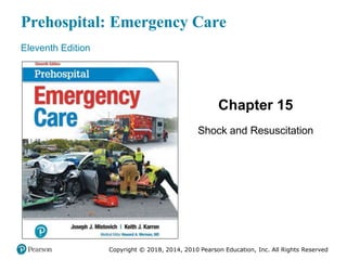
  • 1. Prehospital: Emergency Care Eleventh Edition Chapter 15 Shock and Resuscitation Copyright © 2018, 2014, 2010 Pearson Education, Inc. All Rights Reserved
  • 2. Learning Readiness Copyright © 2018, 2014, 2010 Pearson Education, Inc. All Rights Reserved • EMS Education Standards, text p. 435. • Chapter Objectives, text p. 435. • Key Terms, text p. 436. • Purpose of lecture presentation versus textbook reading assignments.
  • 3. Setting the Stage Copyright © 2018, 2014, 2010 Pearson Education, Inc. All Rights Reserved • Overview of Lesson Topics – Shock – Resuscitation in Cardiac Arrest – Automated External Defibrillation and Cardiopulmonary Resuscitation – Recognizing and Treating Cardiac Arrest – Special Considerations for the AED
  • 4. Case Study Introduction (1 of 2) Copyright © 2018, 2014, 2010 Pearson Education, Inc. All Rights Reserved EMTs Tess Price and Steph Bowman are responding to a report of chest pain, along with the crew from Engine 9. Their general impression is of a man in his 50s who is sitting in a recliner, hand on chest, and in apparent distress. He is pale and extremely sweaty.
  • 5. Case Study Introduction (2 of 2) Copyright © 2018, 2014, 2010 Pearson Education, Inc. All Rights Reserved The EMTs confirm a chief complaint of chest pain, and continue their assessment as they begin treatment. Tess is immediately concerned with the weak, rapid radial pulse and labored respirations.
  • 6. Case Study (1 of 6) Copyright © 2018, 2014, 2010 Pearson Education, Inc. All Rights Reserved • What is the significance of the weak, rapid pulse and labored respirations? • What could explain the the weak, rapid pulse and labored respirations? • What should the EMTs do to manage this patient?
  • 7. Introduction Copyright © 2018, 2014, 2010 Pearson Education, Inc. All Rights Reserved • Shock is a state of inadequate perfusion of the cells that can lead to death. • The body’s attempts to restore homeostasis result in many signs and symptoms of shock. • Resuscitation is the emergency care provided to restore vital body functions.
  • 8. Shock (1 of 38) Copyright © 2018, 2014, 2010 Pearson Education, Inc. All Rights Reserved • Defined as inadequate tissue perfusion or hypoperfusion • Inadequate amounts of oxygen and glucose are available to cells to meet metabolic needs • Inadequate waste product removal
  • 9. Shock (2 of 38) Copyright © 2018, 2014, 2010 Pearson Education, Inc. All Rights Reserved • Shift from aerobic to anaerobic metabolism results in decreased energy production and waste product accumulation. • Cellular sodium/potassium pump fails, leading to cell death. • Cell death results in organ failure.
  • 10. Shock (3 of 38) Copyright © 2018, 2014, 2010 Pearson Education, Inc. All Rights Reserved • Treatment of shock is aimed at restoring perfusion to provide the cells with glucose and oxygen.
  • 11. Shock (4 of 38) Copyright © 2018, 2014, 2010 Pearson Education, Inc. All Rights Reserved • Oxygen delivery to the cells is critical, and requires: – Breathing in an adequate amount of oxygen – Diffusion of oxygen from alveoli to pulmonary capillaries – Oxygen transport to the cellular level – Release of oxygen at the cellular level
  • 12. Shock (5 of 38) Copyright © 2018, 2014, 2010 Pearson Education, Inc. All Rights Reserved • Etiologies of Shock – Poor tissue perfusion is caused by one or more of these problems: ▪ Inadequate volume ▪ Inadequate pump function ▪ Inadequate vessel tone
  • 13. Shock (6 of 38) Copyright © 2018, 2014, 2010 Pearson Education, Inc. All Rights Reserved • Etiologies of Shock – Inadequate Volume ▪ Decreased blood volume decreases preload ▪ Decreased preload causes stroke volume and cardiac output to fall ▪ Decreased cardiac output causes a drop in systolic blood pressure ▪ Decreased systolic blood pressure results in inadequate tissue perfusion
  • 14. Shock (7 of 38) Copyright © 2018, 2014, 2010 Pearson Education, Inc. All Rights Reserved • Etiologies of Shock – Inadequate Volume ▪ May result from loss of whole blood or plasma volume – Bleeding – Vomiting, diarrhea – Excessive urination – Increased capillary leakage
  • 15. Shock (8 of 38) Copyright © 2018, 2014, 2010 Pearson Education, Inc. All Rights Reserved • Etiologies of Shock – Inadequate Volume ▪ Patient requires an increase in blood volume ▪ If red blood cells have been lost, there is a decrease in oxygen-carrying capacity, as well as a decrease in pressure and perfusion
  • 16. Shock (9 of 38) Copyright © 2018, 2014, 2010 Pearson Education, Inc. All Rights Reserved • Etiologies of Shock – Inadequate Pump Function ▪ If the heart fails as a pump, regardless of the blood volume, the delivery of oxygen and glucose to cells decreases
  • 17. Shock (10 of 38) Copyright © 2018, 2014, 2010 Pearson Education, Inc. All Rights Reserved • Etiologies of Shock – Inadequate Pump Function ▪ May occur from myocardial infarction or heart failure, or mechanical obstruction to blood flow – Pericardial tamponade – Tension pneumothorax ▪ Giving fluids may worsen the condition
  • 18. Shock (11 of 38) Copyright © 2018, 2014, 2010 Pearson Education, Inc. All Rights Reserved • Etiologies of Shock – Inadequate Vessel Tone ▪ Blood pressure is a function of cardiac output and Systemic Vascular Resistance (SVR) ▪ If SVR decreases from vasodilation, blood pressure decreases
  • 19. Etiology of Shock: Vasodilation Copyright © 2018, 2014, 2010 Pearson Education, Inc. All Rights Reserved
  • 20. Shock (12 of 38) Copyright © 2018, 2014, 2010 Pearson Education, Inc. All Rights Reserved • Etiologies of Shock – Inadequate Vessel Tone ▪ Massive vasodilation can occur from loss of sympathetic nervous system function or chemicals released within the body. ▪ Treatment includes vasoconstriction and volume restoration.
  • 21. Shock (13 of 38) Copyright © 2018, 2014, 2010 Pearson Education, Inc. All Rights Reserved • Etiologies of Shock – Inadequate Vessel Tone ▪ Treatment of shock depends on underlying cause ▪ Consider requesting ALS, but weigh the benefit against any potential delay in reaching the hospital
  • 22. Categories and Types of Shock Copyright © 2018, 2014, 2010 Pearson Education, Inc. All Rights Reserved
  • 23. Shock (14 of 38) Copyright © 2018, 2014, 2010 Pearson Education, Inc. All Rights Reserved • Categories of Shock – Hypovolemic shock is caused by low blood volume ▪ Hemorrhage is the most common cause of hypovolemic shock ▪ Also caused by burns and dehydration
  • 24. (a) Hemorrhagic Hypovolemia: Loss of Whole Blood; (b) Nonhemorrhagic Hypovolemia: Loss of Plasma Copyright © 2018, 2014, 2010 Pearson Education, Inc. All Rights Reserved
  • 25. Shock (15 of 38) Copyright © 2018, 2014, 2010 Pearson Education, Inc. All Rights Reserved • Categories of Shock – Distributive Shock ▪ Caused by vasodilation leading to a relative reduction in intravascular volume ▪ Can also involve fluid loss from increased capillary permeability
  • 26. Shock (16 of 38) Copyright © 2018, 2014, 2010 Pearson Education, Inc. All Rights Reserved • Categories of Shock – Cardiogenic Shock ▪ Caused by inability of heart to contract effectively ▪ Generally occurs with loss of 40 percent or greater of left ventricular volume ▪ Stroke volume and cardiac output are reduced
  • 27. Heart Attack as a Cause of Cardiogenic Shock Damaged heart muscle results in reduced force of contractions, reduced stroke volume, and reduced cardiac output Copyright © 2018, 2014, 2010 Pearson Education, Inc. All Rights Reserved
  • 28. Shock (17 of 38) Copyright © 2018, 2014, 2010 Pearson Education, Inc. All Rights Reserved • Categories of Shock – Obstructive Shock ▪ Results from a condition that obstructs forward blood flow ▪ Causes include pulmonary embolism, tension pneumothorax, and pericardial tamponade ▪ Treat by relieving the obstruction
  • 29. Causes of Obstructive Shock (a) pulmonary embolism; (b) tension pneumothorax; (c) pericardial tamponade Copyright © 2018, 2014, 2010 Pearson Education, Inc. All Rights Reserved
  • 30. Shock (18 of 38) Copyright © 2018, 2014, 2010 Pearson Education, Inc. All Rights Reserved • Categories of Shock – Metabolic or Respiratory Shock ▪ Some sources list metabolic or respiratory shock as a fifth category ▪ Dysfunction in the ability of oxygen to diffuse into the blood, be carried by hemoglobin, off-load at the cell, or be used effectively by the cell for metabolism
  • 31. Shock (19 of 38) Copyright © 2018, 2014, 2010 Pearson Education, Inc. All Rights Reserved • Specific Types of Shock – Hemorrhagic Hypovolemic Shock ▪ Loss of whole blood from medical or traumatic causes ▪ Results in decreased perfusion pressure and decreased oxygen-carrying capacity ▪ Treat by stopping the bleeding and replacing blood or blood components
  • 32. Shock (20 of 38) Copyright © 2018, 2014, 2010 Pearson Education, Inc. All Rights Reserved • Specific Types of Shock – Nonhemorrhagic Hypovolemic Shock ▪ Loss of fluid without loss of red blood cells ▪ Can result from vomiting, diarrhea, sweating ▪ Administration of IV fluids can be helpful
  • 33. Shock (21 of 38) Copyright © 2018, 2014, 2010 Pearson Education, Inc. All Rights Reserved • Specific Types of Shock – Burn Shock ▪ Is a specific form of nonhemorrhagic hypovolemic shock resulting from a burn injury ▪ Plasma and plasma proteins leak from damaged capillaries ▪ Loss of fluid and plasma oncotic pressure leads to edema
  • 34. Shock (22 of 38) Copyright © 2018, 2014, 2010 Pearson Education, Inc. All Rights Reserved • Specific Types of Shock – Anaphylactic Shock ▪ Chemical substances released in anaphylactic reaction cause massive systemic vasodilation and increased capillary permeability ▪ Epinephrine is the medication of choice
  • 35. Shock (23 of 38) Copyright © 2018, 2014, 2010 Pearson Education, Inc. All Rights Reserved • Specific Types of Shock – Septic Shock ▪ Septic shock is another type of distributive shock ▪ Bacteria and toxins in the blood lead to vasodilation and increased capillary permeability ▪ Patient can benefit from ALS treatment
  • 36. Table 15-1 EMS Screening Tools to Identify Sepsis Copyright © 2018, 2014, 2010 Pearson Education, Inc. All Rights Reserved Robson Screening Tool Sepsis is suspected if two of the following findings are present with a suspected active infection: • Temperature >38.3°C (100.9°F ) or <36°C (96.8°F ) • Heart rate >90 bpm • Respiratory rate >20 breaths/minute • Acute altered mental status • BGL <120 milligram/dL BAS 90-30-90 Scale Sepsis is suspected if one or more of the following findings are present in a patient with a suspected active infection: • Systolic blood pressure <90 millimeterHg • Respiratory rate >30 breaths/minute • SpO2 <90% qSOFA Score Sepsis is suspected if two or more of the following findings are present in a patient with a suspected active infection: •Respiratory rate 22 breaths/minute or greater •Altered mental status (GCS <13) Systolic BP 100 millimeterHg or less
  • 37. Shock (24 of 38) Copyright © 2018, 2014, 2010 Pearson Education, Inc. All Rights Reserved • Specific Types of Shock – Neurogenic Shock ▪ Spinal cord injury can cause loss of sympathetic nerve fiber functions responsible for maintaining blood vessel tone ▪ Loss of systemic vascular resistance
  • 38. Shock (25 of 38) Copyright © 2018, 2014, 2010 Pearson Education, Inc. All Rights Reserved • Specific Types of Shock – Cardiogenic Shock ▪ Causes include acute myocardial infarction, congestive heart failure, abnormal cardiac rhythm, and overdose on certain drugs ▪ Patients can benefit from ALS interventions
  • 39. Click on the Term That Best Describes the Type of Shock That Occurs When There is Massive Systemic Vasodilation Copyright © 2018, 2014, 2010 Pearson Education, Inc. All Rights Reserved A. Obstructive B. Metabolic C. Distributive D. Cardiogenic
  • 40. Shock (26 of 38) Copyright © 2018, 2014, 2010 Pearson Education, Inc. All Rights Reserved • The Body’s Response to Shock – The body attempts to compensate to return perfusion to normal – Many signs and symptoms of shock are related to compensatory mechanisms – Sympathetic nervous system stimulation and the release of hormones are the two major compensatory mechanisms
  • 41. Shock (27 of 38) Copyright © 2018, 2014, 2010 Pearson Education, Inc. All Rights Reserved • The Body’s Response to Shock – Direct Nerve Stimulation ▪ The effects of sympathetic stimulation are immediate – Increase in heart rate – Increase in force of ventricular contraction – Vasoconstriction – Stimulation of the release of epinephrine and norepinephrine from the adrenal gland
  • 42. Shock (28 of 38) Copyright © 2018, 2014, 2010 Pearson Education, Inc. All Rights Reserved • The Body’s Response to Shock – Release of Hormones ▪ The effects of hormones are more sustained – Epinephrine has alpha and beta effects that cause vasoconstriction and increased cardiac output – Norepinephrine has alpha effects that cause vasoconstriction – Other hormones are also released
  • 43. Table 15-2 Effects of Alpha and Beta Stimulation Copyright © 2018, 2014, 2010 Pearson Education, Inc. All Rights Reserved Receptor Stimulatory Effect Sign or Symptom Alpha 1 Contraction of the muscles controlling the iris Dilated pupils Contraction of vascular smooth muscle causing vasoconstriction Pale, cool skin, narrow pulse pressure Stimulation of sweat glands Localized sweating, clammy skin Beta 1 Increased heart rate Tachycardia Increased speed of impulse through conduction system Tachycardia Increased force of contraction Pounding heart Beta 2 Bronchial smooth muscle dilation Decreased resistance in airway Skeletal muscle contractility Tremors
  • 44. Table 15-3 Effects of Hormones Released in Shock (1 of 2) Copyright © 2018, 2014, 2010 Pearson Education, Inc. All Rights Reserved Hormone Effect on Body Sign or Symptom Epinephrine Increased heart rate (beta 1) Tachycardia Increased contractility (beta 1) Pounding heart Vasoconstriction (alpha 1) Pale, cool skin Sweat gland stimulation (alpha 1) Clammy skin Decreased insulin secretion (alpha 2) Increased blood glucose level Conversion of stored glucose in liver to blood glucose Conversion of noncarbohydrates into sugar Iris muscle contraction (alpha 1) Pupillary dilation Norepinephrine Vasoconstriction (alpha 1) Pale, cool skin Sweat gland stimulation (alpha 1) Clammy skin Increased heart rate (beta 1) Tachycardia
  • 45. Table 15-3 Effects of Hormones Released in Shock (2 of 2) Copyright © 2018, 2014, 2010 Pearson Education, Inc. All Rights Reserved Hormone Effect on Body Sign or Symptom Antidiuretic Hormone (Vasopressin) Increased sodium reabsorption in the kidneys Decreased urine output Vasoconstriction Increased blood pressure Angiotensin II Vasoconstriction Pale, cool skin Sodium reabsorption in the kidney Decreased urine output Aldosterone Sodium reabsorption in the kidney Decreased urine output Glucagon Conversion of stored glucose in liver to blood glucose Increased blood glucose level Conversion of noncarbohydrates into sugar Increased heart rate and contractility Tachycardia
  • 46. Shock (29 of 38) Copyright © 2018, 2014, 2010 Pearson Education, Inc. All Rights Reserved • Stages of Shock – The stages of shock are: ▪ Compensatory ▪ Decompensatory
  • 47. The Cycle of Hemorrhagic Shock Copyright © 2018, 2014, 2010 Pearson Education, Inc. All Rights Reserved
  • 48. Shock (30 of 38) Copyright © 2018, 2014, 2010 Pearson Education, Inc. All Rights Reserved • Stages of Shock – Compensatory Shock ▪ The body is able to maintain near-normal blood pressure and perfusion of vital organ ▪ Blood is shunted away from non-vital areas, such as the skin and gastrointestinal tract ▪ Pulse pressure may be narrowed
  • 49. Shock (31 of 38) Copyright © 2018, 2014, 2010 Pearson Education, Inc. All Rights Reserved • Stages of Shock – Decompensatory Shock ▪ Compensatory mechanisms are overwhelmed ▪ The body can no longer maintain a blood pressure and perfusion of the vital organs ▪ Anaerobic metabolism is occurring ▪ Vital organs are not perfused
  • 50. Shock (32 of 38) Copyright © 2018, 2014, 2010 Pearson Education, Inc. All Rights Reserved • Stages of Shock – Multiple Organ Dysfunction Syndrome (MODS) ▪ The stage in which multiple organs begin to fail throughout the body from extreme and prolonged hypoxia, altered metabolism, and elevated carbon dioxide and acid levels ▪ Sometimes referred to as irreversible shock
  • 51. Shock (33 of 38) Copyright © 2018, 2014, 2010 Pearson Education, Inc. All Rights Reserved • Shock Assessment – Signs may be subtle or profound, but rapid recognition of shock is key to treatment – Consider history findings, physical assessment findings, signs of perfusion disturbance, and vital signs – Do not rely on one finding, sign, or symptom
  • 52. Shock (34 of 38) Copyright © 2018, 2014, 2010 Pearson Education, Inc. All Rights Reserved • Shock Assessment – History ▪ Chief complaint ▪ Sample history ▪ Beta blockers and calcium channel blockers can alter the response to shock
  • 53. Shock (35 of 38) Copyright © 2018, 2014, 2010 Pearson Education, Inc. All Rights Reserved • Shock Assessment – Physical exam ▪ Vital signs can appear normal in compensatory shock ▪ Look for signs of poor perfusion
  • 54. Table 15-5 Physical Assessment Indicators of Hypovolemic Shock Copyright © 2018, 2014, 2010 Pearson Education, Inc. All Rights Reserved Vital Signs Signs of Poor Perfusion Decreasing blood pressure Anxiety, anxiousness that Progresses to a decreased mental status Narrowing pulse pressure Pale, cool, clammy skin Tachycardia Delayed capillary refill Tachypnea Weak or absent peripheral pulses Pale, cool, and clammy skin Decreased urine output Unobtainable or poor SpO2 reading
  • 55. Table 15-6 Physical Assessment Indicators of Cardiogenic Shock Copyright © 2018, 2014, 2010 Pearson Education, Inc. All Rights Reserved Vital Signs Signs of Poor Perfusion Decreasing blood pressure Anxiety, anxiousness that progresses to a decreased mental status Narrowing pulse pressure Pale, cool, clammy skin; cyanotic or mottled skin Tachycardia or bradycardia; may be irregular Jugular venous distention and peripheral edema (right-sided heart failure) Tachypnea Weak or absent peripheral pulses Pale, cool, and clammy skin; cyanotic or mottled skin Decreased urine output Decreased SpO2 reading Other sign: Crackles or rales upon auscultation (left-sided heart failure)
  • 56. Table 15-7 Physical Assessment Indicators of Distributive Shock Copyright © 2018, 2014, 2010 Pearson Education, Inc. All Rights Reserved Vital Signs Signs of Poor Perfusion Decreasing blood pressure Anxiety, anxiousness that progresses to a decreased mental status Tachycardia (anaphylactic and septic shock) Relative bradycardia or normal heart rate (shock associated with a spinal cord injury) Mottled, cyanosis (late—sepsis, anaphylactic and neurogenic) Tachypnea with respiratory distress and wheezing (anaphylactic shock) Weak or absent peripheral pulses Tachypnea (septic) Normal respiratory rate (neurogenic) Normal to flushed (early sepsis) Warm, flushed skin (neurogenic) Warm, flushed skin with hives, possible cyanosis (anaphylactic) Mottled, cyanosis (late: sepsis, anaphylactic, and neurogenic) Severely decreased SpO2 reading (anaphylactic) Other signs: Fever (sepsis) Loss of motor/sensory function (neurogenic due to spinal cord injury) Edema (anaphylactic)
  • 57. Table 15-8 Physical Assessment Indicators of Obstructive Shock Copyright © 2018, 2014, 2010 Pearson Education, Inc. All Rights Reserved Vital Signs Signs of Poor Perfusion Decreasing blood pressure Pulsus paradoxus (tension pneumothorax and pericardial tamponade) Anxiety, anxiousness that progresses to a decreased mental status Narrowing pulse pressure Pale, cool, clammy skin; cyanotic or mottled skin Tachycardia Jugular venous distention (pericardial tamponade and tension pneumothorax) Tachypnea Weak or absent peripheral pulses Pale, cool, and clammy skin; cyanotic or mottled skin Decreased urine output Decreased SpO2 reading Severely decreased SpO2 reading (tension pneumothorax and massive pulmonary embolism) Other sign: Severely decreased to absent breath sounds of one hemithorax (tension pneumothorax)
  • 58. Shock (36 of 38) Copyright © 2018, 2014, 2010 Pearson Education, Inc. All Rights Reserved • Age Considerations in Shock – Age may influence the development, presentation, management, and recovery from shock. – Children can compensate well, but deteriorate quickly. – Geriatric patients do not compensate well.
  • 59. Shock (37 of 38) Copyright © 2018, 2014, 2010 Pearson Education, Inc. All Rights Reserved • General Goals of Prehospital Management of Shock – Secure and maintain a patent airway. – Establish and maintain adequate ventilation. – Establish and maintain adequate oxygenation. – Do not hyperventilate the shock patient. – Stop bleeding as quickly as possible.
  • 60. Shock (38 of 38) Copyright © 2018, 2014, 2010 Pearson Education, Inc. All Rights Reserved • General Goals of Prehospital Management of Shock – Splint fractures, don’t delay transport. – Do not remove an impaled object. – Maintain the body temperature. – Keep the patient supine. – Apply PASG, according to protocol. – Rapid transport. – Consider ALS intercept.
  • 61. Case Study (2 of 6) Copyright © 2018, 2014, 2010 Pearson Education, Inc. All Rights Reserved Finding that the patient is confused, the EMTs complete a rapid secondary assessment and obtain a history from the patient's wife, as the engine crew helps them prepare the patient for transport.
  • 62. Case Study (3 of 6) Tess finds that the patient’s respirations are 28 and labored, and that he has crackles in his lungs. His heart rate is 116, with a blood pressure of 92 percent. Copyright © 2018, 2014, 2010 Pearson Education, Inc. All Rights Reserved 100 , 72 and an SpO2 of
  • 63. Case Study (4 of 6) Copyright © 2018, 2014, 2010 Pearson Education, Inc. All Rights Reserved • Is this patient in shock? Explain your answer? • What interventions should the patient be receiving? • What are the potential consequences of failing to intervene appropriately or in a timely manner?
  • 64. Resuscitation in Cardiac Arrest (1 of 12) Copyright © 2018, 2014, 2010 Pearson Education, Inc. All Rights Reserved • Resuscitation means bringing a patient back from a potential or apparent death. • Resuscitation focuses on management of the airway, ventilation, and oxygenation, and restoring adequate circulation.
  • 65. Resuscitation in Cardiac Arrest (2 of 12) Copyright © 2018, 2014, 2010 Pearson Education, Inc. All Rights Reserved • Cardiac arrest occurs when the ventricles of the heart are not contracting or cardiac output is ineffective and no pulses can be felt. – Brain cells begin to die within four to six minutes following cardiac arrest. – Cardiac arrest patients are described as having suffered sudden death when the patient dies within one hour of the onset of the signs and symptoms.
  • 66. Resuscitation in Cardiac Arrest (3 of 12) Copyright © 2018, 2014, 2010 Pearson Education, Inc. All Rights Reserved • A common cause of cardiac arrest is ventricular fibrillation. • Ventricular fibrillation can be treated with defibrillation.
  • 67. Resuscitation in Cardiac Arrest (4 of 12) Copyright © 2018, 2014, 2010 Pearson Education, Inc. All Rights Reserved • Pathophysiology of Cardiac Arrest – The patient goes through three phases of cardiac arrest that lead to biological death ▪ Electrical phase ▪ Circulatory phase ▪ Metabolic phase
  • 68. Resuscitation in Cardiac Arrest (5 of 12) Copyright © 2018, 2014, 2010 Pearson Education, Inc. All Rights Reserved • Pathophysiology of Cardiac Arrest – Electrical Phase ▪ First four minutes. ▪ The heart still has a supply of oxygen and glucose. ▪ Conditions are favorable for resuscitation. ▪ The heart is prepared to respond to defibrillation.
  • 69. Resuscitation in Cardiac Arrest (6 of 12) Copyright © 2018, 2014, 2010 Pearson Education, Inc. All Rights Reserved • Pathophysiology of Cardiac Arrest – Circulatory Phase ▪ Four through ten minutes after cardiac arrest. ▪ Oxygen stores are exhausted; myocardial cells switch to anaerobic metabolism. ▪ CPR is needed to restore a supply of oxygen and glucose to enhance the possibility of successful defibrillation.
  • 70. Resuscitation in Cardiac Arrest (7 of 12) Copyright © 2018, 2014, 2010 Pearson Education, Inc. All Rights Reserved • Pathophysiology of Cardiac Arrest – Metabolic Phase ▪ Begins ten minutes after cardiac arrest. ▪ The heart muscle is acidic and ischemic, and begins to die. ▪ Chances of resuscitation are unfavorable.
  • 71. Resuscitation in Cardiac Arrest (8 of 12) Copyright © 2018, 2014, 2010 Pearson Education, Inc. All Rights Reserved • Terms Related to Out-of-Hospital Cardiac Arrest (OHCA) Resuscitation – Downtime – Total downtime – Return of spontaneous circulation (ROSC) – Survival – Witnessed cardiac arrest – Unwitnessed cardiac arrest
  • 72. Resuscitation in Cardiac Arrest (9 of 12) Copyright © 2018, 2014, 2010 Pearson Education, Inc. All Rights Reserved • Withholding a Resuscitation Attempt – Do not resuscitate orders – Physician orders for life-sustaining treatment – Medical orders for life-sustaining treatment – Injuries incompatible with life
  • 73. Resuscitation in Cardiac Arrest (10 of 12) Copyright © 2018, 2014, 2010 Pearson Education, Inc. All Rights Reserved • The 2015 AHA Chain of Survival – Successful resuscitation depends on a sequence of events ▪ The Chain of Survival is slightly different for pediatric patients.
  • 74. Resuscitation in Cardiac Arrest (11 of 12) Copyright © 2018, 2014, 2010 Pearson Education, Inc. All Rights Reserved • The 2015 AHA Chain of Survival – AHA 2015 OHCA Adult Chain of Survival ▪ Immediate recognition and activation. ▪ Immediate high-quality CPR. ▪ Rapid defibrillation. ▪ Basic and advanced emergency medical services. ▪ Advanced life support and post-arrest care.
  • 75. Resuscitation in Cardiac Arrest (12 of 12) Copyright © 2018, 2014, 2010 Pearson Education, Inc. All Rights Reserved • The 2015 AHA Chain of Survival – AHA 2015 OHCA Pediatric Chain of Survival ▪ Prevention of arrest. ▪ Early high-quality CPR. ▪ Rapid activation of EMS. ▪ Effective advanced life support and rapid transport. ▪ Integrated post-cardiac-arrest care.
  • 76. Automated External Defibrillation and CPR (1 of 9) Copyright © 2018, 2014, 2010 Pearson Education, Inc. All Rights Reserved • Rationale for early defibrillation – The most frequent initial rhythm in sudden cardiac arrest is ventricular fibrillation. – The most effective treatment for ventricular fibrillation is defibrillation. – The probability of successful defibrillation decreases over time.
  • 77. Automated External Defibrillation and CPR (2 of 9) Copyright © 2018, 2014, 2010 Pearson Education, Inc. All Rights Reserved • Rationale for early defibrillation – Successful defibrillation depends on effective CPR; interruptions in chest compressions for defibrillation must be minimized. – Without intervention, ventricular fibrillation degenerates into asystole.
  • 78. Automated External Defibrillation and CPR (3 of 9) Copyright © 2018, 2014, 2010 Pearson Education, Inc. All Rights Reserved • “Push hard and push fast” to provide effective chest compressions in CPR. – Compressions at a rate of at least 100/minute. – Compression-to-ventilation ratio of 30:2. – Minimize interruptions for defibrillation. – Avoid excess ventilation.
  • 79. Automated External Defibrillation and CPR (4 of 9) Copyright © 2018, 2014, 2010 Pearson Education, Inc. All Rights Reserved • Types of Defibrillators – Manual defibrillators require extensive training to use. – Automated defibrillators are simpler to use.
  • 80. Automated External Defibrillation and CPR (5 of 9) Copyright © 2018, 2014, 2010 Pearson Education, Inc. All Rights Reserved • Types of Defibrillators – Advantages of AEDs ▪ Speed of operation ▪ Safer, more effective shock delivery ▪ More efficient monitoring
  • 81. Automated External Defibrillation and CPR (6 of 9) Copyright © 2018, 2014, 2010 Pearson Education, Inc. All Rights Reserved • Types of Defibrillators – Types of AEDs ▪ Fully automated ▪ Semiautomated – AEDs may use a monophasic or biphasic waveform
  • 82. Physio-Control Lifepak® 1000 Defibrillator (© Physio-Control, Inc.) Copyright © 2018, 2014, 2010 Pearson Education, Inc. All Rights Reserved
  • 83. Infant/Child Pads for Physiocontrol Lifepak® 1000 Copyright © 2018, 2014, 2010 Pearson Education, Inc. All Rights Reserved
  • 84. Automated External Defibrillation and CPR (7 of 9) Copyright © 2018, 2014, 2010 Pearson Education, Inc. All Rights Reserved • Analysis of Cardiac Rhythms – Ventricular fibrillation – Ventricular tachycardia – Asystole – Pulseless electrical activity
  • 85. Ventricular Fibrillation is Associated with Chaotic Electrical Discharge in the Ventricles Copyright © 2018, 2014, 2010 Pearson Education, Inc. All Rights Reserved
  • 86. Ventricular Tachycardia Originates in the Conduction System of the Ventricle Copyright © 2018, 2014, 2010 Pearson Education, Inc. All Rights Reserved
  • 87. Asystole, or “Flatline,” is the Complete Absence of Electrical Activity in the Heart Copyright © 2018, 2014, 2010 Pearson Education, Inc. All Rights Reserved
  • 88. Automated External Defibrillation and CPR (8 of 9) Copyright © 2018, 2014, 2010 Pearson Education, Inc. All Rights Reserved • Analysis of Cardiac Rhythms – No one should touch the patient during AED rhythm analysis or shock delivery ▪ Movement interferes with rhythm analysis. ▪ The electrical energy can be transmitted to anyone touching the patient.
  • 89. Automated External Defibrillation and CPR (9 of 9) Copyright © 2018, 2014, 2010 Pearson Education, Inc. All Rights Reserved • When and When Not to Use the AED – AEDs can be used in patients of all ages. – Manual defibrillation is preferred for those less than one year of age. – A dose attenuating system is preferred for use in children. – Use adult pads with children greater than eight years of age.
  • 90. AED Pads Applied to a Pediatric Patient Copyright © 2018, 2014, 2010 Pearson Education, Inc. All Rights Reserved
  • 91. AED Pads Applied to an Infant Copyright © 2018, 2014, 2010 Pearson Education, Inc. All Rights Reserved
  • 92. Recognizing and Treating Cardiac Arrest (1 of 16) Copyright © 2018, 2014, 2010 Pearson Education, Inc. All Rights Reserved • Assessment-Based Approach: Cardiac Arrest – Scene Size-Up and Primary Assessment ▪ If a patient appears unresponsive without signs of life, quickly check for breathing and carotid pulse. ▪ Assess for no longer than ten seconds. ▪ If the patient is apneic or has agonal respirations, begin CPR with a C-A-B approach.
  • 93. Recognizing and Treating Cardiac Arrest (2 of 16) Copyright © 2018, 2014, 2010 Pearson Education, Inc. All Rights Reserved • Assessment-Based Approach: Cardiac Arrest – Patients less than one year of age ▪ If heart rate is >60 with inadequate ventilation, assist or provide ventilations at 12–20 per minute. ▪ If heart rate is <60 or pulse is absent, begin chest compressions.
  • 94. Recognizing and Treating Cardiac Arrest (3 of 16) Copyright © 2018, 2014, 2010 Pearson Education, Inc. All Rights Reserved • Assessment-Based Approach: Cardiac Arrest – Patients less than one year of age ▪ 30 compressions: two ventilations for one EMT performing CPR. ▪ 15 compressions: two ventilations for two EMTs performing CPR.
  • 95. Recognizing and Treating Cardiac Arrest (4 of 16) Copyright © 2018, 2014, 2010 Pearson Education, Inc. All Rights Reserved • Assessment-Based Approach: Cardiac Arrest – Patients one year of age to puberty ▪ 30 compressions: two ventilations for one EMT performing CPR. ▪ 15 compressions: two ventilations for two EMTs performing CPR.
  • 96. Recognizing and Treating Cardiac Arrest (5 of 16) Copyright © 2018, 2014, 2010 Pearson Education, Inc. All Rights Reserved • Assessment-Based Approach: Cardiac Arrest – Adolescents with signs of puberty and adults ▪ 30 compressions: two ventilations ▪ Rate of at least 100/minute. ▪ Compression depth of at least two inches ▪ For an obviously pregnant patient, displace the uterus laterally.
  • 97. Recognizing and Treating Cardiac Arrest (6 of 16) Copyright © 2018, 2014, 2010 Pearson Education, Inc. All Rights Reserved • Assessment-Based Approach: Cardiac Arrest – Secondary Assessment ▪ Attempt to obtain a history from bystanders or relatives, but do not interrupt chest compressions or delay defibrillation.
  • 98. Recognizing and Treating Cardiac Arrest (7 of 16) Copyright © 2018, 2014, 2010 Pearson Education, Inc. All Rights Reserved • Assessment-Based Approach: Cardiac Arrest – Emergency medical care ▪ Continue CPR and defibrillation, as indicated. – Reassessment ▪ Once perfusion is restored, continue reassessment. ▪ Patients may revert into cardiac arrest.
  • 99. Recognizing and Treating Cardiac Arrest (8 of 16) Copyright © 2018, 2014, 2010 Pearson Education, Inc. All Rights Reserved • Performing Defibrillation – Using an AED ▪ Ideally, at least two EMTs are present. ▪ Take standard precautions. ▪ Perform a brief primary assessment. ▪ Perform CPR minimizing breaks. ▪ Prepare and apply the AED. ▪ Analyze the rhythm and defibrillate as recommended by the AED.
  • 100. EMT Skills 15-1 Copyright © 2018, 2014, 2010 Pearson Education, Inc. All Rights Reserved Using a Semiautomated AED
  • 101. In the Unresponsive Patient Suspected of Being in Cardiac Arrest, Quickly Assess for Apnea or Agonal Ventilations and a Pulse Copyright © 2018, 2014, 2010 Pearson Education, Inc. All Rights Reserved
  • 102. One EMT Should Immediately Initiate CPR Beginning with Chest Compressions While the Other EMT Prepares the AED Copyright © 2018, 2014, 2010 Pearson Education, Inc. All Rights Reserved
  • 103. Turn on the AED and Follow the Prompts Copyright © 2018, 2014, 2010 Pearson Education, Inc. All Rights Reserved
  • 104. Apply the Defibrillation Pads While Chest Compressions are Being Performed - Minimize Any Interruption in Chest Compressions Copyright © 2018, 2014, 2010 Pearson Education, Inc. All Rights Reserved
  • 105. Clear the Patient for Rhythm Analysis Copyright © 2018, 2014, 2010 Pearson Education, Inc. All Rights Reserved
  • 106. Deliver a Defibrillation If Advised Copyright © 2018, 2014, 2010 Pearson Education, Inc. All Rights Reserved
  • 107. Immediately Resume CPR Beginning with Chest Compressions Following the Defibrillation Copyright © 2018, 2014, 2010 Pearson Education, Inc. All Rights Reserved
  • 108. After Two Minutes of CPR, Follow the AED Prompts to Check Breathing and Pulse in a Non-Shockable Rhythm Copyright © 2018, 2014, 2010 Pearson Education, Inc. All Rights Reserved
  • 109. Recognizing and Treating Cardiac Arrest (9 of 16) Copyright © 2018, 2014, 2010 Pearson Education, Inc. All Rights Reserved • Performing Defibrillation – Use of the AED by a Single EMT ▪ Simultaneously verify that the patient is unresponsive, with no breathing and no pulse. ▪ Call for additional EMS and the AED. ▪ Immediately begin chest compressions and apply the AED as soon as it is available. ▪ Minimize interruption of compressions.
  • 110. Recognizing and Treating Cardiac Arrest (10 of 16) Copyright © 2018, 2014, 2010 Pearson Education, Inc. All Rights Reserved • Cardiac Arrest in a Pregnant Patient – A pregnant patient in cardiac arrest who is at 20 weeks of gestation or greater, it is necessary to place the patient in a supine position and to manually displace the uterus off the vena cava when doing chest compressions.
  • 111. Recognizing and Treating Cardiac Arrest (11 of 16) Copyright © 2018, 2014, 2010 Pearson Education, Inc. All Rights Reserved • Transporting the Cardiac Arrest Patient – After emergency care procedures and operating the AED, if ALS is not responding to the scene, transport if: ▪ The patient regains a pulse. ▪ Your protocol indicates transport after a prescribed number of shocks or no shock indicated messages.
  • 112. Recognizing and Treating Cardiac Arrest (12 of 16) Copyright © 2018, 2014, 2010 Pearson Education, Inc. All Rights Reserved • Transporting the Cardiac Arrest Patient – Transporting a Patient with a Pulse ▪ Provide oxygen/ventilation as needed. ▪ Have suction ready. ▪ Transfer the patient to the ambulance. ▪ Consider getting ACLS to the patient. ▪ Keep AED attached. ▪ Perform secondary assessment. ▪ Reassess every five minutes.
  • 113. Recognizing and Treating Cardiac Arrest (13 of 16) Copyright © 2018, 2014, 2010 Pearson Education, Inc. All Rights Reserved • Transporting the Cardiac Arrest Patient – Transporting a patient without a pulse ▪ Continue to CPR and defibrillation. ▪ Follow protocol. ▪ Use extreme caution when defibrillating in the ambulance. ▪ Rendezvous with ALS as early as possible.
  • 114. Recognizing and Treating Cardiac Arrest (14 of 16) Copyright © 2018, 2014, 2010 Pearson Education, Inc. All Rights Reserved • Post-Resuscitation Care – Indications that ROSC has occurred ▪ A pulse is felt after the AED indicates a no shock advisory. ▪ Patient regains spontaneous breathing. ▪ The patient begins to move. – Upon ROSC ▪ Assess the patient’s ventilation. ▪ Avoid potential oxygen toxicity issues.
  • 115. Recognizing and Treating Cardiac Arrest (15 of 16) Copyright © 2018, 2014, 2010 Pearson Education, Inc. All Rights Reserved • Providing for Advanced Cardiac Life Support – The AHA’s 2015 Chain of Survival advocates for advanced cardiac life support. – Minimize the time from the delivery of CPR and defibrillation to the arrival of ACLS.
  • 116. Recognizing and Treating Cardiac Arrest (16 of 16) Copyright © 2018, 2014, 2010 Pearson Education, Inc. All Rights Reserved • Summary: Assessment and Care – Review assessment findings that may be associated with cardiac arrest and emergency care for cardiac arrest. Assessment Summary Cardiac Arrest The following are findings that indicate cardiac arest. These findings are obtained during the primary assessment: • Unresponsive • Apneic (not breathing) • Pulseless
  • 117. Emergency Care Protocol: Cardiac Arrest (1 of 3) Copyright © 2018, 2014, 2010 Pearson Education, Inc. All Rights Reserved 1. Take Standard Precautions. 2. Perform a brief primary assesment of the patient. It the patient is unresponsive, is apneic or has agonal ventilation, has no pulse (checked simulataneously with ventilation and for no less than 5 seconds but more than 10 seconds), and no other signs of life, immediately initiate CPR beginning with chest compressions. 3. Immediately intitate CPR beginning with chest compressions (CAB intervention sequence). As soon as the AED is available, apply it while chest compressions are being performed. When the AED is ready to start the rhythm analysis, stop chest compressions and proceed with AED protocol. – If bystanders or first responders are already performing CPR when you arrive, instruct them to continue while you prepare application of the AED.
  • 118. Emergency Care Protocol: Cardiac Arrest (2 of 3) Copyright © 2018, 2014, 2010 Pearson Education, Inc. All Rights Reserved 4. Continue with chest compressions while the AED is readled for operation. 5. Turn on power to the AED. 6. Attach the adhesive monitoring-defibrillation pads to the chest while chest compressions are being performed. Minimize any interruption to chest compressions. 7. Begin analysis of the patient’s cardiac rhythm. 8. If the AED’s analysis indicates a shock, it provides a “deliver shock” message. In that case, proceed with defibrillation by depressing the shock or defibrillation button. If the AED’s analysis determines a nonshockable rhythm, it gives a “no shock” message. In that case, immediately resume CPR beginning with chest compressions. 9. After a shock has been delivered, immediately resume CPR beginning with chest compressions. Perform CPR for approximately 2 minutes.
  • 119. Emergency Care Protocol: Cardiac Arrest (3 of 3) Copyright © 2018, 2014, 2010 Pearson Education, Inc. All Rights Reserved 10. After 2 minutes, the AED reanalyzes the rhythm. If a shock is indicated, proceed with the defibrillation, then immediately resume CPR beginning with chest compressions. If the AED gives a prompt to check breathing and pulse, quickly asses the patient’s breathing and pulse. The AED may indicate the patient now has a pulse or no longer has a shockable rhythm. If the patient is unresponsive, apenic or has agonal ventilations, and has no pulse, immediately resume CPR beginning with chest compressions. Continue to repeat this sequence. If the patient has a pulse, continue with ventilation. Continuously reassess the patient. 11. Follow your local protocol regarding when to transport the patient in cardiac arrest.
  • 120. Click on the Compression-to-Ventilation Ratio Used When Two EMTs Are Performing CPR on a Five- Year-Old Child Copyright © 2018, 2014, 2010 Pearson Education, Inc. All Rights Reserved A. 30:2 B. 15:2 C. 30:1 D. 15:1
  • 121. Case Study (5 of 6) Copyright © 2018, 2014, 2010 Pearson Education, Inc. All Rights Reserved Tess is assisting the patient’s ventilations with a bag-valve- mask and supplemental oxygen as they begin transport to the emergency department. Two minutes into the transport, the patient becomes unresponsive and has agonal respirations. Steph quickly checks for a carotid pulse, but cannot detect one.
  • 122. Case Study (6 of 6) Copyright © 2018, 2014, 2010 Pearson Education, Inc. All Rights Reserved • What is the sequence of steps that Tess and Steph must take to maximize the chances of successful resuscitation? • What safety concerns are there in carrying out those steps?
  • 123. Special Considerations for the AED (1 of 7) Copyright © 2018, 2014, 2010 Pearson Education, Inc. All Rights Reserved • Safety Considerations – Clear everyone from patient before delivering a shock. – Do not defibrillate a patient who is wet. – Use caution defibrillating on metal surfaces. – Remove transdermal medication patches on the chest. – Remove excessive chest hair.
  • 124. Special Considerations for the AED (2 of 7) Copyright © 2018, 2014, 2010 Pearson Education, Inc. All Rights Reserved • AED maintenance – Scheduled maintenance is critical. – Replace batteries on schedule. – Allow machine to perform self-check at the beginning of the shift. – Have extra batteries available.
  • 125. Special Considerations for the AED (3 of 7) Copyright © 2018, 2014, 2010 Pearson Education, Inc. All Rights Reserved • Training and Skills Maintenance – Refresh and maintain skills through continuing education. – Incident review. – Maintain knowledge of protocols.
  • 126. Special Considerations for the AED (4 of 7) Copyright © 2018, 2014, 2010 Pearson Education, Inc. All Rights Reserved • Medical Direction and the AED – AEDs are used under the authority of medical direction. – Medical directors play several roles in an AED program, including ensuring EMTs’ skills and quality improvement.
  • 127. Special Considerations for the AED (5 of 7) Copyright © 2018, 2014, 2010 Pearson Education, Inc. All Rights Reserved • Cardiac pacemakers – Do not place AED pads directly over the pacemaker.
  • 128. An Implanted Pacemaker in an Adult Patient (© Michal Heron) Copyright © 2018, 2014, 2010 Pearson Education, Inc. All Rights Reserved
  • 129. Pacemakers May Also Be Found in Children (© Michal Heron) Copyright © 2018, 2014, 2010 Pearson Education, Inc. All Rights Reserved
  • 130. Special Considerations for the AED (6 of 7) Copyright © 2018, 2014, 2010 Pearson Education, Inc. All Rights Reserved • Automatic Implanted Cardioverter Defibrillators – Detect rhythm disturbances and deliver shocks. – Do not place AED pads over the ICD. – If the ICD is operating, allow 30 to 60 seconds for it to complete its cycle before attaching the AED.
  • 131. Special Considerations for the AED (7 of 7) Copyright © 2018, 2014, 2010 Pearson Education, Inc. All Rights Reserved • Automated Chest Compression Devices – Mechanical piston device – Load-distributing band or vest – Impedance threshold device
  • 132. The Autopulse, a Load-Distributing-Band CPR Device, Compresses the Thorax Copyright © 2018, 2014, 2010 Pearson Education, Inc. All Rights Reserved
  • 133. Case Study Conclusion (1 of 2) Copyright © 2018, 2014, 2010 Pearson Education, Inc. All Rights Reserved Signaling the EMT who is driving to pull to the side of the road, Steph immediately begins chest compressions, as Tess continues airway management. They perform CPR with ratio of 30 compressions to two ventilations, as an EMT from the engine crew attaches the AED.
  • 134. Case Study Conclusion (2 of 2) Copyright © 2018, 2014, 2010 Pearson Education, Inc. All Rights Reserved The AED detects a shockable rhythm and, after clearing the patient, the EMT delivers a shock. Tess immediately resumes chest compressions as they resume transport. Two minutes after defibrillation, Steph stops CPR to check a pulse. The patient has a weak radial pulse. Tess continues ventilations, as Steph gives an update to the receiving hospital.
  • 135. Summary (1 of 2) Copyright © 2018, 2014, 2010 Pearson Education, Inc. All Rights Reserved • Shock is a critical condition related to a decrease in vascular volume, poor cardiac function, or vessel disturbances. • Shock results in a shift to anaerobic cell metabolism. • Shock management focuses on airway, ventilation, oxygenation, circulation, and rapid transport.
  • 136. Summary (2 of 2) Copyright © 2018, 2014, 2010 Pearson Education, Inc. All Rights Reserved • The Chain of Survival from cardiac arrest includes: – Immediate recognition and activation – Early CPR – Rapid defibrillation – Effective ACLS – Integrated post-cardiac arrest care
  • 137. Correct! (1 of 2) Copyright © 2018, 2014, 2010 Pearson Education, Inc. All Rights Reserved When the blood vessels dilate, resulting in inadequate systemic vascular resistance to maintain perfusion, the type of shock is classified as distributive. Anaphylactic, neurogenic, and septic shock are all forms of distributive shock. Click here to return to the program.
  • 138. Incorrect (1 of 5) Copyright © 2018, 2014, 2010 Pearson Education, Inc. All Rights Reserved Obstructive shock results when forward flow of blood through the circulatory system is prevented by an obstruction. Causes include pulmonary embolism, tension pneumothorax, and pericardial tamponade. Click here to return to the quiz.
  • 139. Incorrect (2 of 5) Copyright © 2018, 2014, 2010 Pearson Education, Inc. All Rights Reserved Metabolic shock occurs when something interferes with on- loading of oxygen to hemoglobin, off-loading of oxygen from hemoglobin, or use of oxygen by the cell. Causes include cyanide poisoning and carbon monoxide poisoning. Click here to return to the quiz.
  • 140. Incorrect (3 of 5) Copyright © 2018, 2014, 2010 Pearson Education, Inc. All Rights Reserved Cardiogenic shock results when the heart fails as a pump and cannot maintain adequate cardiac output and blood pressure. Causes include myocardial infarction and heart failure. Click here to return to the quiz.
  • 141. Correct! (2 of 2) Copyright © 2018, 2014, 2010 Pearson Education, Inc. All Rights Reserved The compression-to-ventilation ratio for a 1- to 8-year-old child, with two rescuers performing CPR, is 15:2. Click here to return to the program.
  • 142. Incorrect (4 of 5) Copyright © 2018, 2014, 2010 Pearson Education, Inc. All Rights Reserved 30:2 is the compression-to-ventilation ratio used when one rescuer is performing CPR on a one- to eight-year-old child. Click here to return to the quiz.
  • 143. Incorrect (5 of 5) Copyright © 2018, 2014, 2010 Pearson Education, Inc. All Rights Reserved Neither 15:1 nor 30:1 are compression-to-ventilation ratios used in CPR. Click here to return to the quiz.
  • 144. Copyright Copyright © 2018, 2014, 2010 Pearson Education, Inc. All Rights Reserved