Condylar resorption
•Download as PPTX, PDF•
0 likes•336 views

condylar resorption and condylar sag

Recommended
More Related Content
What's hot
What's hot (20)
Similar to Condylar resorption
Similar to Condylar resorption (20)
Recently uploaded
Recently uploaded (20)
Condylar resorption
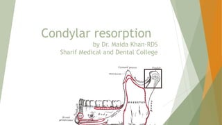
- 1. Condylar resorption by Dr. Maida Khan-RDS Sharif Medical and Dental College
- 2. Condylar resorption Condylar resorption, also called idiopathic condylar resorption, is a temporomandibular joint disorder in which one or both of the mandibular condyles are broken down in a bone resorption process. Also called cheerleader syndrome. Mostly affects condyles symmetrically and bilaterally SYMPTOMS TMJ pain, headache, myofascial pain, clicking and popping, and crepitation
- 3. In bilateral cases symmetric posterior shift of the mandible, development or worsening of the class II occlusal relationship (the lower teeth positioned excessively posteriorly relative to the upper teeth) development of an anterior open bite In unilateral cases the mandibular dental midline and chin shift toward the affected side an ipsilateral class II occlusion, crossbite, and posterior occlusal prematurity develop an open bite develops on the contralateral side of the jaws.
- 4. Causes Idiopathic Local causes parafunctional activity, trauma, orthodontics, or orthognathic surgery Unstable occlusion Systemic causes reactive arthritis rheumatoid arthritis infections (cytokines activates osteoclast releasing MMPs degrades ECM) Metabolic disorder ( decreased estradiol level) Nutritional status ( decreased vit. D)
- 5. Diagnosis 1.Panograms • Flattening of anterior surface of condyle 2. Lateral cephalometric radiograph • skeletal and occlusal class II deformity • anterior open bite, • high occlusal plane angle • , high mandibular plane angle • , decreased vertical height of the ramus, • possible overangulation of the lower incisors
- 6. 3.Cephalometric tomographic evaluation of the TMJs • normal or excessive joint space because of the hyperplasia of the synovial tissues within the joint. • The involved condylar head will appear smaller in size; • The cortical bone on the head of the condyle may lose some integrity. 4. MRI • decreased condylar size and volume; • anterior disc displacement, with or without reduction on opening; • extreme thinness or loss of continuity of cortical bone on the head of the condyle • thick, amorphous-appearing soft tissue occupying the space between the condyle and fossa
- 7. Treatment Non invasive treatment modalities 1. NSAIDS 2. Oral appliance ( occlusal splints) Minimally invasive treatment modalities 1. Arthrocentesis 2. arthroscopy 3. Non surgical orthodontics Invasive modality Orthognathic surgery
- 8. Condylar sag • Immediate / late change in position of condyle in glenoid fossa after surgical establishment of preplanned occlusion and rigid fixation of bone fragments, leading to change in occlusion. • Occurs after BSSO or lefort 1 osteotomy Types Central/ non contact condylar sag Condyle not in contact with glenoid fossa . Soon after the decrease in intraoperative oedema and release of MMF, the condyle moves back to its original position causing malocclusion
- 10. Peripheral/ contact condylar sag
- 11. Methods to avoid condylar sag 1. Condyle is seated with condylar seating tool Light digital pressure at angle Resultant vector is anteroposterior
- 12. 2. Intraoperative Awakening of the Patient During Orthognathic Surgery. immediately after the fixation, IMFs is removed and the occlusions are checked with light digital pressure on the chin Then the patients are rapidly awakened (maintaining the intubation) in a state of conscious analgo-sedation and asked to open and close, and to laterally move the mandible. If clinical examination of the passive and active movements of the mandible was suitable, the anesthesia is reinforced and the operation was concluded.