
Recommended
More Related Content
What's hot
What's hot (20)
Similar to Copd
Similar to Copd (20)
More from Maha Kiran
Recently uploaded
Recently uploaded (20)
Copd
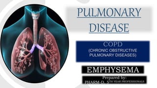
- 1. PULMONARY DISEASE COPD (CHRONIC OBSTRUCTIVE PULMONARY DISEASES) EMPHYSEMA Prepared by: PHARM-D, 5TH YEAR PROFESSIONALS
- 2. 2 GROUP MEMBERS: LARAIB SHAMSI MAHAM UROOJ MAHA KIRAN MAHAM FAYAZ MARIA AKRAM COPD (CHRONIC OBSTRUCTIVE PULMONARY DISEASES)
- 3. 3 COPD(CHRONIC OBSTRUCTIVE PULMONARY DISEASES) A Hypercapnia disease characterized by progressive airflow limitation ,usually both progressive and associated with an abnormal inflammatory response of the lungs to noxious particles or gases
- 4. PATHOPHYSIOLOGY OF COPD 4 ACTIVATION OF WBC’S ACTIVATION OF OXIDATIVE STRESS Etiologic factors like tobacco smoke, noxious gases,etc Activates neutrophils, macrophages and CD8+ lymphocytes Causes release of chemical mediators like tnfα, IL-8, LTB4 Leads to destructive changes in the airways, pulmonary vasculature and lung parenchyma Imbalance between aggressive and protective defense system in lungs Increased oxidants generated by cigarette smoke reacts and damages various proteins and lipids Leading to cell and tissue damage Oxidants also promote inflammation directly and inhibits anti-protease activity Leads to protease-antiprotease imbalance
- 5. “An anatomic alteration of the lung characterized by an abnormal airspace enlargement distal to the terminal bronchioles accompanied by destructive changes of the alveolar walls” EMPHYSEMA
- 6. TYPES OF EMPHYSEMA 6 CENTRIACINAR (CENTRILOBULAR) PANACINAR (PANLOBULAR) DISTAL ACINAR (PARASEPTAL) An abnormal enlargement of airspaces centered on the respiratory bronchiole with coalescence of destroyed lobules An abnormal dilation distributed throughout the pulmonary lobule Emphysematous change adjacent to a pleural surface Central or proximal parts of the acini Uniform injury Effect peripheral parts (dilate) alveolar duct & alveoli Smoking Alpha1-antitrypsin deficiency Unknown (spontaneous pneumothorax is thought to result from rupture of a paraseptal bleb/bulla.)
- 7. ETIOLOGY OF EMPHYSEMA • Age and gender • Smoking • Alpha 1 antitrypsin Deficiency • Exposure to second hand smoke. 7 • Exposure to indoor or outdoor pollution • IV Drug users • Occupational exposure to fumes and dust
- 8. EPIDEMIOLOGY 8 • In US emphysema is estimated to be third leading cause of death. • The prevalence of emphysema in the United States is approximately 14 million which includes 14% white male smokers and 3% white male non- smokers. • Around 90% of COPD deaths occur in low and middle-income countries. • In the most socioeconomically deprived parts of the country, one in 32 people were diagnosed with COPD, compared with one in 98 in the most affluent areas. • 25 million people may have COPD if currently undiagnosed cases are included.
- 9. • Doctor will start diagnosis by medical examination , asking about history and symptoms and observing signs as well. CLINICAL MANIFESTATION: • Two key symptoms are chronic cough and SOB. • These symptoms appear in early stages. As disease progress following symptoms also observed: • Wheezing, having hard time getting air out of lungs • Dyspenoea , difficulty in catching breaths • Morning headaches 9 DIAGNOSIS
- 10. • Tachypnoea with prolonged expiration, inadequate O2 due to lung infection • Use of accessory muscles for ventilation , malfunctioning of lungs) • Barrel chest (rounding of chest), Overinflated lungs with air • Pursed lips to prevent expiratory airway collapse • Cardiac enlargement due inadequate oxygen supply • Clubbing of nails in which end of the fingers appear as upside down spoon • Sleep disturbance 10 DIAGNOSIS Cont….
- 11. DIAGNOSIS (LAB TEST) 11 X-RAY & CT-SCAN Chest X-rays & CT-scan can help confirm a diagnosis of emphysema and rule out other lung conditions. Lungs are large and hyper inflated. Signs of hyperinflation are: •Low set diaphragm •Hyper lucent lung fields Signs of hyperinflation can be seen in emphysema
- 12. 12 COMPLETE BLOOD COUNT In emphysema , your body produces more RBCs to makeup for decreased oxygen. These cells carry oxygen. WBCs higher than normal. ARTERIAL BLOOD GAS The test measures the oxygen level in your blood and carbon dioxide (CO2) is removed properly. It can also determine the acidity (pH) of your blood. Imbalances in the amount of oxygen, carbon dioxide, or pH can serve as a way to evaluate emphysema. • In emphysema, the blood is more acidic, as the pH levels are low and the PaCO2 levels are above than normal. DIAGNOSIS (LAB TEST) Cont....
- 13. 13 DIAGNOSIS (LAB TEST) Cont.... SPUTUM EXAMINATION SPIROMETRY Analysis of cells in your sputum can help determine the cause of some lung problems It is a simple test that is often done to monitor emphysema. It measures the amount and how fast you can breathe in and breathe out.
- 14. 14 DIAGNOSIS (LAB TEST) Cont.... PULMONARY FUNCTIONING TEST HIGH RESOLUTION COMPUTED TOMOGRAPHY (HRCT) This test involves a series of breathing maneuvers that measure the airflow and volume of air in our lungs. It is used in the diagnosis of various health problems, though most commonly for lung disease, by assessing the lung parenchyma.
- 15. 15 ASSESSMENT
- 16. TREATMENT
- 18. MEDICATIONS • Bronchodilators. These drugs can help relieve coughing, shortness of breath and breathing problems by relaxing constricted airways. • Steroids. Corticosteroid drugs inhaled as aerosol sprays reduce inflammation and may help relieve shortness of breath. • Antibiotics. If you have a bacterial infection, like acute bronchitis or pneumonia, antibiotics are appropriate.
- 19. BRONCHODILATORS 19 Short-acting beta-agonist bronchodilators – called “SABAs” SABAs are the most common type of rescue inhaler. SABAs can start providing relief for symptoms in 3 to 5 minutes, but are only effective for about 4 to 6 hours. Short-acting anti muscarinic bronchodilators – called “SAMAs” SAMAs start working a little more slowly than SABAs do. They take about 15 minutes to start working, so they cannot be used as a rescue inhaler but are used as a maintenance therapy for COPD Long-acting beta-agonist bronchodilators – called “LABAs” Long-acting bronchodilators are often used as maintenance therapy for COPD patients in later stages. Long-acting muscarinic agonists bronchodilators – called “LAMAs” Patients need to take long- acting bronchodilators every day in order for them to work well. Having a regular amount of the drug in the patient’s body all the time helps to provide more constant relief for COPD symptoms. 1 2 3 4
- 22. 22 CORTICOSTERIODS Several types of corticosteroids are available. Some are inhalable and should be used every day as directed. They’re usually prescribed in combination with a long-acting COPD drug. Other corticosteroids are injected or taken by mouth. These forms are used on a short-term basis when your COPD suddenly gets worse. • Fluticasone (Flovent), which comes as an inhaler that you use twice daily. Side effects can include headache, sore throat, voice changes, nausea, cold-like symptoms. • Budesonide (Pulmicort), which comes as a handheld inhaler or for use in a nebulizer. Side effects can include colds. • Prednisolone, which comes as a pill, liquid. It’s usually given for emergency rescue treatment. Side effects can include headache, muscle weakness, upset stomach, and weight gain.
- 23. OTHER THERAPY • Pulmonary rehabilitation: Include breathing exercises and techniques that may help reduce your breathlessness. • Nutrition therapy: Give advice about proper nutrition. • Oxygen supplements: Using oxygen regularly at home and when you exercise may provide some relief.
- 24. SURGERY • Lung volume reduction surgery: Surgeons remove small wedges of damaged lung tissue. Removing the diseased tissue helps the remaining lung tissue expand and work more efficiently and helps improve breathing. • Lung transplant: Lung transplantation is an option if you have severe lung damage.
- 25. HOME REMEDIES • Stop smoking. • Avoid other respiratory irritants. • Exercise regularly. • Protect yourself from cold air. • Get recommended vaccinations. • Prevent respiratory infections.
- 26. MONITORING 26 Mild stable COPD patients may be followed up at 6- month intervals, while patients with severe and recently hospitalized patients need follow-up at 2- week to 1-month intervals. PFTs should be monitored at least every 3 years, to evaluate response to therapy and possible need for change in medications. 2 1
- 27. 27 Oxygen saturation should be monitored and patients evaluated periodically for the need of supplemental oxygen. 3 MONITORING
- 28. PROGNOSIS 28 MILD EMPHYSEMA 80% of patients are alive after 4 years. MODERATE EMPHYSEMA 60-70% of patients are alive after 4 years. SEVERE EMPHYSEMA 50% are alive after 4 years. VERY SEVERE EMPHYSEMA short life expectancy.
- 29. REFERENCES: 29 • https://www.mayoclinic.org/diseases-conditions/emphysema/diagnosis-treatment/drc-20355561 • https://www.ucsfhealth.org/conditions/emphysema/treatment.html • Roger-Walker Clinical and Therapeutics 5th Editions • https://www.ncbi.nlm.nih.gov/pmc/articles/PMC3882898/ • file:///C:/Users/Faizan%20M.Sharif/Downloads/copd-130709072917-phpapp01.pdf • https://bestpractice.bmj.com/topics/en-us/7/monitoring • Robert AKinsmanPh. D.*R.AYaroushPh.D.*EnriqueFernandezM.D.*Jerald F.DirksPsy. D.*MarshaSchocketM.S.W.*JoleneFukuharaB.A • RA Kinsman, E Fernandez, M Schocket… - Journal of behavioral , 1983 – Springer • https://www.slideshare.net/kaugustine66/presentation-adv-practice-1 • https://www.physio-pedia.com/Emphysema • www.ncbi.nlm.nih.gov • https://en.wikipedia.org/wiki/Chronic_obstructive_pulmonary_disease#Epidemiology • http://ksumsc.com/download_center/Archive/1st/434/3- Respiratory%20Block/Teams%20Work/Pathology/2.ObstructiveLungDiseases.pdf • https://www.slideshare.net/imangalal/chronic-obstructive-pulmonary-disease-33386223 • https://www.webmd.com > lung > copd • https://my.clevelandclinic.org > disease • https://www.webmd.com/lung/copd/emphysema-diagnosis-and-treatments#2 • https://www.ucsfhealth.org/conditions/emphysema/diagnosis.html • http://www.healthcommunities.com/emphysema/treatment.shtml • https://bmjopen.bmj.com/content/3/11/e003541
- 30. 30 THANK YOU