
Recommended
More Related Content
What's hot
What's hot (20)
Similar to Acute Appendicitis.pptx
Similar to Acute Appendicitis.pptx (20)
Recently uploaded
Recently uploaded (20)
Acute Appendicitis.pptx
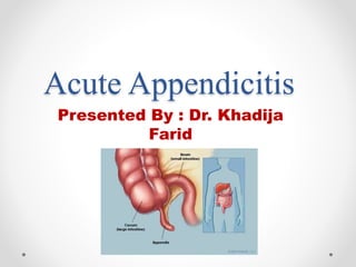
- 1. Acute Appendicitis Presented By : Dr. Khadija Farid
- 2. Case Presentation : • A 15 year-old boy with no past medical history presents to the SER with 24 hours of abdominal pain. The day prior to presentation, he developed diffuse, vague abdominal discomfort , he lost his appetite and went to bed early secondary to malaise. The following morning, the pain worsened in intensity, became sharp, and localized to the right lower quadrant. • The patient is febrile, with increased pulse rate. On examination, he is focally tender to palpation in the right lower quadrant with voluntary guarding. Palpation of the left lower quadrant reproduces pain on the right. Scrotum is normal , His lab work is unremarkable with the exception of a mild leukocytosis to 13. • Provisional Diagnosis : Acute Appendicitis
- 3. Definition : • Appendicitis is defined as an inflammation of the inner lining of the vermiform appendix that spreads to its other parts , despite diagnostic and therapeutic advancement in medicine, appendicitis remains a clinical emergency and one of the most common cause of acute abdominal pain – Acute Abdomen.
- 4. • Blood supply : appendicular artery (ileocolic artery ) • Venous drainage : appendicular vein ( post ceceal vein ) • Lymphatic drainage : lymphatics drain into ileocaecal lymph nodes • Types: • 1) Acute catarrhal (non obstructive) appendicitis • 2) Acute obstructive appendicitis
- 6. Presentation • The classic history of anorexia and periumblical pain followed by nausea , RLQ pain , and vomiting occurs in 50% of cases. Features include the following :- • Abdominal pain (colicky or dull periumblical pain that shift to right iliac fossa of the abdomen , pain exacerbated on coughing or laughing • associated with: • Nausea • Anorexia • Vomiting ( nearly always follows the onset of pain ; vomiting that precedes pain suggests intestinal obstruction) • Diarrhea or constipation
- 7. Clinical Signs : • Pointing Sign Rovsing’s Sign • Obturator and Psoas Sign Markle Sign or Jar tend.
- 10. Investigation : • CBC : WBC > 14,000 / microliter Neutrophilia • C-reactive Protein : CRP > 1mg/dl Very high levels of CRP in appendicitis indicate gangrenous evolution of the diseases , esp if it is associated with leukocytosis and neutrophilia. • Urinary 5-HIAA : Its level increases significantly in acute appendicitis and decreases when inflamation shift to necrosis of appendix , therefore , such decrease could be an early warning sign of perforation of appendix. • Pregnancy test • Urea and electrolytes
- 11. Imaging : • Ultrasonography : (of abdomen and pelvis) • Pain abdominal Xray • CT scan (contrast enhanced ) • MRI
- 12. Alvardo Scoring System :
- 13. Management : In SER : • Establish IV access and administer aggressive crystalloid therapy to patients with clinical signs of dehydration or septecemia. • Keep patients NPO • Administer analgesic and antiemetic • If there is appendiceal mass :- • Phlegmon or small abscess : after IV antibiotic therapy interval appendectomy performed 4- 6 weeks later. • Larger well-defined abscess : after percutaneous drainage and IV antibiotics administration , patient can be discharged with catheter in place , interval appendicectomy can be performed after fistula is closed.
- 14. • Multicompartmental abscess : These patients require early surgical drainage. • Antibiotics : Antiobiotic prophylaxis should be administered before every appendecectomy. • Broad spectrum gram negative and anaerobic coverage is indicated. • Cefotetan and cefoxitin seem to be the best choices of antibiotic • In penicillin allergic patients , carbapenems are good option. • Pregnant patients should receive Category A or B antibiotics.
- 15. Surgical Treatment : Appendectomy : 1- Conventional Open Appendectomy : Incision :- • Gridiron incision / Mc Burney’s incision • Transverse Skin crease (Lanz) incision • Rutherford Morison’s incision • Lower midline incision
- 16. • Procedure :- • There are three muscle layers in the lateral abdominal wall. As these are encountered when entering the abdomen, these are the external oblique, the internal oblique, and the transversus abdominis muscles. • Each muscle aponeurosis is cut in the direction of the muscle fibers. • A muscle-splitting technique is used to spread apart each muscle layer along the orientation of the muscle fibers until the peritoneum is reached
- 17. • The peritoneum is then grasped with forceps in order to assure no bowel is adherent and is incised with sciccors to enter abdomimal cavity • An appropriate retractor is placed to enhance operative exposure. • After opening the periton -eal cavity , any serous exudates or pus around appendix is sucked and pack is inserted in wound on medial side to push the loops of small intestine medially.
- 18. Exposure of the Appendix • After the peritoneum is entered, the cecum is identified. Sponge sticks can be helpful to sweep the small bowel in a lateral to medial direction in order to expose the cecum. • Once the cecum is identified, the anterior taenia is identified , the cecum is then mobilized, following the anterior taenia to its confluence with the appendiceal base. • The convergence of all three teniae coli allows for the correct identification of the base of the appendix.
- 19. • Inflamatory adhesions of appendix, if present are divided and appendix is delivered into the wound with the help of Babcock forceps. • The mesoappendix is ligated and divided, • This is critical to ensure that the entire appendix is removed.Failure to remove the base of the appendix may cause a closed loop obstruction between a persistent fecalith at the base of • the appendix and the stump staple line. This may lead to an appendiceal stump blowout postoperatively. • In cases of retrocecal appendicitis the cecum will need to be fully mobilized in a lateral to medial fashion so that it is completely reflected from the retroperitoneum in order to find the appendix.
- 21. • The completely free appendix is crushed near its junction with cecum with artery forceps , artery forceps is removed and reapplied just distal to the crushed portion. • An absorbable ( vicryl 3/0 ) ligature is tied around the crushed porion and the appendix is amputated between the artery forceps and the ligature.
- 22. • Inversion of the appendiceal stump may be performed if the surgeon desires. Commonly, a “Z-stitch” is used for this purpose. • In cases of severe appendiceal stump edema and inflammation, a gastrointestinal stapler may be used to transect the base of the appendix, even including a segment of healthy cecal base in the resection; be careful to avoid impingement of the ileocecal valve when firing the stapler. • All three muscle layers are closed separately with running absorbable suture. No drain is indicated in simple appendicitis.
- 23. •
- 24. Closure :- • Hemostasis is checked , peritoneum is closed with vicryl 3/0 ( depends on surgeon’s decesion) • Internal oblique and transverse abdominis muscle are approximated with vicryl no1 in an interrupted manner andn external oblique aponeurosis is closed in continuous manner. • Skin can be closed by few interuppted stitches.
- 25. 2- Laproscopic Appendectomy • Pneumoperitoneum is crceated either by closed or open technique through a small incision at umblicus. • Laproscope is then passed through 10mm umblical port . • Thorough peritoneal examination is done , diagnosis confirmedd , appendix is identified.
- 26. • Then two 5mm working ports are inserted , one at suprapubic and other at right lumbar area in ant. Axillary line. • Mesoappendix is clipped or cauterized. • Appendicular base is clipped or ligated by intra-corporeal suture. Appendix removed through lumbar port.
- 27. Complications: Per operative :- • Injury to cecum and ileum • Bleeding from appendicular artery • Urinary bladder injury with suprapubic port Post – Operatively :- • Appendicular stump leakage / Wound dehiscence • Adhesive intestinal ostruction • Abdominal / pelvic abscess formation • Wound infection • Portal pyema ( pylephlebitis ) • Stump appendicitis • Ileus • Venous thrombosis and embolism • Fecal fistula
- 28. • Check List for Unwell patient following appendicectomy :- • Examine the wound and abdomen for abscess. • Consider a pelvic abscesss , and perform a DRE. • Examine the lungs – pneumonitis or collapse. • Examine the legs – consider venous thrombosis. • Examine the conjuctivae for an icteric tinge and the liver for enlargement , and enquire wether the patient has had rigours ( pylephlebitis ) • Examine the urine for organisms ( pylephlebitis ). • Suspect subphrenic abscess.
- 29. THANK YOU !!!