
Recommended
More Related Content
What's hot
What's hot (20)
Similar to Mast cells in health and disease final dr karishma
Similar to Mast cells in health and disease final dr karishma (20)
Recently uploaded
Recently uploaded (20)
Mast cells in health and disease final dr karishma
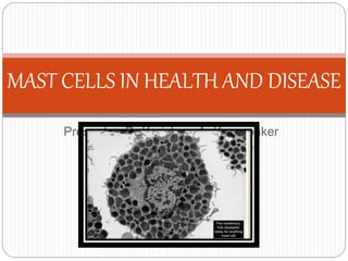
- 1. Presenter :Dr Karishma A. Korgaonker Moderator : Dr . Gauri Metkar MAST CELLS IN HEALTH AND DISEASE
- 2. Introduction Activation of mast cells Mast cell mediators Mast cell in health >Wound healing >Angiogenesis Mast cell in disease >Immediate hypersensitivity reaction >Infection >Inflammation • Mast cell disorders • Summary
- 3. Introduction Dr Paul Ehrlich in 1878 Large mononuclear cells Cytoplasmic membrane bound granules - contain a variety of biologically active mediators and sulphated glycosaminoglycans Bone marrow–derived cells-- originate from CD34+/CD117+ multipotent hematopoietic progenitor They are abundant near blood vessels and nerves and in subepithelial tissues
- 5. Mast cells function Participate in defence against bacterial and parasitic infection Develop immediate hypersensitivity reactions Take part in reparation of tissues - angiogenesis, production of intercellular substances
- 6. Introduction Activation of mast cells Mast cell mediators Mast cell in health >Wound healing >Angiogenesis Mast cell in disease >Immediate hypersensitivity reaction >Infection >Inflammation • Mast cell disorders • Summary
- 7. Activation of mast cells Cross-linking of high-affinity IgE Fc receptors Complement components C5a and C3a Mast cell secretagogues include some chemokines (e.g., IL-8), drugs such as codeine and morphine, adenosine, mellitin (present in bee venom), and physical stimuli (e.g., heat, cold, sunlight).
- 8. Activation of mast cells Mast cells express a high-affinity receptor, called FcεRI, that is specific for the Fc portion of IgE, and avidly binds IgE antibodies. In the first step in this sequence, antigen (allergen) binds to the IgE antibodies previously attached to the mast cells The bridging of the Fcε receptors activates signal transduction pathways from the cytoplasmic portion of the receptors.
- 9. Activation of mast cells These signals lead to mast cell degranulation with the discharge of preformed (primary) mediators that are stored in the granules, and de novo synthesis of secondary mediators
- 13. Introduction Activation of mast cells Mast cell mediators Mast cell in health >Wound healing >Angiogenesis Mast cell in disease >Immediate hypersensitivity reaction >Infection >Inflammation • Mast cell disorders • Summary
- 16. Mast cell mediators • Vasoactive amines: The most important mast cell–derived amine is histamine. = intense smooth muscle contraction, =increased vascular permeability, and =increased mucus secretion by nasal, bronchial, and gastric glands.
- 17. Mast cell mediators • Enzymes: = contained in the granule matrix = include neutral proteases (chymase, tryptase) and several acid hydrolases. = cause tissue damage and lead to the generation of kinins and activated components of complement (e.g., C3a)
- 18. Mast cell mediators Proteoglycans: = include heparin (anticoagulant) and chondroitin sulfate. = serve to package and store the amines in the granules.
- 19. Lipid Mediators. are synthesized by sequential reactions in the mast cell membranes = lead to activation of phospholipase A2, an enzyme that acts on membrane phospholipids to yield arachidonic acid. =This is the parent compound from which leukotrienes and prostaglandins are derived by the 5-lipoxygenase and cyclooxygenase pathways
- 20. Lipid Mediators. Leukotrienes: C4 and D4 --the most potent vasoactive and spasmogenic agents known. B4 --is highly chemotactic for neutrophils, eosinophils, and monocytes.
- 21. Lipid Mediators. Prostaglandin D2 : = most abundant mediator produced in mast cells by the cyclooxygenase pathway. = intense bronchospasm as well as increased mucus secretion.
- 22. Lipid Mediators. • Platelet-activating factor (PAF): = platelet aggregation, release of histamine, bronchospasm, increased vascular permeability, and vasodilation. =chemotactic for neutrophils and eosinophils, =at high concentrations it activates the inflammatory cells, causing them to degranulate.
- 23. Cytokines. play an important role in immediate hypersensitivity reactions. The cytokines include: >TNF, IL-1, and chemokines - promote leukocyte recruitment >IL-4- which amplifies the TH2 response.
- 24. Introduction Activation of mast cells Mast cell mediators Mast cell in health >Wound healing >Angiogenesis Mast cell in disease >Immediate hypersensitivity reaction >Infection >Inflammation • Mast cell disorders • Summary
- 25. Woundhealingandangiogenesis MC’s mediator influence- All phase of wound healing Acute inflammatory Promote influx of inflammatory cells to injury site Proliferative phase Re-epithelialization & angiogenesis Release many angiogenic factors to induce revasculaization of damage tissue Heparin from MC stimulates endothelial cell migration to form new blood vessel
- 26. Woundhealingandangiogenesis MC tryptase stimulates vessel tube formation & enhances growth of microvascular endothelial cells MC chymase promotes angiogenesis through effects of angiotensin II Generate growth factors : fibroblast growth factor, vascular endothelial growth factor (VEGF), platelet-derived growth factor (PDGF), nerve growth factor (NGF) all growth factors induce proliferation of epithelial cells & fibroblasts
- 27. Woundhealingandangiogenesis Remodeling phase As fibroblast expand in previous phase, they deposit collagen & other extracellular matrix proteins molded and remodeled into scar tissue Hair follicle recycling MC histamine, TNF, substance P involved are thought to contribute in hair follicle recycling
- 28. Woundhealingandangiogenesis Bone remodeling MCs are source of osteopontin, glycoprotein component of bone matrix, that contributes to bone resorption & calcification In human systemic mastocytosis, bone turnover is accelerated enhance bone loss
- 29. Introduction Activation of mast cells Mast cell mediators Mast cell in health >Wound healing >Angiogenesis Mast cell in disease >Immediate hypersensitivity reaction >Infection >Inflammation • Mast cell disorders • Summary
- 31. Immediate hypersensitivityreactions Histamine and leukotrienes, are released rapidly from sensitized mast cells and are responsible for the intense immediate reactions characterized by edema, mucus secretion, and smooth muscle contraction
- 32. Mast Cells vs. Allergens Allergen detection leads to mast cell histamine release In skin, this leads to wheal and flare reactions. In the gut ,leads to intestinal hyper-permeability, smooth muscle contraction, altered water and ion transport, and intestinal symptoms. • In the lungs, this leads to smooth muscle contraction, mucus production, and airway remodeling.
- 33. Introduction Activation of mast cells Mast cell mediators Mast cell in health >Wound healing >Angiogenesis Mast cell in disease >Immediate hypersensitivity reaction >Infection >Inflammation • Mast cell disorders • Summary
- 34. Mast Cells vs. Parasites Mast cells offer a first line of defense against parasitic pathogens. They are preferentially located in organs targeted by parasites, where they release proteases, recruit neutrophils, and regulate vessel permeability. Mast cells release antimicrobial peptides.
- 35. mastcell in infection Helminth infection MCs are activated by helminth & MC hyperplasia is observed in helminth infection
- 36. Mast Cells vs. Bacteria Mast cells express most TLRs on their membrane surfaces. TLR binding triggers distinctive patterns of mast cell activation including the release of antimicrobial peptides and the production of complement-mediated membrane attack complexes.
- 37. mastcell in infection Bacterial infection MC-derived TNF, together with LTC4 & LTB4 contributed to recruitment neutrophils to clearance Klebsiella pneumoniae, Listeria monocytogenes, Pseudomonas aeruginosa
- 38. Mast Cells vs. Yeast and Fungal Forms Parasite antigen stimulation of TLRs) and C-type lectin receptors (CLRs) initiates a wide range of mast cell IgE receptors resulting in the release of pro- inflammatory cytokines, chemokines, and other pre-stored mediators of inflammation. The release of pre-formed mediators of inflammation is
- 39. mastcell in infection Fungal infection Human MCs respond to zymosan, but not peptidoglycan, Trichoderma viridae, indoor fungus, induce MC degranulation (high dose) but low dose enhance histamine secretion from MCs Aspergillus fumigatus induce IgE-independent MC degranulation
- 40. Mast Cells vs. Viruses Upper respiratory viral infections worsen allergic asthma, suggesting increased activation of mast cells during infection. Mast cells recognize the presence of double-stranded viral RNA and respond by releasing: IL-29, interferon- alpha, interferon-beta, and tumor necrosis factor alpha. Virally infected mast cells respond by releasing the anti-viral proteins.
- 41. mastcell in infection 41 Viral infection This aspect is emerging field Report from HIV-infected patients Increased serum IgE predict worse prognosis Israël-Biet D et al.JACI 1992 Jan;89:68-75
- 42. mastcell in infection Viral infection Dengue virus can activate MCs to release IL-1, IL-6, RANTES, MIP-1 and MIP-1 Respiratory syncytial virus (major cause of LRT in infant & associated with development of asthma later in life Airway MC numbers increase in parainfluenza infection
- 43. Mast Cells vs. Toxins Mast cells are known to initiate an immediate IgE- dependent hypersensitivity response to venoms. Mast cells can also be activated by various endotoxins and exotoxins. It is not known whether mold or bacterial toxins activate mast cell responses, but mast cells release VEGF and MMP-9, markers affected in biotoxin-mediated
- 44. Introduction Activation of mast cells Mast cell mediators Mast cell in health >Wound healing >Angiogenesis Mast cell in disease >Immediate hypersensitivity reaction >Infection >Inflammation • Mast cell disorders • Summary
- 45. Mast cell- skin inflammation Skin mast cells are located close to sensory nerve endings and triggered by neuropeptides released by dermal neurons Acute stress releases CRH in the skin inducing local response , increases vascular permeability Acute stress induces redistribution of leukocytes from the sytemic circulation into the skin and excerbates skin delayed hypersensitivity reactions
- 46. Mast cell – inflammatory arthritis Fluid aspirated from joints of patients with arthrosynovitis contains RANTES and MCP- 1(chemoattractants) Mast cells in the joints of RA patients express CRH receptors CRH and CRH receptors are increased in the joints of inflammatory and RA patients symptoms of which worsen by stress
- 47. Mast cell – Coronary inflammation Cardiac mast cells participate in the development of artherosclerosis , coronary inflammation, and cardiac ishemia Mast cells are prominent in coronary arteries during spasm The human mast cell proteolytic enzyme chymase is the main cardiac source of coronary constrictor angiotensin II Cardiac mast cell derived histamine constrict the coronary arteries
- 48. Mast Cells and Coronary Artery Disease People with mastocytosis or myalgic encephaplitis/chronic fatigue syndrome are particularly prone to coronary hypersensitivity reactions. • Stress precipitates and worsens activation of mast cells via activation of surface receptors by corticotropin releasing hormone (CRH) and other hypothalamic neuropeptide regulators.
- 50. Mast cells and Irritable Bowel Syndrome Mast cell activation plays a role in post-infectious IBS. • Mast cell degranulation associates with cytokine alterations and intestinal hyperpermeability in patients with IBS. • Mast cell hyperplasia is seen in inflammatory bowel disorders.
- 51. Mast Cells and the Blood-Brain Barrier Acute stress triggers CRH production which activates histamine release by mast cells, which leads to increased blood-brain barrier permeability. This implicates mast cells in neuroinflammatory disorders including multiple sclerosis.
- 52. 57 TC Moon et al.Mucosal Immunology 2010; 3(2):111- 128 mast cell homeostasis
- 53. Rolein cancer In connective tissue of certain tumours mast cells occur in great quantities Cutaneous carcinomas especially basal cell cancers Enormous accumulation of mast cells is in cutaneous papilloma these are precancerous lesions The moment tumour grow malignant the mast cells disappear and escape detection Mast cells and their products play a part in the local tissue resistance against development and growth of the tumour
- 54. Mast cells in vaccines Enhancing adaptive immune response Adjuvant activity Mast cell activator – humoral immunity IgA production, mucosal immunity
- 55. Introduction Activation of mast cells Mast cell mediators Mast cell in health >Wound healing >Angiogenesis Mast cell in disease >Immediate hypersensitivity reaction >Infection >Inflammation • Mast cell disorders • Summary
- 56. MASTOCYTOSIS A rare group of disorders Pathological increase in mast cells in tissues Mastocytosis can form part of other haematological disorders, including myelodysplastic syndromes, myeloproliferative disorders or AML. Cutaneous mastocytosis: involve just the skin Systemic mastocytosis: involve multiple tissues and are associated with systemic symptoms
- 57. MASTOCYTOSIS A c-kit point mutation leads to a single amino acid substitution (Asp816Val) . This mutation leads to ligand-independent phosphorylation of c-Kit and a consequent clonal expansion of mast cells.
- 60. MASTOCYTOSIS Cutaneous manifestations Urticaria pigmentosa is the usual presenting feature in children and adults. Yellowish-brown lesions,usually macular and sometimes papular, appear in a patchy distribution. Pruritus is common, as is flushing, and some cases develop haemorrhagic bullous disease. Whealing of lesions upon rubbing is known as Darier’s sign.
- 61. MASTOCYTOSIS Systemic disease Systemic manifestations are very heterogeneous and are secondary to mast cell mediator release. Episodes of flushing, angioedema, or even anaphylaxis with or without any specific trigger, can arise as a result of systemic histamine release. Gastrointestinal symptoms include abdominal pain,diarrhoea, nausea and vomiting. Gastritis and peptic ulceration may occur secondary to hyperhistaminaemia and severe cases may develop malabsorption.
- 62. MASTOCYTOSIS Osteoporosis leading to pathological fractures. Peripheral blood cytopenias. Hepatosplenomegaly. Fever, fatigue and weight loss Symptoms of organ failure due to organ infiltration are characteristic of aggressive systemic mastocytosis.
- 63. Investigations Diagnosis usually requires histological and biochemical confirmation Routine investigations should include a full blood count, liver function tests and a random serum tryptase. Plasma levels of soluble CD25 and CD117 (kit) have shown promise as novel markers of mast cell disease.
- 64. Investigations Bone marrow aspiration and trephine biopsy Mast cell aggregates can be visualized on conventional haematoxylin and eosin-stained sections but stand out clearly with stains such as toluidine blue . Immunochemistry using antitryptase antibodies highly specific for mast cells. Flow cytometry to look for expression of CD2 and CD25 in bone marrow mast cells may be useful as this phenotype is not seen in normal mast cells.
- 65. Investigations Abdominal ultrasound or computerized tomography (CT) scanning should be performed to look for hepatosplenomegaly and lymphadenopathy. Plain radiography and bone densitometry can be used to assess bone involvement and the presence of osteoporosis. Endoscopy and biopsy can be useful if gut involvementis suspected
- 66. Treatment No curative treatment exists for mastocytosis, The management of which remain symptomatic
- 67. Prognosis Age and disease category are the most important determinants of outcome. The most benign syndrome is paediatric mastocytoma,which disappears with time in over 50% of cases. Paediatric urticaria pigmentosa also has a good prognosis and resolves in about one-half of the cases. Indolent systemic mastocytosis carries a favourable prognosis and usually persists as a chronic low-grade disorder
- 68. other Mast Cells Disorders The Most Common Forms of Mast Cell Activation Syndrome ===Idiopathic (non-clonal) mast cell activation syndrome (also known as nc-MCAS): - occurs more often in women MC triggers are usually unknown. Idiopathic anaphylaxis and mastocytosis should be ruled out.
- 69. other Mast Cells Disorders Monoclonal mast cell activation syndrome (MMAS): ---- systemic reactions to hymenoptera stings or -- unexplained anaphylaxis with high baseline tryptase. Bone marrow biopsy shows monoclonal MCs but not mastocytosis. Thought to be rare.
- 70. Histamine-Overdrive Mast Cell Disorders Histaminosis: commonly induced by exposure to histamine from foods; rarely, an acute syndrome caused by scromboid poisoning (due to seafood high in histamine). • Histamine intolerance: sensitivity to histamine from foods and/or from reduced histamine breakdown.
- 71. Histamine Intolerance Reduced central or peripheral breakdown of histamine can lead to: Rashes Headaches Fatigue Hives or flushing Allergies Dysmenorrhea Estrogen dominance GI disturbances Dysrhythmias Arthritis Central, peripheral and/or autonomic neurologic
- 72. Diet and Nutrition in the Treatment of Mast Cell Disorders Some high histamine foods Pickles , beer, mayonnaise, nuts, wine ,chocolate, champagne ,vinegar ,cocoa, shellfish ,dried fruits processed meats, tofu, cheese, yeast, canned vegetables, mushrooms food additives, egg whites, aged cheeses , tomatoes ,spices ,spinach
- 73. Diet and Nutrition in the Treatment of Mast Cell Disorders Apples: rich in quercitin Carrots: rich in vitamin A Watercress: inhibits histamine release Broccoli: H2 receptor antagonist Ginger: H2 receptor antagonist Thyme: anaphylaxis inhibitor Fennel: antioxidants, antihistamine, anti- inflammatory Turmeric: stabilizes mast cells, inhibits histamine release me foods with anti-histamine effects
- 74. Introduction Activation of mast cells Mast cell mediators Mast cell in health >Wound healing >Angiogenesis Mast cell in disease >Immediate hypersensitivity reaction >Infection >Inflammation • Mast cell disorders • Summary
- 75. Mast Cells: The Good, the Bad, and the Ugly The Good An essential player in the body’s innate immune response. They patrol the front lines: skin, connective tissues and mucus membranes. They release chemical alarms to summon defenses against pathogens, allergens, toxins, and other noxious incitants.
- 76. Mast Cells: The Good, the Bad, and the Ugly The Bad Over-activation of mast cells can lead to: • Allergic disease/Asthma/Anaphylaxis • Eczema/atopic dermatitis • Migraines/neurological disorders • Autoimmune disorders • Gastrointestinal disorders • Unexplained multi-symptom illnesses Histamine is only one of many inflammatory elements released by mast cells. Excess release of mast cell-derived chemicals results in mast cell-related inflammation.
- 77. Mast Cells: The Good, the Bad, and the Ugly The Ugly Abnormal mast cell proliferation results in mastocytosis, which can lead to mast cell tumors and severe forms of cutaneous or systemic disease. Systemic mastocytosis commonly results in an unexplained multi-system, multi-symptom illness with damage to multiple organ systems that can go unrecognized in multiple medical settings.
- 78. Take Home Message Mast cells are functionally diverse cells that have a constitutive presence at mucosal surfaces and elaborate impressive array of mediators, making them an attractive therapeutic target. Mast cell proteases,their well-documented ability to alter intestinal permeability, is a key factor in several GI autoimmune/inflammatory pathologies
- 79. Take Home Message Mast cells have emerged as unique immune cells that can be activated by many immune and nonimmune triggers, including acute stress through CRH Inhibition of mast cell activation by CRH, therefore, is a novel target for the development of new treatments for inflammatory and autoimmune disease
- 80. References Robbins and Cotran pathologic basis of disease , Eighth Edition p.198-202 A. Victor Hoffbrand ,Daniel Catovsky MD, Edward G.D. Tuddenham MD, Postgraduate Haematology, Fifth edition 2005;p-775-778 Beaven M. Our perception of the mast cell from Paul Ehrlich to now. European Journal of Immunology. 2009 Jan;39(1):11-25. Moon TC, et al. Advances in mast cell biology: new understanding of heterogeneity and function.Mucosal Immunology. 2010;3:111-128.
- 81. References Metz M, Maurer M. Mast cells: key effectors in immune response. Trends in Immunology. 2007;28:234-241. Theoharis C. Theoharides,and Dimitrios Kalogeromitros The Critical Role of Mast Cells in Allergy and InflammationAnn. N.Y. Acad. Sci. 1088: 78–99 (2006). C 2006 New York Academy of Sciences.doi: 10.1196/annals.1366.025 Gustav Asboe-hansen Mast cells in health and disease New Understanding Of Mast Cell surasarit Khawlaor
Editor's Notes
- (production of IgE antibodies in response to an antigen, binding of IgE to Fc receptors of mast cells, cross-linking of the bound IgE by the antigen and release of mast cell mediators)
- . Although the production of PAF is also triggered by the activation of phospholipase A2, it is not a product of arachidonic acid metabolism
- The inflammatory cells that are recruited by mast cell–derived TNF and chemokines are additional sources of cytokines and of histamine-releasing factors that cause further mast cell degranulation.
- As is evident from this figure, mast cells are capable of signalling to all other immune cells and other cells, the presence of a pathogen.
- Mast cells are highly adaptible in nature. Very heterogeous and phenotypically malleable. Originally classified based on staining patterns as mucosal or connective tissue mast cells. These so called types actually differ in their phenotype, their granule composition. For eg. Mucosal mast cells show a different range of proteases than connective tissue mast cells. This composition is further affected based on the stimuli for eg. A cytokine signal that it receives. And based on this stimuli, the response is also quite differently modulated. These features allow mast cells to respond to different pathogens entering different tissues in different ways.
- Mast cells functions by virtue of their mediators include increasing vascular permeability and oedema, immune cell recruitment like neutrophils and eosinophils, DCs, T cells and others. Producing antimicrobial peptides and reactive o2 species that have direct bactericidal properties. By its abbility to interact with smooth muscle cells, nerve cells and mucosal cells it can impede colonization of pathogens, and even its physical expulsion for eg. Diarrhoea in response to bacterial toxins. They further activate and modulate Ag presentation of DCs. Mast cells also influence cell trafficking to draining lymph nodes and lymphocyte retention, thus increasing the chances of induction of rare Ag-specific lymphocytes.
- With all these functions that mast cells are capable of, they become an attractive addition to vaccines since the clearly enhance the asaptive response by serving as an adjuvant. It was shown 48/80.