
Recommended
More Related Content
Similar to Appendicular.pptx
Similar to Appendicular.pptx (20)
Recently uploaded
Recently uploaded (20)
Appendicular.pptx
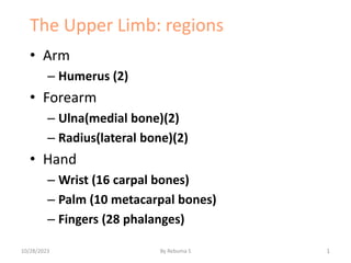
- 1. The Upper Limb: regions • Arm – Humerus (2) • Forearm – Ulna(medial bone)(2) – Radius(lateral bone)(2) • Hand – Wrist (16 carpal bones) – Palm (10 metacarpal bones) – Fingers (28 phalanges) 1 10/28/2023 By Rebuma S
- 3. THE UPPER LIMB • The upper limb is characterized by its mobility and ability to grasp, strike, and conduct fine motor skills (manipulation). • The upper limb consists of four segments : Shoulder: which includes the pectoral, scapular, . Arm : the part between the shoulder and the elbow . Forearm: the part between the elbow and the wrist and contains the ulna and radius 10/28/2023 By Rebuma S 3
- 4. HANDE Hand : the part distal to the forearm and contains the carpus, metacarpus, and phalanges. It is composed of the wrist, palm, dorsum of hand, and fingers . 10/28/2023 By Rebuma S 4
- 5. BONES OF UPPER LIMB • The pectoral girdle and bones of the free part of the upper limb form the superior Appendicular skeleton, articulates with the axial skeleton only at the sternoclavicular joint. • The pectoral girdle is supported and stabilized by axioappendicular muscles, which attach to the ribs, sternum, and vertebrae of the axial skeleton. 10/28/2023 By Rebuma S 5
- 6. Pectoral girdle 10/28/2023 By Rebuma S 6
- 7. The Pectoral Girdle • Consists of: – 2 clavicles – 2 scapulae • Connects with the axial skeleton only at the manubrium (sternoclavicular joint) 10/28/2023 By Rebuma S 7
- 8. The Clavicles • Also called collarbones • Long, S-shaped bones • Originate at the manubrium (sternal end) • Articulate with the scapulae (acromial end) • It articulates medially with the manubrium of the sternum at (SC) joint & laterally with the acromion of the scapula at (AC) joint 10/28/2023 By Rebuma S 8
- 9. cont,,, 10/28/2023 By Rebuma S 9
- 10. SCAPULA 10/28/2023 By Rebuma S 10
- 11. SCAPULA • The scapula (shoulder blade) . • a triangular , flat, & broad bone . • have small supraspinous fossa and a much larger infraspinous fossa. • Have Superior &inferior angle • Have medial & lateral border. 10/28/2023 By Rebuma S 11
- 12. HUMERUS • Also called arm bone. • the largest & longs bone in the upper limb. • articulates with the scapula at the glenohumeral joint and the radius and ulna at the elbow joint . -GHJ allows flexion/ Extention, abduction/adduction, medial & lateral rotation and circumduction of the arm 10/28/2023 By Rebuma S 12
- 13. humerus bone 10/28/2023 By Rebuma S 13
- 14. ulna and radius o The ulna: , is the medial and longer of the two forearm bones -Its proximal end has two prominent projections, the olecranon posteriorly & coronoid process anteriorly w/c forms trochlear notch. • The radius: is the lateral and shorter of the two forearm bones. Its proximal end consists of a cylindrical head, a short neck, and a projection from the medial surface (the radial tuberosity) . -It flipped over the adjacent head of medial bone , ulna. b/c the hand is articulated with radius, it can efficiently move from a palm-anterior position to palm-posterior position by crossing distal end of radius over ulna. 10/28/2023 By Rebuma S 14
- 15. Cont… 10/28/2023 By Rebuma S 15
- 16. The Wrist or carpal bones –8 carpal bones: •4 proximal carpal bones •4 distal carpal bones •allow wrist to bend and twist 10/28/2023 By Rebuma S 16
- 17. cont… • From lateral to medial, the four bones in the proximal row of carpals are , Scaphoid : a boat-shaped bone that has a prominent scaphoid tubercle. Lunate : a moon-shaped bone that is broader anteriorly than posteriorly Triquetrum : a pyramidal bone on the medial aspect of the carpus. Pisiform : a small, pea-shaped bone that lies on the palmar surface the triquetrum 10/28/2023 By Rebuma S 17
- 18. the proximal surfaces of distal row of carpals articulates with proximal row of carpals & their distal surfaces articulate with metacarpals From lateral to medial, the four bones in the distal row of carpals are: • Trapezium : a four-sided bone, on the lateral side of the carpus. • Trapezoid: a wedge-shaped bone, that resembles a trapezium. • Capitate : the head-shaped bone that is the largest bone in the carpus. • Hamate: a wedge-shaped bone, which has a hooked process that extends anteriorly. 10/28/2023 By Rebuma S 18
- 19. Cont… 10/28/2023 By Rebuma S 19
- 20. 20 10/28/2023 By Rebuma S
- 21. Metacarpal Bones • The 5 long bones of the hand • Numbered I–V from lateral (thumb) to medial • Articulate with proximal phalanges 10/28/2023 By Rebuma S 21
- 22. Phalanges of the Hands • Thumb: –2 phalanges (proximal, distal) • Fingers: –3 phalanges (proximal, middle, distal) 10/28/2023 By Rebuma S 22
- 23. Conti….. 10/28/2023 By Rebuma S 23
- 24. The Lower Limb: regions Thigh – Femur(2) Patella (knee cap)(2) Leg – Tibia( medial bone)(2) – Fibula(lateral bone)(2) Ankle (14 Tarsal bones) Foot (10 Metatarsal bones) Toes (28 Phalanges) 24 10/28/2023 By Rebuma S
- 25. The Lower Limbs • Functions: –weight bearing –motion Note: leg = lower leg; thigh = upper leg 10/28/2023 By Rebuma S 25
- 26. Bones of the Lower Limbs • Femur (thigh) • Patella (kneecap) • Tibia and fibula (leg) • Tarsals (ankle) • Metatarsals (foot) • Phalanges (toes) 10/28/2023 By Rebuma S 26
- 27. Femur • The femur is the longest and heaviest bone in the body • linea aspera :a prominent double-edge ridge on its posterior aspect of shaft of femur. • The proximal end of the femur consists of a head, neck, and two trochanters 10/28/2023 By Rebuma S 27
- 28. 28 10/28/2023 By Rebuma S
- 29. The Femur (longest, heaviest ) Figure 8–11 10/28/2023 By Rebuma S 29
- 30. The proximal end of the femur • the fovea capitis: medially placed depression or pit of head of femur. • lesser trochanter: extends medially from the posteromedial part of the junction of the femoral neck and shaft. • greater trochanter: is a large, laterally placed mass that projects superomedially where the neck joins the shaft. • intertrochanteric line: is a roughened ridge running from the greater to the lesser trochanter anteriorly. • intertrochanteric crest :smoother ridge posteriorly. 10/28/2023 By Rebuma S 30
- 31. The distal end of the femur • femoral condyles (medial and lateral): spirally curved articular surfaces , articulate with the tibial condyles to form the knee joint. • The medial epicondyle: is a rounded eminence on the medial condyle and the lateral epicondyle, on lateral surface of the lateral condyle. • Just posterosuperior to the medial epicondyle, there is the adductor tubercle. • Patellar surface: depression anteriorly b/n condyles. • Intercondylar notch: found posteriorly. 10/28/2023 By Rebuma S 31
- 32. 10/28/2023 By Rebuma S 32
- 33. Anterior Femur head fovea capits neck patellar surface 10/28/2023 By Rebuma S 33
- 34. Posterior Femur greater trochanter lesser trochanter lateral condyle medial condyle gluteal tuberosity linea aspera lateral epicondyle medial epicondyle Intercondylar fossa 10/28/2023 By Rebuma S 34
- 35. Bones of the Leg • The leg is the part of the lower limb between the knee joint and the ankle joint. • Includes : Patella, Tibia & Fibula 10/28/2023 By Rebuma S 35
- 36. 36 10/28/2023 By Rebuma S
- 37. Patella • Is the largest sesamoid bone (i.e., A bone that develops within the tendon of the quadriceps femoris muscle in front of the knee joint) • It is triangular, and its apex lies inferiorly being connected to the tuberosity of the tibia by the ligamentum patellae. • The posterior surface articulates with the condyles of the femur. • It is separated from the skin by an important subcutaneous bursa. 10/28/2023 By Rebuma S 37
- 38. tibia The tibia is the medial and larger of the two bones in the leg . is the only bone that articulates with the femur ( knee joint ) and head of fibula proximally & with the distally with the fibula . It has expanded upper end(weight bearing ),smaller lower end and the shaft. consists of a flattened medial and lateral condyles separated by an intercondylar region (intercondylar eminence). Medial malleolus: medial projection at ankle 10/28/2023 By Rebuma S 38
- 39. Tibia medial condyle lateral condyle anterior crest tibial tuberosity medial malleolus 10/28/2023 By Rebuma S 39
- 40. Fibula • The slender lateral bone of the leg • Takes no part in the articulation at the knee joint, but below it forms the lateral malleolus of the ankle joint • It takes no part in the transmission of body weight, but it provides attachment for muscles • Has an expanded upper end, a shaft, and a lower end • The upper end, or head, possesses an articular surface for articulation with the lateral condyle of the tibia. 10/28/2023 By Rebuma S 40
- 41. 10/28/2023 By Rebuma S 41
- 42. Bones of the Foot • Are the tarsal bones, the metatarsals, and the phalanges Tarsal Bones • Are the calcaneum, the talus, the navicular, the cuboid, and the three cuneiform bones • Only the talus articulates with the tibia and the fibula at the ankle joint 10/28/2023 By Rebuma S 42
- 43. Bones of the foot 10/28/2023 By Rebuma S 43
- 44. Ankle - tarsus consists of 7 tarsal bones Figure 8–14a Talus: carries weight from tibia across trochlea & its head articulate anterior with navicular. Calcaneus: (heel bone): transfers weight from talus to ground & attaches Achilles tendon. - largest & strongest bone & articulate talus superiorly and cuboid anteriorly. Cuboid bone: articulates with calcaneus posteriorly & most lateral bone in the distal row of the tarsus. Navicular bone: a flattened ,boat shaped bone w/c located b/n talar head and cuneiform bones. :articulates with talus and 3 cuneiform bones. Cuneiform bones: they are three in number 1, Medial cuneiform 2, Intermediate cuneiform 3, Lateral cuneiform 10/28/2023 By Rebuma S 44
- 45. Figure 8–14a 5 metatarsal bones long bones of foot numbered I–V, medial to lateral Articulate with toes phalanges bones of the toes hallux: big toe, 2 phalanges (distal, proximal) Other 4 toes: 3 phalanges (distal, medial, proximal) Feet 10/28/2023 By Rebuma S 45
- 46. Foot Tarsals (7) Metatarsals (5) Calcaneus Talus Digits (5) 10/28/2023 By Rebuma S 46
- 47. 3 Hallux (Great Toe) 1 10/28/2023 By Rebuma S 47
- 48. 48 10/28/2023 By Rebuma S
- 50. Introduction • Joints or articulations are sites where two or more bones meet. • Functions – provide skeletal mobility – hold the skeleton together • Weakest parts of the skeleton but have ability to resist the forces that tear them apart. 50 10/28/2023 By Rebuma S
- 51. Classification of Joints • Structural classification – focuses on the material binding the bones together and whether or not there is a joint cavity • Functional classification – based on the amount of movement allowed at the joint 51 10/28/2023 By Rebuma S
- 52. Functional Classification • Synarthroses – Immovable joints • Amphiarthroses – Slightly movable joints • Diarthroses – Freely movable joints 52 10/28/2023 By Rebuma S
- 53. Structural Classification • Fibrous – Joined by fibrous tissue • Cartilaginous – Joined by cartilage • Synovial – Joined and surrounded by a joint cavity 53 10/28/2023 By Rebuma S
- 54. Synovial Joints • Articulating bones are located within a fluid containing joint cavity. • Permit substantial range of motion 54 10/28/2023 By Rebuma S
- 55. Structures of Synovial Joint • Articular cartilage – Hyaline cartilage on opposing bone surfaces • Joint (synovial) cavity – Space filled with fluid • Articular capsule – Capsule to confine fluid • Synovial fluid – Fluid to lubricate joints • Reinforcing ligaments – Maintain joint alignment 55 10/28/2023 By Rebuma S
- 56. Articular Cartilage • Hyaline cartilage covers the bone surfaces • Cartilage keeps the bone ends from being crushed 56 10/28/2023 By Rebuma S
- 57. Synovial cavity • unique to synovial joints • filled with synovial fluid 57 10/28/2023 By Rebuma S
- 58. Articular capsule • The joint cavity is enclosed by a double layered articular capsule – The external layer is fibrous capsule – The inner synovial membrane 58 10/28/2023 By Rebuma S
- 59. Synovial Fluid • lubricates joint • nourishing cells 59 Synovial Fluid 10/28/2023 By Rebuma S
- 60. Reinforcing ligaments • Ligaments reinforce joint – Extracapsular – Intracapsular 60 Extracapsular Ligament Intracapsular Ligament 10/28/2023 By Rebuma S
- 61. Movements Allowed by Synovial Joints • Nonaxial: no rotation around an axis • Uniaxial: motion is within a single plane • Biaxial: allow movement in two planes • Multiaxial: movement is possible in all planes 61 10/28/2023 By Rebuma S
- 62. Types of Synovial Joints • Based on the shape of their articular surfaces there are six major categories of synovial joints –Plane –Hinge –Pivot –Condyloid –Saddle –ball and socket 62 10/28/2023 By Rebuma S
- 63. Plane Joint • Articular surfaces are essentially flat • Allow only short slipping or gliding movements • nonaxial joint • Examples – Intercarpals – Intertarsals – Vertebrae 63 10/28/2023 By Rebuma S
- 64. Hinge Joint • a cylindrical shaped projection of bone fits into a trough shaped surface of another bone. • Motion is within a single plane • Example – elbow joint 64 10/28/2023 By Rebuma S
- 65. Pivot Joint • The rounded end of a bone protrudes into a ring of bone and ligaments on another bone • Only rotational movement is allowed around long axis • Example – proximal radioulnar joint 65 10/28/2023 By Rebuma S
- 66. Condyloid Joints • The oval articular surface of one bone fits into a complementary concavity in another • biaxial joints • Example – Metacarpo- phalangeal joints 66 10/28/2023 By Rebuma S
- 67. Saddle Joints • Each surface has both a concave and a convex surface that fit together • Example – carpometacarpal 67 10/28/2023 By Rebuma S
- 68. Ball and Socket Joint • The spherical head of one bone articulates with the cuplike socket of another • Multiaxial • The most freely moving synovial joint • Examples – Shoulder joint – Hip joint 68 10/28/2023 By Rebuma S