
Recommended
More Related Content
What's hot
What's hot (13)
Similar to Combination therapy for Alzheimer’s Disease, Lithium + a Beta-Secretase (BACE) Inhibitor, Memantine + Levetiracetam + a BACE Inhibitor, Levetiracetam + a BACE Inhibitor
Similar to Combination therapy for Alzheimer’s Disease, Lithium + a Beta-Secretase (BACE) Inhibitor, Memantine + Levetiracetam + a BACE Inhibitor, Levetiracetam + a BACE Inhibitor (20)
Recently uploaded
Recently uploaded (20)
Combination therapy for Alzheimer’s Disease, Lithium + a Beta-Secretase (BACE) Inhibitor, Memantine + Levetiracetam + a BACE Inhibitor, Levetiracetam + a BACE Inhibitor
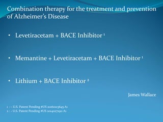
- 1. James Wallace Combination therapy for the treatment and prevention of Alzheimer’s Disease 1 : - U.S. Patent Pending #US 20160213645 A1 2 : - U.S. Patent Pending #US 20140271911 A1 • Levetiracetam + BACE Inhibitor 1 • Memantine + Levetiracetam + BACE Inhibitor 1 • Lithium + BACE Inhibitor 2
- 2. Words of Caution This presentation is for educational purposes only. Please work with a knowledgeable physician before beginning any type of supplement or medication protocol.
- 3. I developed the hypothesis that simultaneously targeting neuronal hyperactivity and the enzymatic cleavage of Amyloid Precursor Protein (APP), prior to the onset of symptoms during the long preclinical phase, will prevent or reduce the severity of Alzheimer’s disease by specifically targeting region- specific brain areas that are vulnerable to pathology.
- 4. Hypothesis APP CleavageNeuronal Hyperactivity Alzheimer’s Dementia Mediated by Ca2+ signaling Mediated by β- and γ- secretase
- 5. Linking Neuronal Hyperactivity with Calcium Signaling, Amyloid, Mitochondrial Dysfuntion, ER Stress, and Neurodegeneration Increased Activity Is Paired With Calcium Signaling and Calcium Influx Metabolic Stress and ROS 1,2 Calcium Derangements 3,4 Increased Synaptic Exocytosis 5,6 • Progressive Oxydative Damage • Mitochondrial Dysfunction • Diminished Metabolic Capacity • ER Stress/UPR/Inflam./Misfolding • Excitotoxic Damage • ER Stress/UPR/Inflam./Misfolding • Synaptic Dysfunction • Interstitial Amyloid • Amyloid Deposits • Synaptic Dysfunction 1 – Celsi F. et al., “Mitochondria, calcium and cell death: a deadly triad in neurodegeneration” Biochim. Biophys. Acta., Vol. 1787, 2009, 335-344. 2 - Calì T. et al., “Mitochondrial Ca2+ and neurodegeneration.” Cell Calcium, vol. 52, 2012, 73-85. 3 – Szydlowska K. and Tymianski M., “Calcium, ischemia and excitotoxicity” Cell Calcium, vol. 47, 2010, 122-129. 4 – Zhang H. et al., “Calcium signaling, excitability, and synaptic plasticity defects in a mouse model of Alzheimer's disease” J. Alzheimers Dis., vol. 45, 2015, 561-580. 5 – Cirrito J.R. et al., "Synaptic activity regulates interstitial fluid amyloid-beta levels in vivo.", Neuron, vol. 48, 2005, 913-922. 6 - Bero A.W. et al. “Neuronal activity regulates the regional vulnerability to amyloid-β deposition” Nat. Neurosci., vol 14, 2011, 750-766.
- 6. In AD: Post–Alzheimer’s Disease Milieu •Synaptic dysfunction •Autophagy dysfunction •Increased resting Ca2+ levels •Reduced metabolic functioning •Decreased # of mitochondria •ER stress/Inflammation •Amyloid/Tau
- 7. BACE Inhibitors in Late-Stage Testing: LY3202626 - Eli Lilly E2609 (Elenbecestat) – Eisai / Biogen CNP520 – Novartis / Amgen Clinical Trials Levetiracetam: The HOPE4MCI Phase 3 clinical trial to evaluate a low dose form of levetiracetam for Alzheimer’s disease has received support from the National Institutes of Health (NIH) (expected to start in 2018). Lithium: The Lithium As a Treatment to Prevent Impairment of Cognition in Elders (LATTICE) is Phase 4 clinical trial to evaluate lithium for impeding cognitive decline.
- 8. Levetiracetam: Levetiracetam for Alzheimer's Disease- Associated Network Hyperexcitability (LEV-AD) Clinical Trials ID: NCT02002819 Phase 2 Lithium: Lithium As a Treatment to Prevent Impairment of Cognition in Elders (LATTICE) Clinical Trials ID: NCT03185208 Phase 4
- 9. AGB-101 AGB-101 is a long-acting form of levetiracetam developed by AgeneBio in partnership with Johns Hopkins University. AGB-101 (Hope4MCI)
- 10. As described in the Amyloid hypothesis, Alzheimer’s disease is preceded by, and in some unproven manner is initiated by, Amyloid. 1-3 By disrupting the cleavage of Amyloid Precursor Protein (APP), beta- secretase inhibitors can potentially reduce the generation of Amyloid and reduce Amyloid deposits 4,5, and thus, beta-secretase inhibitors, may potentially impede the downstream effects from Amyloid and the progression of AD, prior to the onset of symptoms. Proposed Mechanism of Action for BACE Inhibition 1 - Hardy J. and Allsop D., “Amyloid deposition as the central event in the aetiology of Alzheimer's disease”, Trends Pharmacol. Sci., vol. 12, 1991, 383-388. 2 - Karran E. et al., "The amyloid cascade hypothesis for Alzheimer’s disease: an appraisal for the development of therapeutics", Nat. Rev. Drug Discov., vol. 10, 2011, 698-712.. 3 - Sperling R.A. et al., "Toward defining the preclinical stages of Alzheimer's disease: recommendations from the National Institute on Aging-Alzheimer's Association workgroups on diagnostic guidelines for Alzheimer's disease”, Alzheimers Dement., vol. 7, 2011, 280-292.. 4 - Ghosh A.K. et al., “Beta-Secretase as a therapeutic target for Alzheimer's disease”, Neurotherapeutics, vol. 5, 2008, 399-408. 5 - Vassar R. et al., “Beta-secretase cleavage of Alzheimer's amyloid precursor protein by the transmembrane aspartic protease BACE”, Science, vol. 286, 1999, 735-741.
- 11. A coding substitution on Amyloid Precursor Protein (APP) adjacent to the beta-secretase cleavage site is associated with protection against Alzheimer’s disease. The A673T Substitution Jonsson T. et al., "A mutation in APP protects against Alzheimer’s disease and age-related cognitive decline", Nature, 2012, vol. 488, 96–99.
- 12. Long before the onset of neurodegeneration, brain regions with higher rates of activity and metabolism in young adults - overlap regions observed to have significant quantities of amyloid depositi0n in subjects with Alzheimer’s disease - in the default mode network (DMN). 1,2 In transgenic Tg2576 mice, higher concentrations of interstitial amyloid and higher quantities of amyloid deposition have been observed in brain regions with higher rates of activity. 3 It has been proposed that neurons with increased activity rates have higher rates of synaptic vesicle release and higher rates of synaptic amyloid release resulting in increased concentrations of interstitial amyloid. 4 In Tg2576 mice, higher concentrations of interstitial amyloid have been shown to influence amyloid deposition. 3 Does region-specific neuronal hyperactivity influence and promote region-specific amyloid deposition? 1 - Buckner R.L. et al.,“Molecular, structural, and functional characterization of Alzheimer's disease: evidence for a relationship between default activity, amyloid, and memory”, J. Neurosci., vol. 25, 2005, 7709-7717. 2 - Buckner R.L. et al., “The brain's default network: anatomy, function, and relevance to disease”, Ann. NY Acad. Sci., vol. 1124, 2008, 1-38. 3 - Bero A.W. et al., "Neuronal activity regulates the regional vulnerability to amyloid-β deposition." Nat. Neurosci., vol. 14, 2011, 750-756. 4 - Cirrito J.R. et al., "Synaptic activity regulates interstitial fluid amyloid-beta levels in vivo.", Neuron, vol. 48, 2005, 913-922. Roselli F. and Caroni P., "From Intrinsic Firing Properties to Selective Neuronal Vulnerability in Neurodegenerative Diseases" Neuron, vol. 85, 2015, 901-910. Busche M.A. and Konnerth A., "Neuronal hyperactivity - A key defect in Alzheimer's disease?", Bioessays, 2015, Epub ahead of print.
- 13. Increased Neuronal Activity Increased Interstitial Amyloid Amyloid Deposits Neuronal Hyperactivity (1-4) 1 - Bero A.W. et al., "Neuronal activity regulates the regional vulnerability to amyloid-β deposition“, Nat. Neurosci., vol. 14, 2011, 750-756. 2 - Cirrito J.R. et al., "Synaptic activity regulates interstitial fluid amyloid-beta levels in vivo", Neuron, vol. 48, 2005, 913-922. 3 - Roselli F. and Caroni P., "From Intrinsic Firing Properties to Selective Neuronal Vulnerability in Neurodegenerative Diseases“, Neuron, vol. 85, 2015, 901-910. 4 - Busche M.A. and Konnerth A., "Neuronal hyperactivity - A key defect in Alzheimer's disease?", Bioessays, 2015, Epub ahead of print. Higher Rates of Synaptic Vesicle and Synaptic Amyloid Exocytosis
- 14. The Default Mode Network and Amyloid Buckner R.L. et al.,“Molecular, structural, and functional characterization of Alzheimer's disease: evidence for a relationship between default activity, amyloid, and memory”, J. Neurosci., vol. 25, 2005, 7709-7717
- 15. Linking Neuronal Hyperactivity with Calcium Signaling, Amyloid, Mitochondrial Dysfuntion, ER Stress, and Neurodegeneration Increased Activity Is Paired With Calcium Signaling and Calcium Influx Metabolic Stress and ROS 1,2 Calcium Derangements 3,4 Increased Synaptic Exocytosis 5,6 • Progressive Oxydative Damage • Mitochondrial Dysfunction • Diminished Metabolic Capacity • ER Stress/UPR/Inflam./Misfolding • Excitotoxic Damage • ER Stress/UPR/Inflam./Misfolding • Synaptic Dysfunction • Interstitial Amyloid • Amyloid Deposits • Synaptic Dysfunction 1 – Celsi F. et al., “Mitochondria, calcium and cell death: a deadly triad in neurodegeneration” Biochim. Biophys. Acta., Vol. 1787, 2009, 335-344. 2 - Calì T. et al., “Mitochondrial Ca2+ and neurodegeneration.” Cell Calcium, vol. 52, 2012, 73-85. 3 – Szydlowska K. and Tymianski M., “Calcium, ischemia and excitotoxicity” Cell Calcium, vol. 47, 2010, 122-129. 4 – Zhang H. et al., “Calcium signaling, excitability, and synaptic plasticity defects in a mouse model of Alzheimer's disease” J. Alzheimers Dis., vol. 45, 2015, 561-580. 5 – Cirrito J.R. et al., "Synaptic activity regulates interstitial fluid amyloid-beta levels in vivo.", Neuron, vol. 48, 2005, 913-922. 6 - Bero A.W. et al. “Neuronal activity regulates the regional vulnerability to amyloid-β deposition” Nat. Neurosci., vol 14, 2011, 750-766.
- 16. Epilepsy TBI Other conditions with a known association of regional neuronal over-stimulation + regional amyloid accumulations:
- 17. Extracellular/Interstitial Amyloid has been proposed to exert toxicity on cell membranes and neurotransmitter receptors, contributing to synaptic failure 1 - 3 Intracellular Amyloid has been proposed to disrupt the intracellular milieu of neurons, contributing to organelle pathology, including mitochondrial damage, and ER Stress. 4, 5 Extracellular and Intracellular Amyloid 1 - Nixon R.A., "Autophagy, amyloidogenesis and Alzheimer disease“, J. Cell Sci., vol. 120, 2007, 4081-91. 2 - Nixon R.A., "Alzheimer neurodegeneration, autophagy, and Abeta secretion: the ins and outs (comment on DOI 10.1002/bies.201400002)", Bioessays, vol. 36, 2014, 547. 3 - Wang Y. et al., "Multiple effects of β-amyloid on single excitatory synaptic connections in the PFC”, Front Cell Neurosci., vol 7, 2013: 129 4 - Umeda T., "Intraneuronal amyloid β oligomers cause cell death via endoplasmic reticulum stress, endosomal/lysosomal leakage, and mitochondrial dysfunction in vivo", J. Neurosci. Res., vol. 89, 2011, 1031-1042. 5 - Wirths O., "Intraneuronal Aβ accumulation and neurodegeneration: lessons from transgenic models“, Life Sci., vol. 91, 2012, 1148-1152.
- 18. Increased Neuronal Activity Increased Interstitial Amyloid Neuronal Hyperactivity and Amyloid Higher Rates of Synaptic Vesicle and Synaptic Amyloid Exocytosis Synaptic Dysfunction + Extracellular Amyloid Accumulations (early symptoms) Cellular Dysfunction/Intracellular Pathology - Intracellular Amyloid Accumulations + Tau + Impaired Autophagy (moderate - late disease stages)
- 19. Autophagy mediated extrusion of amyloid (which has been shown to be less impaired during preclinical stages) might influence and contribute to extracellular amyloid accumulations – particularly during early phases of disease. 1 - 2 Autophagy impairments (which have been shown to become significant during the later stages of Alzheimer’s disease) might influence and contribute to intracellular amyloid accumulations – particularly during later phases of disease. 3 Potential Role of Autophagy in Amyloid Processing - Extracellular and Intracellular Amyloid 1 - Nilsson P. et al., "Aβ secretion and plaque formation depend on autophagy", Cell Rep., vol 5, 2013, 61-69. 2 - Nilsson P., Saido T.C., "Dual roles for autophagy: degradation and secretion of Alzheimer's disease Aβ peptide", Bioessays, vol. 36, 2014, 570-578. 3 - Nixon R.A., "Autophagy, amyloidogenesis and Alzheimer disease" J. Cell Sci., vol. 120, 2007, 4081-91.
- 20. Recent Evidence and Discussion of Region-Specific Dysfunction in AD – 2018 Chhatwal J.P. et al., "Preferential degradation of cognitive networks differentiates Alzheimer's disease from ageing." Brain, 2018. Gordon B.A. et al., "Spatial patterns of neuroimaging biomarker change in individuals from families with autosomal dominant Alzheimer's disease: a longitudinal study." Lancet Neurol., v. 17, 2018, 241-250.
- 21. Recent Evidence and Discussion of Region-Specific Dysfunction in AD – 2018 Zott B, Busche MA, Sperling RA, Konnerth A. “What Happens with the Circuit in Alzheimer's Disease in Mice and Humans?” Annu Rev Neurosci., v. 41, 2018, 277-297.
- 22. Amyloid Accumulations (regional) Dementia Amyloid -> Dementia Hypometabolism (regional) Atrophy (regional) Gordon B.A. et al., "Spatial patterns of neuroimaging biomarker change in individuals from families with autosomal dominant Alzheimer's disease: a longitudinal study." Lancet Neurol., v. 17, 2018, 241-250.
- 23. Two regions in the DMN marked by early amyloid accumulations upstream of symptom onset are the Precuneus and the Posterior Cingulate Gyrus. The Precuneus and Posterior Cingulate Gyrus, along with the hippocampus, may have significance in both the pathogenesis and early identification of AD. Metabolism in the Precuneus has been shown to decrease an average of 18.8 years prior to estimated onset of symptoms in DIAN subjects. 1 Atrophy in the Precuneus has been shown to begin an average of 13 years prior to the estimated onset of symptoms in DIAN subjects. 1 1-Gordon B.A. et al., "Spatial patterns of neuroimaging biomarker change in individuals from families with autosomal dominant Alzheimer's disease: a longitudinal study." Lancet Neurol., v. 17, 2018, 241-250. Precuneus and the Posterior Cingulate Gyrus
- 24. Precuneus + the DMN
- 25. Precuneus + the DMN 1 - Utevsky A.V., Smith D.V., Huettel S.A., "Precuneus is a functional core of the default-mode network." J Neurosci., vol. 34, 2014, 932-940. The Precuneus has been described as the functional core of the Default Mode Network. 1 Over-activity of the DMN, and specifically, over-activity in the Precuneus, may potentially contribute to the early deterioration of the Precuneus, and DMN degradation more broadly, observed in subjects who subsequently develop AD.
- 26. Can levetiracetam influence the Precuneus? * Press D., "The effect of acute administration of levetiracetam on cerebral perfusion in Alzheimer's disease as measured by arterial spin labeling MRI.", Alzheimer's and Dementia, vol.9, 2013, p.894. “The acute administration of i.v. levetiracetam lead to a pattern of relative decreased perfusion in posterior parietal regions and relative increased perfusion in anterior temporal lobe regions. The presence of significant changes in CBF after drug administration suggests that LEV is modifying neuronal activity. The relative CBF increase in anterior temporal lobe and decreases in posterior cingulate/precuneus regions could represent differential effects directly on hyperexcitability vs. indirect changes related to reduced inhibitory network activity.” *
- 27. Lerdkrai C. et al. “Intracellular Ca2+ stores control in vivo neuronal hyperactivity in a mouse model of Alzheimer's disease.” Proc Natl Acad Sci U S A. vol. 115, 2018. E1279-E1288. Intracellular Ca2+ and Neuronal Hyperactivity
- 28. In neurons, Ca2+ in the cytoplasm mediates both Long-Term Potentiation (LTP) and Long-Term Depression (LTD). 1 Resting intraneuronal calcium levels in the cytoplasm ~ 100 nM. 2 Increased intracellular calcium concentrations have been observed in neurons from 3xTg-AD mice. 3 Models of Alzheimer’s disease suggest that increased resting intracellular calcium levels contributes to the disruption of memory consolidation. 4 Intracellular Calcium 1 - Malenka R.C. and Bear M.F., "LTP and LTD: an embarrassment of riches", Neuron, vol. 44, 2004, 5–21. 2 - Berridge M.J et al., "The versatility and universality of calcium signaling", Nat. Rev. Mol. Cell Biol., vol. 1, 2000, 11–21. 3 - Lopez J.R. et al., “Increased intraneuronal resting [Ca2+] in adult Alzheimer's disease mice”, J. Neurochem., vol. 105, 2008, 262-271. 4 - Bezprozvanny I. and Hiesinger P.R., “The synaptic maintenance problem: membrane recycling, Ca2+ homeostasis and late onset degeneration”, Mol. Neurodegener., vol. 8, 2013, 23.
- 29. Lithium has been shown to antagonize NMDA receptors -Nonaka et al. EC50 of Li – 1.3 mEq/L, 6–7 days of Li required for maximum effect, 24 h of Li was ineffective; Hashimoto et al. EC 50 of Li – 0.4 mEq/L, At 1.0 mEq/L 5–6 days required for maximum effect, 1 h of Li was ineffective. 1,2 Lithium has been shown to inhibit Inositol Monophosphatase (IMP) and reduce Inositol Triphosphate (IP3) -In vitro experiments by Hallcher et al. (1980) and Berridge et al.(1982) have shown that lithium inhibits IMP half-maximally at 0.80 mEq/L and 1.0 mEq/L respectively. 3,4 -Lithium has been shown to dose-dependently reduce carbachol-stimulated IP3 accumulation at concentrations as low as 0.1 mEq/L, and half-maximally at 1 mEq/L. 5 Lithium has been shown to reduce intracellular calcium ion concetrations This effect was observed in a 7 day protocol of lithium at 1.0 mEq/L. 6 How can lithium influence calcium signaling? Therapeutic Concentrations of Lithium 0.6 – 1.2 mEq/L (FDA) 1 - Nonaka S. et al., “Chronic lithium treatment robustly protects neurons in the central nervous system against excitotoxicity by inhibiting N- methyl-D-aspartate receptor-mediated calcium influx”, Proc. Natl. Acad. Sci. U. S. A., vol. 95, 1998, 2642-2647. 2 - Hashimoto R. et al., “Lithium protection against glutamate excitotoxicity in rat cerebral cortical neurons: involvement of NMDA receptor inhibition possibly by decreasing NR2B tyrosine phosphorylation”, J. Neurochem., vol. 80., 2002, 589-597. 3 - Hallcher L.M. and Sherman W.R., “The effects of lithium ion and other agents on the activity of myo-inositol-1-phosphatase from bovine brain”, J. Biol. Chem., vol. 255, 1980, 10896-10901. 4 - Berridge M.J. et al., “Lithium amplifies agonist-dependent phosphatidylinositol responses in brain and salivary glands”, Biochem. J., vol. 206, 1982, 587. 5 - Batty I. and Nahorski S.R., "Differential effects of lithium on muscarinic receptor stimulation of inositol phosphates in rat cerebral cortex slices", J. Neurochem., vol. 45, 1985, 1514-1521. 6 - Sourial-Bassillious N. et al., "Glutamate-mediated calcium signaling: a potential target for lithium action." Neuroscience. vol. 161, 2009, 1126-1134.
- 30. Mg2+ and Li+ have similar ionic radii and charge (Li+ has a slightly smaller ionic radius compared the Mg2+, allowing Li+ to fit into Mg2+ substrates). Given the similarities between the two ions, Li + is implicated in cross- reacting with substrates regulated by Mg2+. 1-4 Mg2+ sensitive targets including IMP and NMDA receptor channels – and potentially, hundreds of other biological substrates regulated by Mg2+, are potential targets of lithium ions. Mg2+ also has similar physical properties with Ca2+ – and this might account for the inhibitory effects of Mg2+, and Li+, on NMDA receptor channel mediated Ca2+ influx, and might also account for the possible influence of Li+ on other calcium signaling pathways. Lithium, Magnesium, and Calcium 1 - Birch N.J., “Letter: Lithium and magnesium-dependent enzymes. Lancet”, vol. 304, 1974, 965-966. 2 - Amari L., et al., “Comparison of fluorescence, (31)P NMR, and (7)Li NMR spectroscopic methods for investigating Li(+)/Mg(2+) competition for biomolecules.” Anal. Biochem., vol. 272, 1-7. 3 - Amari L., "Competition between Li+ and Mg2+ in neuroblastoma SH-SY5Y cells: a fluorescence and 31P NMR study", Biophys. J., vol. 76, 1999, 2934-2942. 4- Pasquali L. et al., "Intracellular pathways underlying the effects of lithium", Behav. Pharmacol., vol 21, 2010, 473-492.
- 31. PHYSICOCHEMICAL PROPERTIES OF SOME ALKALI AND ALKALINE EARTH ELEMENTS * (Li) (Mg) (Ca) Atomic Radius 1.33 1.36 1.74 Crystal Ionic Radius 0.60 0.65 0.99 Corrected Hydrated Radius 3.40 4.65 3.21 Electronegativity 1.0 1.2 1.0 Polarizing Power 2.8 4.7 2.05 * Stern, K. H.; Amis, E. S., Chem. Rev., 1959, 59, 1. Lithium, Magnesium, and Calcium
- 32. Feed-Forward Interactions between Intracellular Calcium and Amyloid Ca2+ Amyloid 1 - Green K.N. and LaFerla F.M., "Linking calcium to Abeta and Alzheimer's disease", Neuron, vol. 59, 2008, 190-194. 2 - Demuro A. et al., "Calcium signaling and amyloid toxicity in Alzheimer disease", J. Biol. Chem., vol. 285, 2010, 12463-12468. 3 - De Caluwé J. and Dupont G., "The progression towards Alzheimer's disease described as a bistable switch arising from the positive loop between amyloids and Ca(2+)", J. Theor. Biol., vol. 331, 2013, 12-18. 4 - Texidó L. et al., “Amyloid β peptide oligomers directly activate NMDA receptors” Cell Calcium, vol. 49, 2011, 184-190. 5 - Jensen L.E. et al., "Alzheimer's disease-associated peptide Aβ42 mobilizes ER Ca(2+) via InsP3R- dependent and -independent mechanisms.“, Front. Mol. Neurosci., vol. 6:36, 2013.
- 33. Abnormal Ca2+ signaling through plasma membrane channels Extracellular Amyloid Intracellular Amyloid Abnormal Ca2+ signaling through endoplasmic reticulum channels Calcium Microdomains
- 34. Lithium Lithium NMDA Antagonism IMP Inhibition Lowered Intracellular Calcium Intrusion from External Stores Reduced IP3 Lowered Intracellular Calcium Intrusion from Internal Stores ER Ca2+External Ca2+
- 35. Lithium BACE Inhibitor Calcium Signaling (NMDA Receptor Antagonism) (IMP Inhibition/IP3 lowered) Stabilized Intracellular Calcium Reduced Amyloid (Aβ) Lithium ER Ca2+External Ca2+ Amyloid (Aβ) Beta-Secretase Inhibition
- 36. In the past decade, two observational studies, one in Brazil, and one in Denmark, have shown that subjects with bipolar disorder who are treated with lithium have a reduced prevalence of dementia. While this data suggests that continued use of lithium may reduce the risk for developing Alzheimer’s disease in asymptomatic adults, confounding factors could have affected the results. 1,2 1 - Nunes P.V. et al., "Lithium and risk for Alzheimer's disease in elderly patients with bipolar disorder", Br. J. Psychiatry, vol. 190, 2007, 359-360. 2 - Kessing L.V. et al., "Does lithium protect against dementia?", Bipolar Disord., vol. 12, 2010, 87-94. Repositioning Lithium for the Prevention of Neurodegenerative Disease
- 37. 2015 – The British Journal of Psychiatry An observational study involving over 40,000 U.S. subjects in 8 states age ≥ 50 showed that subjects with bipolar disorder who received 301–365 days of lithium had a reduced risk of developing dementia. (hazard ratio = 0.77, 95% CI 0.60-0.99) Gerhard T. et al.., "Lithium treatment and risk for dementia in adults with bipolar disorder: population-based cohort study”, Br. J. Psychiatry, 2015.
- 38. Differential responses to lithium in hyperexcitable neurons from patients with bipolar disorder. (Mertens J et al.) – Nature 2015. Neurons derived from patients with bipolar disorder divide into intrinsically different sub-populations of neurons, predicting the patients' responsiveness to lithium. (Stern S et al.) – Molecular Psychiatry 2017 Lithium and Neuronal Hyperactivity
- 39. ‘Extensive functional analysis showed that intrinsic cell parameters are very different between the two groups of BD neurons, those derived from lithium (Li)-responsive (LR) patients and those derived from Li-non- responsive (NR) patients, which led us to partition our BD neurons into two sub-populations of cells and suggested two different subdisorders. Training a Naïve Bayes classifier with the electrophysiological features of patients whose responses to Li are known allows for accurate classification with more than 92% success rate for a new patient whose response to Li is unknown. Despite their very different functional profiles, both populations of neurons share a large, fast after- hyperpolarization (AHP). We therefore suggest that the large, fast AHP is a key feature of BD and a main contributor to the fast, sustained spiking abilities of BD neurons. Confirming our previous report with fibroblast- derived DG neurons, chronic Li treatment reduced the hyperexcitability in the lymphoblast-derived LR group but not in the NR group, strengthening the validity and utility of this new human cellular model of BD.’ (Stern S et al.) – Molecular Psychiatry 2017 Lithium and Neuronal Hyperactivity
- 40. 1. The membrane voltage-gated calcium ion channel CALHM1 has been linked to intracellular Ca2+ regulation and maintaining Ca2+ homeostasis in neurons - in response to extracellular Ca2+ concentration variations. 2. Mg2+ has been shown to influence CALMH1 gating (IC50 = 3.26 mM; Hill coefficient = 2.3), but at a lower affinity compared to Ca2+ (Ma Z et al. 2012); - according to the authors, the dose response relation for Mg2+ is similar in shape to that of Ca2+ at the same holding potential, suggesting that both cations bind to the same CALHM1 sites and regulate CALHM1 channel gating by a similar mechanism. 3. CALHM1 receptors have been shown to be similarly regulated by Ca2+ and Mg2+ (Tanis JE et al. 2013). 4. Given the influence by Ca2+ and Mg2+ on CALHM1, CALHM1 might represent an additional candidate membrane target of Li+ that could potentially influence neuronal calcium signaling by binding to and antagonizing the flow of Ca2+ into neurons through CALHM1 channels, lowing intracellular Ca2+ levels, and modulating neuronal hyperexcitability. Calcium Homeostasis Modulator 1 (CALHM1) • Ma Z. et al., “Calcium homeostasis modulator 1 (CALHM1) is the pore-forming subunit of an ion channel that mediates extracellular Ca2+ regulation of neuronal excitability” Proc. Natl. Acad. Sci. U. S. A., vol. 109, 2012, E1963-71. • Ma Z. et al, “Calcium homeostasis modulator (CALHM) ion channels” Pflugers Arch., 2016. • Tanis J.E. et al, “CLHM-1 is a functionally conserved and conditionally toxic Ca2+-permeable ion channel in Caenorhabditis elegans” J. Neurosci., vol. 33, 2013, 12275-86.
- 41. In a clinical trial, subjects with amnesic mild cognitive impairments treated with levetiracetam showed: (1) improvements in memory task performance (2) reduced rates of elevated hippocampal dentate gyrus/CA3 activation. Levetiracetam and Neuronal Hyperactivity Bakker A. et al., “Response of the medial temporal lobe network in amnestic mild cognitive impairment to therapeutic intervention assessed by fMRI and memory task performance”, Neuroimage Clin., vol 7, 2015, 688-698.
- 42. Levetiracetam and Calcium Signaling Nagarkatti N. et al., “Levetiracetam inhibits both ryanodine and IP3 receptor activated calcium induced calcium release in hippocampal neurons in culture”, Neurosci. Lett., vol. 436, 2008, 289-293. Fukuyama K. et al., “Levetiracetam inhibits neurotransmitter release associated with CICR” Neurosci Lett., vol. 518, 2012, 69-74. By inhibiting IP3R and RyR calcium induced calcium release, Levetiracetam can reduce calcium ion influx from internal ER stores. 1,2
- 43. Levetiracetam + BACE inhibitor * * U.S. Patent Pending #US 20160213645 A1 IP3 receptor CICR Inhibition Ryanodine receptor (RyR) CICR Inhibition Beta-Secretase Targets Include:
- 44. Levetiracetam BACE Inhibitor Calcium Signaling (IP3R CICR Inhibition) (RyR CICR Inhibition) Decreased Calcium Induced Calcium Release (CICR) from ER stores Reduced Amyloid Amyloid (Aβ)ER Ca2+ Beta-Secretase Inhibition Stabilized Intracellular Calcium
- 45. Memantine + Levetiracetam + BACE Inhibitor * Combining Memantine + Levetiracetam + BACE inhibitor: A. to influence calcium signaling from both external and internal calcium stores – for a lithium-like effect with an improved side-effect profile, and reduced monitoring B. to inhibit the cleavage of Amyloid Precursor Protein (APP) by beta-secretase * U.S. Patent Pending #US 20160213645 A1
- 46. Targets Include: NMDA Receptors IP3 receptor CICR Inhibition Ryanodine receptor (RyR) CICR Inhibition Beta-Secretase
- 47. By antagonizing NMDA receptors, Memantine can inhibit the stimulation of neurons and inhibit external calcium ion influx. Memantine, NMDA Receptors, and External Calcium Influx
- 48. Memantine - 2018 Kodis, Erin J. et al. “N-methyl-D-aspartate receptor–mediated calcium influx connects amyloid-β oligomers to ectopic neuronal cell cycle reentry in Alzheimer's disease.” Alzheimer's & Dementia, 2018. A study that tested the hypothesis that Aβ oligomer (AβO)-stimulated calcium entry also drives neuronal cell cycle re- entry (CCR), a prelude to neuron death in AD. ‘Results: In cultured neurons, Aβ oligomer (AβO)-stimulated cell cycle re-entry (CCR) was blocked by NMDAR antagonists, total calcium chelation with 1,2-Bis(2-aminophenoxy)ethane- N,N,N′,N′-tetraacetic acid tetrakis (acetoxymethyl ester) (BAPTA-AM), or knockdown of the NMDAR subunit, NR1. NMDAR antagonists also blocked the activation of calcium- calmodulin-dependent protein kinase II and treatment of Tg2576 AD model mice with the NMDAR antagonist, memantine, prevented CCR. Discussion: This study demonstrates a role for AβO-stimulated calcium influx via NMDAR and CCR in AD and suggests the use of memantine as a disease-modifying therapy for presymptomatic AD. ’
- 49. Levetiracetam BACE Inhibitor Calcium Signaling (NMDAR Ca2+ influx antagonism) + (IP3R + RyR CICR Inhibition) Stabilized Intracellular Calcium Reduced Amyloid Memantine ER Ca2+External Ca2+ Amyloid (Aβ) Beta-Secretase Inhibition
- 50. Alzheimer's Association National Plan Milestone Workgroup - 2014 PubMed ID 25341459 - http://www.ncbi.nlm.nih.gov/pubmed/25341459
- 51. Alzheimer's Association National Plan Milestone Workgroup - 2014 Milestone A: Convene an advisory meeting of experts from the pharmaceutical industry, government, academia, the FDA, and the nonprofit sector to advance rational drug repositioning and combination therapy based on translational bioinformatics and network pharmacology approaches and to explore opportunities for new public-private partnerships to facilitate drug rescue/repurposing and combination therapy. Timeline: 1 yr - 2014
- 52. Alzheimer's Association National Plan Milestone Workgroup - 2014 Milestone B: Initiate research programs for translational bioinformatics and network pharmacology to support rational drug repositioning and combination therapy from discovery through clinical development. Timeline: 3–5 yrs - 2015–2019
- 53. Alzheimer's Association National Plan Milestone Workgroup - 2014 Milestone C: Initiate early clinical development for at least 6 existing drugs or drug combinations for the treatment or prevention of AD. Timeline: 2-4 yrs - 2018–2021
- 54. Wallace J., “Calcium dysregulation, and lithium treatment to forestall Alzheimer's disease - a merging of hypotheses”, Cell Calcium, vol. 55, 2014, 175-81. Additional Reading