G ferretti pe imaging what’s new jfim hanoi 2015
•
1 like•17,564 views

pulmorary embolism imaging what’s new

Recommended
Recommended
More Related Content
What's hot
What's hot (20)
Viewers also liked
Viewers also liked (20)
Similar to G ferretti pe imaging what’s new jfim hanoi 2015
Similar to G ferretti pe imaging what’s new jfim hanoi 2015 (20)
More from JFIM - Journées Francophones d'Imagerie Médicale
More from JFIM - Journées Francophones d'Imagerie Médicale (20)
Recently uploaded
Recently uploaded (20)
G ferretti pe imaging what’s new jfim hanoi 2015
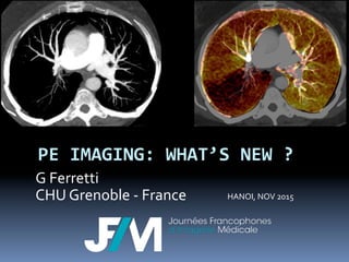
- 1. PE IMAGING: WHAT’S NEW ? G Ferretti CHU Grenoble -‐ France HANOI, NOV 2015
- 2. 1992 – 2014 PE diagnosis has been revolutionized by Spiral CT scan ú Non invasive technique ú Quick to acquired ú High reproducibility ú High accuracy for diagnosing PE and differential diagnosis ú Availability 24/365 ú Low cost vs VQ scintigraphy or P angiography Remy-‐Jardin M. Radiology 2007;245:315-‐29.
- 3. Recent technical inovations § Slice thickness decreased from 5 to <1 mm § Speed of acquisition decreased from 30 sec to 1-‐2 sec: dyspneic patients § contrast media administration decreased from de 120 ml to 50 ml § 100 Kv is optimal for image contrast § RX dose can be optimized Ghaye B, Radiology. 2011;219:629-‐36. 94% of 5th order P arteries and 74% d of 6th order P arteries are visualized MDCT: Se 83 to 100%, Sp: 89 to 97%
- 4. Increased use of CT has enabled earlier recognition of PE Wittram C. J Thorac Imaging 2004;19(3):164 –70. But, controversy exists 1. Do all identified patients with PE had clinically significant lesions requiring anticoagulation? Should isolated sub segmental PE be treated? 2. Does CT show to much clinically significant incidental finding (24%) and too frequent alternative diagnosis (33%) 3. Was CT adopted too quickly to diagnose PE without considering the risks and benefits?
- 11. D dimers § Level of positivity should be adapted to the age in patients > 50 yo § In case of low or intermediate probability, the level of positivity = age X 10 (microg/ml) § Such age-‐level increases the number of PE that are eliminated in patients > 75yo from 6% using the conventional level to 30% using the new level (based on 800 patients study) Righini JAMA 2014;311,1117-‐24
- 12. Possible role of ECG-‐gated CT scanner § Does it help to see the pulmonary arteries? ú Decreases motion artifacts in the lung in contact with the heart (lingula, LLL). ú Increases the dose delivered to the patient as well as the time of acquisition ú Only 1% of segmental arteries are impacted Ghaye B Ragiology 1997
- 13. Possible role of ECG-‐gated CT scanner § Functional evaluation of the heart? ú In particular the right heart function § However, analysis of the cavities of the heart on axial CT seems to do as well as ECG gated CT Abel E. Acta Radiol. 2012 1;53:720-‐7. Kamel EM, J Comput Assist Tomogr. 2008;32:438-‐43.
- 15. Possible role of ECG-‐gated CT scanner § triple rule-‐out in case of atypical chest pain in order to eliminate the “big three” ú PE ú Myocardial infarction ú Aortic dissection § This technique is still controversial Branch KR, PLoS One. 2013 16; 8:e61121.
- 16. Radiation Dose and CT for PE § Risk are individual and collective ú Due to the increased used of CT for suspicion of PE ú While the rate of positive CT is declining from 25% in 2000 to 4-‐6% nowadays. § In 2010, a monocentric study showed that ú 90% of 2003 CT were negative 93.6% in patients referred from the ER 86.5% for hospitalized patients Mamlouk MD, Radiology. 2010;256:625-‐32
- 17. PE and radiation dose ? § Great question: appropriate pre CT selection of the patients that should be send to CT??? ú Lot of papers and algorithm… few clinical applications § Optimization of CT dose delivered to patients ú 100 kV instead of 120 kV in thin to normal BMI patients Dose reduction: 40% to 20% Increases quality of angiogram ú iterative reconstruction : dose reduction 30 to50% Kubo T, AJR Am J Roentgenol. 2008;190:335-‐43. Pontana F, Radiology. 2013;267:609-‐18
- 19. PE signs were described in 1992, but have no wrinkle in 2015 § Filling defect within an opacified PA: low density matérieal surrounded by contrast media § Complete occlusion of the PA is often associated with an enlargement of the artery Remy-‐Jardin M, Radiology. 1992;185: 381-‐7.
- 20. Causes of false + and -‐ False positive False negative § Hilar or bronchopulmonary lymph nodes § Partial opacification of pulmonary veins or arteries § Partial volume effect § Use of high spatial frequency reconstruction algorithm § Inadequate opacification of the PA § Sub segmental PE § Motion artifacts § Partial volume effect § Low signal to noise Quality of CT should be verified for every patient, if inadequate, re scan the patient
- 21. Indirect signs of PE § Pulmonary infarction, pleural effusion § GGO surrounded by pulmonary consolidation § Angio CT remains necessary to detect PE Coche EE, Radiology. 1998;207:753-‐8. Revel MP, Radiology. 2007;244:875-‐82.
- 25. pneumonia infarction Chest pain with pulmonary consolidation Pneumonia vs. Infarction
- 26. Sub segmental PE § No treatment is a good option if: ú Respiratory function is preserved ú No deep venous thrombosis ú The clinical condition that is a high risk for thrombosis is canceled ú No history of central venous catheter ú Follow up by US of the legs possible ú The patients should be informed and agree
- 27. Spiral of severity in PE
- 28. Clinical severity score: PESI
- 29. Severity of PE: dysfunction of RV or embolic burden? § presence of RVD on echocardiography is associated with an increased risk for in-‐hospital mortality (risk ratio, 2.5; 95% CI, 1.2-‐5.5). § Correlation between clot burden and early mortality is still debated § The importance of preexisting alteration of RV function has certainly an important role to explain the discordance § Research: estimation of the pulmonary perfusion alteration studied with double energy CT scan in patients with PE Moroni AL, Eur J Radiol 2011;79:452–8. Apfaltrer P, Eur J Radiol. 2012;81:3592-‐7. Remy-‐Jardin M, Radiol Clin North Am. 2014;52:183-‐93.
- 30. Severity of PE: dysfunction of RV or embolic burden? § Axial CT allows to systematically assess signs related to RVD ú Enlargement of the right ventricle as compared to the left ventricle ú RV/LV ration > 1 ú Ventricular septal bowing ú reflux in the IVC Kamel EM, J Comput Assist Tomogr. 2008;32:438-‐43. Kumamaru KK. Int J Cardiovasc Imaging. 2012;28 :965-‐73.
- 31. Severity of PE: dysfunction of RV or embolic burden? § The RV/LV ratio is correlated to ú Hemodynamic severity of PE ú Intra hospital morbidity and mortality of PE ú Mortality prédiction at 3 months: 226 patients with and initial stable clinical condition: RV/LV>1 is a predictif sign of mortality when the clot burden is < 40% § a prospective study including 848 patients with clinical stable PE showed that the size of RV (RV/LV>0,9on CT did not correlate to prognostic at 30 days Jiménez D, Thorax. 2014;69:109-‐15. Contractor S, J Comput Assist Tomogr. 2002;26:587-‐91 Collomb D, Eur Radiol 2003; 13:1508 –1514 Ghaye B, Radiology. 2006;239:884-‐91.
- 33. CAD for PE detection? § CAD par rapport à la lecture classique 197 patients (159 sans embolie et 38 avec) par 6 lecteurs d’expérience variable. § La référence était établie par 2 lecteurs indépendants (35). ú la sensibilité de détection des EP variait Sans CAD, de 68% à 100% avec CAD de 76% à 100% (p<0,001), sans perte de spécificité lecteur dépendante (p<0,001). réduction significative de la durée de lecture (24-‐208 sec sans CAD versus 17-‐196 sec avec CAD) accroissement significatif de la confiance diagnostique § Une autre étude soulignait que le CAD améliorait la sensibilité de détection des EP des lecteurs inexpérimentés au prix d’une augmentation importante des faux positifs Wittenberg R. J Thorac Imaging. 2013;28:315-‐21 Blackmon KN, Eur Radiol. 2011;21:1214-‐23.
- 34. Double energy CT § A dream: ú Morphology of the arteries ú Function of the parenchyma / perfusion § However perfusion defects are not specific for PE and may be related to small airway disease Lu GM, Zhao Y, Zhang LJ, Schoepf UJ. Dual-energy CT of the lung. AJR Am J Roentgenol. 2012;199(5 Suppl):S40-53. Lu GM, Zhao Y, Zhang LJ, Schoepf UJ. Dual-energy CT of the lung. AJR Am J Roentgenol. 2012;199(5 Suppl):S40-53. Lu GM, AJR. 2012;199(5 Suppl):S40-53. Kang MJ, Radiographics. 2010;30:685-‐98
- 35. PE and pregnancy § Radiation dose ú Angio CT: 3 to 10 mSv ú (natural radiation: 3 mSv/year ) § Mammary dose : ú 50-‐90 mGy (2 breasts) ú (mammography 2 incidences : 3 mGy) § Higher risk of breast cancer ? Sadigh G AJR 2011; 196:497-‐515
- 38. PE and pregnancy In pregnant women with suspected PE § R1. we suggest that D-‐dimer not be used to exclude PE (weak recommendation). § R2. in presence of signs and symptoms of DVT, we suggest performing bilateral CUS of lower extremities followed by anticoagulation treatment, if positive and further testing, if negative (weak recommendation, very-‐low-‐ quality evidence). § R3. if no signs and symptoms of DVT, we suggest performing studies of the pulmonary vasculature rather than CUS of the lower extremities
- 39. PE and pregnancy In pregnant women with suspected PE § R4. we recommend a CXR as the first radiation-‐ associated procedure in the imaging work-‐up (strong recommendation, low-‐quality evidence). § R5. if CXR is normal , we recommend lung scintigraphy as the next imaging test rather than CTPA (strong recommendation, low quality evidence) § R5. if V/ Q scan is nondiagnostic , we suggest further diagnostic testing rather than clinical management alone . we recommend CTPA rather than DSA (strong recommendation, very-‐low-‐quality evidence). § R6. If CXR is abnormal , we suggest CTPA as the next imaging test rather than lung scintigraphy (weak recommendation, very-‐low-‐quality evidence)
- 41. PE and pregnancy § Pregnancy modifies hemodynamic parameters, therefore it is recommended to increase: ú the rate of CM Injection (6 cc/sec) ú the concentration of CM (350mg/ml) ú the volume if injected CM (90cc vs 70cc) ú during an apnea but not deep Hartmann IJ, Eur J Radiol. 2010;74:40-‐9.
- 42. MR and PE § Many drawbacks of MRI ú Length of imaging ú difficulty to take care of an emergency in MR ú limited spatial resolution ú Limited sensitivity ú limited availability ú complex technique imposing trained and experimented technicians and radiologists ú higher cost than CT
- 43. MRI ú Steady-‐state-‐free-‐precession ú Contrast enhanced angio MR ú Parenchymal perfusion § Compared with contrast-‐enhanced angiographic sequences, unenhanced sequences demonstrate lower sensitivity, except for proximal PE, but high specificity and agreement. § The negative predictive value of perfusion sequences was insufficient to safely rule out PE. Revel MP, Eur Radiol. 2013;23:2374-‐82 Hosch W, Emerg Radiol. 2014;21:151-‐8. Kluge A, AJR Am J Roentgenol 2006; 187: W7–W14
- 44. MRI § Expert centers results ú Sensitivity : 71-‐100% ú specificity : 92 à 100% § PIOPED III Sensitivity : 78 à 90% specificity : 99% à 100% NPV 97% at 3 months in case of technically optimal MRI BUT MRI were technically optimal in 48 to 89 % of cases (mean 75%) Biederer J, Insights Imaging 2012; 3: 373–86. Pleszewski B, Clin Imaging 2006; 30:166–172 Stein PD, (PIOPED III). Ann Intern Med 2010; 152: 434–43,
- 45. MRI in clincal practice § Reserved to ú Expert centers ú Patients intolerance to iodine CM, young patients, pregnancy Ersoy H, AJR Am J Roentgenol 2007;188:1246–54. Kalb B, Radiology. 2012;263:271-‐8.
- 46. Diagnosis of pulmonary embolism in patients with cancer § A negative D-‐dimer test has the same diagnostic value as in non-‐cancer patients. § On the other hand, D-‐dimer levels are non-‐specifically increased in many patients with cancer § In one study, raising the D-‐dimer cut-‐off level to 700 mg/ L, or using age-‐dependent cut-‐off levels, increased the proportion of cancer patients in whom PE could be ruled out from 8.4 to 13% and 12%, respectively; the corresponding false-‐negative rates appeared acceptable. Douma RA, Thromb Haemost 2010;104(4):831–836. Dentali F,. Thromb Res 2010;125(6):518–522.
- 47. Take home messages § PE without or with hypotension: CTA § D dimer related to the age if patient > 50ans § Favor exploration with 100kVp § Severity: RV larger than left ventricle § PE and pregnancy ú CXR first If unremarkable: VQ scan If abnormal : CTA first