
Recommended
Recommended
More Related Content
What's hot
What's hot (20)
Similar to Discitis and osteomyelitis in Sickle cell disease
Similar to Discitis and osteomyelitis in Sickle cell disease (20)
More from Jayanth Hiremagalur
More from Jayanth Hiremagalur (20)
Recently uploaded
Recently uploaded (20)
Discitis and osteomyelitis in Sickle cell disease
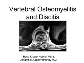
- 1. Vertebral Osteomyelitis and Discitis Rose-Krystel Hegngi MS 3. Jayanth H.Keshavamurthy M.D.
- 2. Case Presentation • 14-year-old male with hemoglobin SS disease, maintained on Hydroxyurea 800 mg daily, was recently admitted to the hospital on 11/14/2017 for one day due to acute pain crisis with microocclusion/vasooclusive of the back. He had been having low back pain since 09/27/17 located at the upper lumbar area, radiating up his spine. The pain is worsened with breathing and movement and relieved with laying down and taking Lortab 5.0. He is not able to sleep well at night and walking has become slightly more difficult due to the back pain. Mom reports good compliance with his Hydroxyurea 800mg Qday, Amoxicillin 250mg BID, folic acid, and Lortab 5.0 every 3-4 hours for pain.
- 3. MRI 11/14/17 • Impression 1. Pattern of chronic extensive bone marrow changes associated to sickle cell anemia with combination of old bone infarcts and bone marrow hematopoietic recruitment throughout thoracic segments. 2. Chronic appearing compression deformities with typical fish mouth deformity of vertebra, more prominent at T4, T9 and T10 as well as upper endplates of L2 on L3. Axial T2
- 4. MRI 11/14/17 • Impression 1. Pattern of chronic extensive bone marrow changes associated to sickle cell anemia with combination of old bone infarcts and bone marrow hematopoietic recruitment throughout thoracic segments. 2. Chronic appearing compression deformities with typical fish mouth deformity of vertebra, more prominent at T4, T9 and T10 as well as upper endplates of L2 on L3. SAG T2 SAG T1
- 5. Case Continued • The patient came in for a follow up visit 3 weeks after MRI. Since his last visit, he has been at home, not moving around much, but the pain has been manageable. He currently wears a back brace as the pain is still worsened with movements, at a 6/10 at its worst. His appetite has increased and he is beginning to gain back some weight. Mom reports good compliance with his Hydroxyurea 800mg Qday, Amoxicillin 250mg BID, folic acid, and vitamin D. TDC on 9/6/2017 was normal. Mom was told by Orthopedics that IR will perform bone biopsy of the spine. She will follow up after the biopsy.
- 6. CT Guided Aspiration/Biopsy/Injection • IMPRESSION 1. Successful CT-guided T10 paraspinal mass biopsy and T10 vertebral body bone biopsy. 2. As compared with recent MR 11/4/2017, there is progressive kyphotic deformity and vertebral body compression at T10-T11, with with early compression of T9.
- 7. Biopsy Results • Specimin-Vertebra, Cul Aer/Ana/+Gm • Prelim: • 1+ Yellow Colony Further studies in progress • 1+ White Colony Further studies in progress • Anaerobe culture in progress • Gram_Stn_B • Moderate neutrophils • No squamous epithelial cells • Very rare gram positive cocci • Specimin-Vertebra, Cul Fungus • Specimen received; culture in progress. • Presumtive Diagnosis: Vertebral Osteomyelitis/Discitis
- 8. Differential Diagnosis of Vertebral Osteomyelitis and Discitis • Other infections: • Septicemia • Cellulitis • Septic arthritis- given the early decrease in splenic function, bacterial infections of bone and joints are common in children with sickle cell disease • Deep abscess • Non-Infectious conditions: • Chronic non-bacterial osteomyelitis, • Vertebral compression fracture, • Vaso-occlusive pain episode- local warmth, tenderness, and swelling are common in both vaso-occlusive pain episodes and osteomyelitis. • Malignancy • Vitamin C deficiency, • Gaucher disease
- 9. Vertebral Osteomyelitis and Discitis: Radiographic Mimics • (1)Fibrous dysplasia • (2)Osteoid osteoma and osteoblastoma • (3)Eosinophilic granuloma and other forms of histiocytosis https://www.google.com/search?q=fibrous+dyspla sia+spine&source=lnms&tbm=isch&sa=X&ved=0 ahUKEwjQ5NKdsIfYAhWokOAKHdIHAIMQ_AUI CigB&biw=2100&bih=1303#imgrc=ZzEyGr3CV4C -aM:&spf=1513181465605 https://www.google.com/search?biw=2100&b ih=1303&tbm=isch&sa=1&ei=G1ExWonRMo m0_Absha6IAw&q=osteoid+osteoma+spine+ radiology&oq=osteod+osteoma+spine&gs_l= psy- ab.1.1.0i13k1l4.99094.105104.0.107102.24. 21.2.1.1.0.326.2534.9j10j0j1.20.0....0...1c.1. 64.psy- ab..1.23.2700...0j0i67k1j0i10k1j0i8i13i30k1.0 .ass7ZA96- qk#imgrc=tnZH73NCZ6HkeM:&spf=1513181 573645 https://www.google.com/search?biw=2100&bih= 1303&tbm=isch&sa=1&ei=sFIxWuyrJOKkggfXkK sg&q=eosinophilic+granuloma+vertebra+plana& oq=eosi&gs_l=psy- ab.1.0.0i67k1l2j0l2j0i67k1j0l5.9797.10252.0.124 65.4.4.0.0.0.0.156.492.2j2.4.0....0...1c.1.64.psy- ab..0.4.486....0.NPT07aukngM#imgrc=ENYYUy8 FpA-CjM:&spf=1513181883790 3.1. 2.
- 10. Vertebral Osteomyelitis and Discitis: Pathophysiology • 3 mechanisms • Hematogenous spread from a distant site or focus of infection • Most common cause of vertebral osteomyelitis • Direct inoculation from trauma/invasive spinal diagnostic procedures or surgery • Contiguous spread from soft tissue infections • Location • Vertebral Bodies-involved in approximately 4% of cases of osteomyelitis in children (80% occur in the long bones). • Intervertebral Discs-usually in children younger than 5, occurs almost exclusively in the lumbar region.
- 11. Vertebral Osteomyelitis and Discitis: Clinical Features • Signs and Symptoms • Localized neck or back pain, exacerbated by movement and palpation • Toxic appearance • Chronic low-grade fever • Lab • Elevated or normal WBC count • Elevated ESR and CRP in more than 80% of patients
- 12. Vertebral Osteomyelitis and Discitis: Diagnosis • Diagnosis is based on positive culture obtained from computed tomography (CT)-guided biopsy of the involved vertebra(e) and/or disc space. • Microbiology Cultures • Blood cultures are positive in up to 50% of cases • Radiographic Imaging • MRI: most sensitive radiologic technique in the diagnosis of vertebral osteomyelitis. It is also useful in differentiating between pyogenic, tuberculous, and fungal infections, and a neoplastic process. • CT: acceptable alternative to MRI, optimal for biopsies. • Plain radiograph: Often normal in early infection. *Chest radiography is warranted in the clinical suspicion of tuberculosis. • Radionuclide scanning: useful adjunct to plain film and CT when there is high suspicion for osteomyelitis in the setting of absent radiographic changes
- 13. Treatment • Combination of antimicrobial therapy and percutaneous drainage of abscess(es) if present. Routine treatment consists of a minimum of 6 weeks. • Pathogen-directed therapy (based on blood culture results) • Staphylococcal spp: if MSSA tx. nafcillin or oxacillin, if MRSA tx vancomycin • Streptococcal spp: if fully sensitive to penicillin, tx. ceftriaxone or high dose penicillin • Gram-negative bacilli: 3rd or 4th generation cephalosporin, or fluoroquinolone • Empiric therapy • Patients with Gram negative stain and culture results should be treated empirically, ie. vancomycin + one of the following: cefotaxime, a 3rd or 4th generation cephalosporin, or ciprofloxacin • Surgery • Reserved for patients with neurologic deficits, evidence of epidural/paravertebral abscess, and or spinal cord compression
- 14. Works Cited • Gaillard, F. (2017). Spondylodiscitis | Radiology Reference Article | Radiopaedia.org. [online] Radiopaedia.org. Available at: https://radiopaedia.org/articles/spondylodiscitis [Accessed 13 Dec. 2017]. • Uptodate.com. (2017). Bone and joint complications in sickle cell disease. [online] Available at: https://www.uptodate.com/contents/bone-and-joint-complications-in-sickle- cell- disease?source=see_link§ionName=OSTEOMYELITIS%20AND%20SEPTIC%20A RTHRITIS&anchor=H12#H12 [Accessed 13 Dec. 2017]. • Uptodate.com. (2017). Hematogenous osteomyelitis in children: Epidemiology, pathogenesis, and microbiology. [online] Available at: https://www.uptodate.com/contents/hematogenous-osteomyelitis-in-children- epidemiology-pathogenesis-and-microbiology?source=see_link [Accessed 13 Dec. 2017]. • Uptodate.com. (2017). Vertebral osteomyelitis and discitis in adults. [online] Available at: https://www.uptodate.com/contents/vertebral-osteomyelitis-and-discitis-in- adults#H11 [Accessed 13 Dec. 2017].