
Recommended
More Related Content
Similar to acutesevereasthmapicumanagement-150809163311-lva1-app6892-1.pptx
Similar to acutesevereasthmapicumanagement-150809163311-lva1-app6892-1.pptx (20)
Recently uploaded
Recently uploaded (20)
acutesevereasthmapicumanagement-150809163311-lva1-app6892-1.pptx
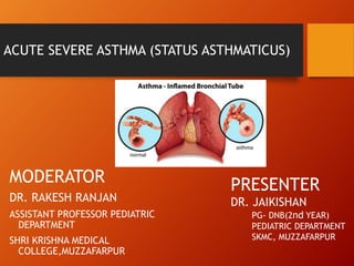
- 1. ACUTE SEVERE ASTHMA (STATUS ASTHMATICUS) MODERATOR DR. RAKESH RANJAN ASSISTANT PROFESSOR PEDIATRIC DEPARTMENT SHRI KRISHNA MEDICAL COLLEGE,MUZZAFARPUR PRESENTER DR. JAIKISHAN PG- DNB(2nd YEAR) PEDIATRIC DEPARTMENT SKMC, MUZZAFARPUR
- 3. DEFINITION OF ASTHMA • It is a chronic inflammatory condition of the lung airway resulting in episodic airflow obstruction. This chronic inflammation heightens the twitchiness of the - airways hyperresponsiveness (AHR) - to common provocative exposure
- 4. ETIOLOGY
- 5. PATHOPHYSIOLOGY Airway inflammation & hyper- reactivity Smooth muscle spasm Mucosal edema & plugging
- 6. CLINICAL MANIFESTATION • Intermittent dry coughing and EXPIRATORY WHEEZING. • Shortness of breath and chest congestion and tightness. • Respiratory symptoms worse at night, especially during prolonged exacerbation triggered by RESPIRATORY INFECTIONS or INHALANT ALLERGENS (pollens, malts, dust mites, animal dander). • Day time symptoms are often linked with physical activities (exercise or play induced) • Asthma symptoms can be triggered by numerous common events or exposure – Physical exertion and hyper ventilation, cold or dry air, and airway irritants. • Common respiratory pathogens – Rhinovirus, RSV, Influenza and parainfluenza, myco plasma pneumonia has induced airway inflammation.
- 7. Initial treatment of Asthma
- 8. GINA 2020
- 9. 1. Remove cap and shake inhaler in vertical direction 2. Breathe out gently 3. Put mouth piece in mouth. At start of inspiration that should be slow and deep, press canister down and continue to inhale deeply. 4. Hold breath for 10 seconds or as long as possible then breathe out slowly. 5. Wait for a few seconds before repeating steps 2-4. A) METERED DOSE INHALER B) METERED DOSE INHALER WITH SPACER 1. Remove cap, shake inhaler and insert into spacer device 2. Place mouth piece of spacer in mouth. 3. Start breathing in and out gently and observe movement of valve. 4. Once breathing pattern is established press canister and continue to breathe 5 -10 times (Tidal Breathing) 5. Remove the device from mouth and wait for 30 seconds before repeating steps 1-4.
- 10. C) METERED DOSE INHALER WITH SPACER AND FACEMASK 1. Attach baby mask to the mouth end of spacer 2. Shake MDI; insert it in the MDI end of spacer device. 3. Cover baby’s mouth and nose with baby mask 4. Press canister and encourage the child to take tidal breathing with mouth open (if possible) 5-10 times. 5. Remove baby mask and wait for 30-60 seconds before repeating step 1-4. D) ROTAHALER 1. Hold rotahaler vertically and insert capsule (clear end first) into square hole, make sure that the capsule is level with top of hole 2. Hold rotahaler horizontally, twist barrel in clockwise and anti-clockwise directions, this will split the capsule into two. 3. Breath out gently, put mouth end of rotahaler in mouth and take deep inspiration. 4. Remove rotahaler from mouth, hold breath for 10 seconds.
- 11. E) NEBULIZER 1. Connect output of compressor to nebulizer chamber by the tubing provided with the nebulizer. 2. Put measured amount of drug in nebulizer chamber, add normal saline to make the total volume 2.5 – 3 ml 3. Switch on the compressor and look for aerosol coming out form the nebulizer chamber. 4. Attach facemask to the nebulizer chamber, ensure appropriate fit to cover nose and mouth to the child. 5. Encourage child to take tidal breathing to open mouth. Children < 4-year old: MDI with spacer with facemask Children > 4-year old: MDI with spacer preferred Children > 12 year old: MDI used directly. Use of spacer improves drug deposition.
- 12. Definition of Status Asthmaticus • A severe exacerbation of asthma that does not improve with standard therapy is termed as status asthmaticus. • Clinical Definition - Severe asthma that failsto respond to inhaled β2 agonists, oral or IV steroids, and O2, and that requires admission to the hospital for treatment
- 13. Pathophysiology ofstatus asthmaticus Pathologic changes in the airway airflow obstruction premature airway closure on expiration dynamic hyperinflation hypercarbia Dynamic hyperinflation or “air-trapping” also leads to ventilation / perfusion (V/Q) mismatching causing hypoxemia
- 14. Presentation Varies by severity, asthmatic trigger, and patient age. ▶ Cough ▶ Wheezing ▶ Increased work of breathing. ▶ The noisy chest The degree of wheezing does not correlate well with severity of the disease.
- 15. Assessment Primary assessment ▶ Airway ▶ Breathing ▶ Circulation ▶ Disability ▶ Exposure Secondary assessment (Focused history and examination)
- 16. Prognosis High risk factors for asthma severity and fatality ▶ Previous severe sudden deterioration, ▶ Past PICU admissions ▶ Previous respiratory failure ▶ Need formechanical ventilation.
- 17. Presentation ‘Red-alerts’ Severe respiratory compromise: ▶ ‘Silent Chest’ with increased respiratory efforts usually precede respiratory failure. ▶ Agitation or dyspnea ▶ Altered consciousness ▶ Inability to speak >1-2 words at a time ▶ Central cyanosis ▶ Diaphoresis ▶ Inability to lie down ▶ Pulsusparadoxus > 25 mmHg ▶ PaCO2 normalization or hypercapnia (ominous) ▶ Bradycardia ▶ Severe Hypoxia
- 18. Assessmentof severity BeckerAsthma score The assessment of severity of acute severe asthma is based on clinical observation of the child. Becker Asthma score is a quick assessment of severity, by using respiratory rate, wheezing, inspiratory, expiratory ratio and accessory muscles use.
- 19. Oxygen Therapy ▶ 100% oxygen ▶ Oxygen saturation monitoring ▶ Other monitors
- 20. Pulsus Paradoxus
- 21. Cardiopulmonary Interactions ▶ Severe the attack, more negative intrapleural pressure ▶ Increased left ventricular afterload ▶ Increased transcapillary filtration of edema fluid into airspaces resulting in a high risk for pulmonary edema. ▶ Overhydration increasesmicrovascular hydrostatic pressure and furtherworsens pulmonary edema. ▶ High right ventricularafter load due to ▶ Hypoxic pulmonary vasoconstriction, ▶ Acidosis ▶ Increased lung volume.
- 22. Chest Radiography Limited role but indicated in- ▶ First time wheezers ▶ Clinical evidence of parenchymal disease ▶ Those requiring admission to PICU. ▶ Suspected airleak orpneumonia ▶ When the underlying cause of wheezing is in doubt
- 23. Arterial blood gas ▶ In all children at baseline ▶ Subsequently as indicated ▶ Hypocarbia in early stage ▶ Normalization of CO2 with persistent respiratory distress indicates impending respiratory failure. ▶ A PaO2<60 mm Hg and a normal or increased PaCO2 (>45 mm Hg) indicates the presence of respiratory failure
- 24. PICUAdmission ▶ Comfortable environment ▶ IV access ▶ Maintain euvolemia ▶ Continuous cardio-respiratory monitoring ▶ Avoid sedation ▶ Monitorpotassium ▶ Antibiotics, if indicated ▶ If ventilated -arterial and central venous access
- 25. Fluid ▶ Restoration of euvolemia ▶ Isotonic fluid like normal saline or Ringer’s lactate ▶ Fluid balance ▶ Avoid overhydration;Riskof pulm edema ▶ Serum potassium monitoring
- 26. Antibiotics ▶ Not routinely indicated ▶ Reserved forchildren with evidence of bacterial infection ▶ High fever ▶ Consolidation on X ray film or ▶ Very high leucocyte counts
- 27. Pharmacologic Targets ▶ Improving oxygen delivery ▶ Relaxation of bronchial smooth muscles ▶ B2 receptors ▶ M1 receptors ▶ Attenuating underlying inflammation
- 32. Adverse effectsof β2- agonists ▶ Cardiovascularsystem ▶ Tachycardia ▶ Increased QTc interval ▶ Dysarrhythmia ▶ Hypertension ▶ Diastolic hypotension. ▶ Excessive CNSstimulation ▶ Hyperactivity, ▶ Tremors ▶ Nausea with vomiting ▶ Hypokalemia ▶ Hyperglycemia
- 33. Corticosteroids: Side effects ▶ Short-term use of high-dose steroids ▶ Hyperglycemia ▶ Hypertension ▶ Acute psychosis ▶ Prolonged steroid ▶ Immunosuppression ▶ Hypothalamic-pituitary-adrenal axis suppression, ▶ Osteoporosis ▶ Myopathy ▶ Weakness
- 34. Magnesium Sulfate ▶ Mechanism: ▶ InhibitsCa2+ influxintocytosol smooth muscle relaxant ▶IncreasesB2agonist affinity for itsreceptor, thereby potentiating itseffect ▶ Inhibitshistaminerelease frommastcells ▶ 50mg/kg IV over20-30minwithmax of 2gm ▶ Repeat once ortwice after4–6h. SIDEEFFECTS ▶ Hypotension ▶ CNSdepression, ▶ Muscle weakness ▶ Flushing ▶ Cardiac arrhythmia/ complete heartblock, ▶ Respiratoryfailuredue to severe muscle weakness
- 35. Mechanical Ventilation Indications ▶ Poorresponse to initial therapy ▶ Severe hypoxia ▶ Rapid deterioration in mental state ▶ Rising PCO2 ▶ Cardiopulmonary arrest
- 36. Intubation Tips ▶ Preoxygenate with 100%oxygen ▶ Anticipate hypotension ▶ Cuffed ETtube with the largest appropriate diameter ▶ Avoid histamine-producing agents like morphine or atracurium ▶ Ketamine: preferred induction agent due to its bronchodilatory action. ▶ Use atropine, Benzodiazepam and by a rapid- acting muscle relaxant (vecuronium).
- 37. Complications ▶ Hypotension ▶ Oxygen desaturation ▶ Pneumothorax/ subcutaneousemphysema, ▶ Cardiac arrest ▶ Suspect tension pneumothorax and treat promptly
- 38. Chest Physiotherapy ▶ Useful in children with segmental or lobar atelectasis. ▶ In others no therapeutic benefit in the critically ill patient with status asthmaticus.
- 39. Summary
- 40. Thankyou