
Recommended
More Related Content
What's hot
What's hot (20)
Similar to Fibroid Uterus: Symptoms, Diagnosis and Treatment
Similar to Fibroid Uterus: Symptoms, Diagnosis and Treatment (20)
More from hemachandra59
More from hemachandra59 (20)
Recently uploaded
Recently uploaded (20)
Fibroid Uterus: Symptoms, Diagnosis and Treatment
- 2. INTRODUCTION Fibroid is the commonest benign tumor of the uterus and also the commonest benign solid tumor in female. Histologically, this tumor is composed of smooth muscle and fibrous connective tissue, so named as uterine fibromyoma.
- 3. Definition 1. Uterine fibroids are the most common benign pelvic tumors in women of reproductive age. They affect 20–40% of those women but are found in 75% of women. This is due to the fact that most fibroids are asymptomatic. 2. Fibroids are common benign uterine smooth muscle tumours that may occur singly or multiply and vary greatly in size. 3. Uterine fibroids or leiomyoma are benign tumors of the uterine muscle, called myometrium. They contain receptors for female reproductive hormones (estrogen and progesterone) and other enzyme receptors related to estrogen production .
- 4. Etiological factors Age, Any hormonal disturbances Hereditary history Any previous history of uterine disorder
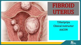
- 5. Types of fibroid uterus 1. Sub-serosal Fibroids are found superficially under the outer lining of the uterus, the serosa. They can grow to the interior part of the wall or completely under the serosa. 2. Intramural Fibroids are the most common. They are situated in the middle layer of the uterine muscle. 3. Submucosal Fibroids grow in the myometrium near the inner lining of the uterus, called endometrium. they can become pedunculated and protrude into the uterine cavity
- 6. Cont. 4. Pedunculated Fibroids are benign (noncancerous) growths in the uterus. These fibroids are attached to the uterine wall. These fibroids can grow both inside and outside the uterus. Inside the uterus, this type of growth is called a pedunculated submucosal fibroid. Outside the uterus it’s called a pedunculated subserosal fibroid.
- 8. Clinical manifestations Increase in size and number. A uterine tumor rapidly growing after menopause is unlikely to be a fibroid Menorrhagia (prolonged and heavy bleeding within normal cycle). Infertility. Secondary dysmenorrhea (new onset of period pain). Pressure symptoms from the bowel and bladder, e.g. constipation, frequency, chronic urinary tract infections (UTIs). Chronic pelvic pain, dyspareunia.
- 9. Cont. Pregnancy-associated symptoms: Spontaneous abortion Recurrent abortion Abdominal pain and pressure signs in pregnancy Premature rupture of membranes Dystocia( Difficulty or obstructed labor Post-partum hemorrhage. Less common symptoms include: For submucosal, pedunculated fibroids: protrusion through cervical Os with pain and bleeding. separation from the uterus.
- 10. Diagnostic findings Complete history collection Physical examination Speculum examination reveals cervical mucus or discharge and ulcerations Ultrasound is very helpful in differentiating it from other conditions such as ovarian tumor. Laboratory investigations ,CBC is done to determine if the pt have iron deficiency anemia because of chronic blood loss. Urinalysis to detect UTI. Endometrial biopsy is preformed by taking tissue sample from uterus.
- 11. Special diagnostic studies Hysterosalpingogram ( an ultrasound exam is done while contrast fluid is injected into the uterus from cervix Hysteroscopy (HSC) ( to look at the uterus by passing a small fiberoptic camera through the opening of the cervix determines fibroid is presented r not
- 13. Medical management Symptomatic treatment Analgesics. Iron and folic acid to correct anaemia Tranexamic acid or non-steroidal anti-inflammatory drugs Gonadotropin-releasing hormone analogs Progesterone antagonists
- 14. Surgical Management Abdominal hysterectomy(A hysterectomy is the removal of the uterus with or without the ovaries and the tubes. Myomectomy (Myomectomy means the excision of fibroids from the myometrium without removing the uterus) Laparoscopic myomectomy or Hysterectomy
- 15. Cont. A myomectomy is a surgical procedure used to remove non-cancerous growths, also called uterine fibroids or leiomyomas, that are found growing in the uterus of women who are of childbearing age. The advantage of laparoscopic surgery is faster recovery with a shorter hospital stay and fewer complications.
- 17. Preoperative Nursing Management 1.Pain Management 2.Fluid Replacement 3.Bleeding Control 4.Patient Education
- 18. Cont. ♣The nurse assesses the intensity of the patient’s pain and assists the patient with analgesia as prescribed. ♣The nurse counts the perineal pads used, assesses the extent of oxygen saturation with blood, and monitors vital signs. ♣Abdominal dressing is monitored for drainage if an abdominal surgical technique was used. ♣Nurse should instruct to contact the nurse or obstetrician if bleeding is excessive. ♣The patient is encouraged and assisted to change positions frequently. ♣Nurse helps the patient to ambulate early in the postoperative period.
- 19. Cont. ♣Intake and output chart is monitored. ♣Explain the patient about follow up visits. ♣Administer iron and folic acid as prescribed. ♣Encourage verbalization of feelings of patient ♣Monitor active fluid loss from wound drainage, bleeding. ♣Encourage patient to drink plenty of oral fluid ♣Monitor serum electrolytes.
- 20. Cont. ♣Observations of changes in mental status, behavior or level of consciousness. ♣Note the catheter patency was settled (when using catheter) ♣Assess nutritional status, including weight, history of weight loss and serum albumin status. ♣Encourage intake of protein and calorie-rich foods. ♣Help in developing effective coping strategies.
- 21. Complications Degeneration Necrosis Infection Hemorrhage In pregnant mother; Necrosis Spontaneous abortion. Premature labor and delivery. Abruption of placenta. Post-partum hemorrhage.
- 22. Nursing diagnosis 1. Impaired urinary incontinence related to presence of local tissue edema or hematoma. 2. Ineffective Tissue Perfusion related to postoperative tissue inflammation. 3. Sexual dysfunction related to change in sexual response pattern. 4. Constipation or Diarrhoea related to manipulation of the bowel or weakening of abdominal musculature. 5. Knowledge deficit related to Information misinterpretation. 6. Risk for low self esteem related to effect on the sexual relationship.