
Recommended
Recommended
More Related Content
What's hot
What's hot (20)
Similar to Amplificación de ácidos nucleicos
Similar to Amplificación de ácidos nucleicos (20)
Recently uploaded
Recently uploaded (20)
Amplificación de ácidos nucleicos
- 1. | 1Nucleic Acid Amplification PROTOCOLS & APPLICATIONS GUIDE CONTENTS I. Introduction 1 D. Tli DNA Polymerase 16 A. Basic PCR 1 E. Pfu DNA Polymerase 16 B. RT-PCR 2 V. Reverse Transcriptases 17 C. Hot-Start PCR 3 A. AMV Reverse Transcriptase 17 D. Long PCR 4 B. M-MLV Reverse Transcriptase 17 E. Quantitative Endpoint PCR 5 C. M-MLV Reverse Transcriptase, RNase H Minus 17 F. Quantitative Real-Time PCR 5 VI. Example of a PCR Protocol 18 G. Rapid Amplified Polymorphic DNA Analysis 7 VII. Example of an RT-PCR Protocol 19 H. Rapid Amplification of cDNA Ends (RACE) 7 A. Access RT-PCR Protocol 19 I. Differential Display PCR 8 B. ImProm-II™ Reverse Transcription System Protocol 19 J. In situ PCR 9 K. High-Fidelity PCR 9 VIII.Troubleshooting PCR and RT-PCR 22 L. PCR and DNA Sequencing: Cycle Sequencing 9 IX. References 23 M. Cloning PCR Products 9 II. General Considerations for PCR Optimization 10 A. Magnesium Concentration 10 B. Buffer Considerations 11 C. Enzyme Concentration 11 D. PCR Primer Design 12 E. Template Quality 12 F. Template Quantity 12 G. Cycling Parameters 12 H. PCR Enhancers and Additives 13 I. Nucleic Acid Cross-Contamination 13 III. General Considerations for RT-PCR 14 A. Overview of the Access and AccessQuick™ RT-PCR Systems 14 B. Template Considerations 14 C. Reverse Transcription Primer Design 14 D. Cycle Parameters 15 IV. Thermostable DNA Polymerases 15 A. Taq DNA Polymerase 15 B. Tfl DNA Polymerase 16 C. Tth DNA Polymerase 16 Protocols & Applications Guide www.promega.com rev. 3/09
- 2. | 1Nucleic Acid Amplification PROTOCOLS & APPLICATIONS GUIDE I. Introduction optimum for the DNA polymerase. For most thermostable DNA polymerases, this temperature is in the range of The polymerase chain reaction (PCR) is a relatively simple 70–74°C. The extension step lasts approximately 1–2 technique that amplifies a DNA template to produce minutes. The next cycle begins with a return to 94°C for specific DNA fragments in vitro. Traditional methods of denaturation. cloning a DNA sequence into a vector and replicating it in a living cell often require days or weeks of work, but Each step of the cycle should be optimized for each template amplification of DNA sequences by PCR requires only and primer pair combination. If the temperature during hours. While most biochemical analyses, including nucleic the annealing and extension steps are similar, these two acid detection with radioisotopes, require the input of steps can be combined into a single step in which both significant amounts of biological material, the PCR process primer annealing and extension take place. After 20–40 requires very little. Thus, PCR can achieve more sensitive cycles, the amplified product may then be analyzed for detection and higher levels of amplification of specific size, quantity, sequence, etc., or used in further sequences in less time than previously used methods. These experimental procedures. features make the technique extremely useful, not only in An animated presentation (www.promega.com basic research, but also in commercial uses, including /paguide/animation/selector.htm?coreName=pcr01) genetic identity testing, forensics, industrial quality control illustrating the PCR process is available. and in vitro diagnostics. Basic PCR has become commonplace in many molecular biology labs where it is Additional Resources for Basic PCR used to amplify DNA fragments and detect DNA or RNA Technical Bulletins and Manuals sequences within a cell or environment. However, PCR has evolved far beyond simple amplification and detection, TB254 GoTaq®PCR Core Systems Technical Bulletin and many extensions of the original PCR method have been (www.promega.com/tbs/tb254/tb254.html) described. This chapter provides an overview of different 9PIM750 PCR Master Mix Promega Product types of PCR methods, applications and optimization. A Information detailed treatment of these methods is beyond the scope (www.promega.com of this publication. However, an extensive bibliography is /tbs/9pim750/9pim750.html) provided in the References section for researchers who 9PIM300 GoTaq® DNA Polymerase Promega Product require more comprehensive information. Information A. Basic PCR (www.promega.com /tbs/9pim300/9pim300.html) The PCR process was originally developed to amplify short segments of a longer DNA molecule (Saiki et al. 1985). A 9PIM829 GoTaq® Flexi DNA Polymerase Promega typical amplification reaction includes the target DNA, a Product Information thermostable DNA polymerase, two oligonucleotide (www.promega.com primers, deoxynucleotide triphosphates (dNTPs), reaction /tbs/9pim829/9pim829.html) buffer and magnesium. Once assembled, the reaction is Promega Publications placed in a thermal cycler, an instrument that subjects the PN091 GoTaq® Green Master Mix: From reaction to a series of different temperatures for set amounts Amplification to Analysis of time. This series of temperature and time adjustments (www.promega.com is referred to as one cycle of amplification. Each PCR cycle /pnotes/91/12972_13/12972_13.html) theoretically doubles the amount of targeted sequence PN083 Introducing GoTaq® DNA Polymerase: (amplicon) in the reaction. Ten cycles theoretically multiply Improved amplification with a choice of the amplicon by a factor of about one thousand; 20 cycles, buffers by a factor of more than a million in a matter of hours. (www.promega.com Each cycle of PCR includes steps for template denaturation, /pnotes/83/10492_21/10492_21.html) primer annealing and primer extension (Figure 1.1). The PN078 Performance advantages designed into initial step denatures the target DNA by heating it to 94°C Promega's PCR Master Mix or higher for 15 seconds to 2 minutes. In the denaturation (www.promega.com process, the two intertwined strands of DNA separate from /pnotes/78/9186_09/9186_09.html) one another, producing the necessary single-stranded DNA More (www.promega.com/pnotes/apps/pcr/) template for replication by the thermostable DNA publications polymerase. In the next step of a cycle, the temperature is Online Tools reduced to approximately 40–60°C. At this temperature, Amplification Product Selector (www.promega.com the oligonucleotide primers can form stable associations /selectors/amplification/) (anneal) with the denatured target DNA and serve as primers for the DNA polymerase. This step lasts approximately 15–60 seconds. Finally, the synthesis of new DNA begins as the reaction temperature is raised to the Protocols & Applications Guide www.promega.com 1-1 rev. 3/09
- 3. | 1Nucleic Acid Amplification PROTOCOLS & APPLICATIONS GUIDE Target Region Unamplified DNA 5′ 3′ 3′ 5′ Cycle 1 5′ 3′ Denature and anneal primers 3′ 5′ Extend primers 5′ 3′ 3′ 5′ 5′ 3′ 3′ 5′ Cycle 2 5′ 3′ Denature and 3′ 5′ anneal primers 5′ 3′ 3′ 5′ Extend primers 5′ 3′ 3′ 5′ 5′ 3′ 3′ 5′ 5′ 3′ 3′ 5′ 5′ 3′ 3′ 5′ Cycle 3 5′ 3′ 3′ 5′ 5′ 3′ Denature and 3′ anneal primers 5′ 3′ 3′ 5′ 5′ 3′ 3′ 5′ Extend primers 5′ 3′ 3′ 5′ 5′ 3′ 3′ 5′ 5′ 3′ =Short "target" product 3′ 5′ 5′ 3′ 3′ 5′ 5′ 3′ 3′ 5′ 5′ 3′ 3′ 5′ 5′ 3′ 3′ 5′ 5′ 3′ =Long product 3′ 5′ Cycles 4-30 Amplification of short "target" product Figure 1.1. Schematic diagram of the PCR process. Citations by agarose gel electrophoresis followed by ethidium Bermejo-Alvarez, P. et al. (2008) Can bovine in bromide staining. vitro-matured oocytes selectively process X- or Y-sorted PubMed Number: 18579751 sperm differentially? Biol. Reprod. 79, 594–7. To determine whether oocytes are able to select X-bearing B. RT-PCR or Y-bearing spermatozoa, the authors performed in vitro The thermostable DNA polymerases used in the basic PCR fertilization of bovine oocytes with X-sorted semen, process require a DNA template, and as such, the technique Y-sorted semen, a mixture of X- and Y-sorted semen, and is limited to the analysis of DNA samples. Yet numerous unsorted semen. The gender of the resulting embryos was instances exist in which the amplification of RNA would determined by amplifying two DNA targets: a Y be preferred. To apply PCR to the study of RNA, the RNA chromosome-specific target for gender assignment and a sample must first be reverse transcribed to cDNA to provide bovine-specific satellite sequence as a control. PCRs were the necessary DNA template for the thermostable performed using GoTaq® Flexi DNA Polymerase (1 unit polymerase (Figure 1.2). This process is called reverse per 25μl reaction), and amplified products were analyzed transcription (RT), hence the name RT-PCR. Protocols & Applications Guide www.promega.com 1-2 rev. 3/09
- 4. | 1Nucleic Acid Amplification PROTOCOLS & APPLICATIONS GUIDE Avian myeloblastosis virus (AMV) or Moloney murine PN079 Using ImProm-II™ Reverse Transcription leukemia virus (M-MLV or MuLV) reverse transcriptases System for coupled RT-PCR are generally used to produce a DNA copy of the RNA (www.promega.com template using either random primers, an oligo(dT) primer /pnotes/79/9492_15/9492_15.html) or sequence-specific primers. Alternatively, some thermostable DNA polymerases (e.g., Tth DNA polymerase) PN078 Technically speaking: Promega RT-PCR possess a reverse transcriptase activity, which can be systems explained activated by using manganese instead of magnesium as a (www.promega.com cofactor (Myers and Gelfand, 1991). After this initial reverse /pnotes/78/9186_21/9186_21.html) transcription step has produced the cDNA template, basic PN073 Using the Access RT-PCR System: PCR is carried out to amplify the target sequence. Reaction parameters that affect efficient amplification The quality and purity of the starting RNA template is (www.promega.com crucial to the success of RT-PCR. Total RNA or poly(A)+ /pnotes/73/8235_14/8235_14.html) RNA can be used as the starting template, but both must be intact and free of contaminating genomic DNA. Specific More (www.promega.com capture of poly(A)+ RNA will enrich a targeted message publications /pnotes/apps/rt_cdna/) so that less of the reverse transcription reaction is needed Citations for the subsequent amplification. The efficiency of the Nanashima, N. et al. (2008) The hairless phenotype of the first-strand synthesis reaction, which can be related to the Hirosaki hairless rat is due to the deletion of an 80-kb quality of the RNA template, will also significantly impact genomic DNA containing five basic keratin genes. J. Biol. the results of the subsequent amplification. Chem. 283, 16868–75. First Strand Synthesis: The mutation responsible for the hairless phenotype was linked to a 80kb deletion of chromosome 7q36. Because Random primer many basic keratin genes are located at 7q36, the authors mRNA 5′ AAAAAAAA 3′ first-strand examined keratin gene expression in the Hirosaki rat using N6 N6 N6 N6 N6 N6 cDNA RT-PCR. Expression of kb21, kb23 and kb26 were not detected, whereas other basic keratin genes were expressed. Oligo(dT) primer mRNA RT-PCR was performed using 0.5μg of total RNA isolated 5′ AAAAAAAA 3′ first-strand 3′ T T T T T T T T 5′ cDNA PCR from rat skin for 21–30 cycles. PubMed Number: 18420582 Sequence-specific primer ( ) mRNA Capozzo, A.V. et al. (2003) Development of DNA vaccines 5′ AAAAAAAA 3′ against hemolytic-uremic syndrome in a murine model. 1439MA04_6A first-strand 3′ 5′ cDNA Infect. Immun. 71, 3971–8. Figure 1.2. Schematic diagram of RT-PCR. Researchers used the pGEM®-T Vector System to clone the entire 1.4kb Shiga toxin type 2 gene (Stx2) from E. coli Additional Resources for RT-PCR O157-H7 C600 (933W). The resultant construct, named pGEMTStx2, was used as a template in PCR to amplify Technical Bulletins and Manuals each region of the gene corresponding to Shiga toxin type TB220 Access RT-PCR System Technical Bulletin 2 subunits A and B. Each PCR product was digested with (www.promega.com/tbs/tb220/tb220.html) BamHI and EcoRI, then ligated into pCDNA 3.1+ to create TM236 ImProm-II™ Reverse Transcription System pStx2ΔA and pStx2B. Mice were then immunized with Technical Manual either one or both of these constructs and another construct (www.promega.com expressing murine granulocyte-macrophage /tbs/tm236/tm236.html) colony-stimulating factor. Expression of each subunit in TB099 Reverse Transcription System Technical mouse tissue was verified by RT-PCR using specific primers Bulletin and the AccessQuick™ RT-PCR System. (www.promega.com/tbs/tb099/tb099.html) PubMed Number: 12819084 9PIA170 AccessQuick™ RT-PCR System Promega Product Information C. Hot-Start PCR (www.promega.com Hot-start PCR is a common technique to reduce nonspecific /tbs/9pia170/9pia170.html) amplification due to the assembly of amplification reactions Promega Publications at room temperature. At these lower temperatures, PCR PN079 AccessQuick™ RT-PCR System: Simple, primers can anneal to template sequences that are not stable and sensitive perfectly complementary. Since thermostable DNA (www.promega.com polymerases have activity at these low temperatures /pnotes/79/9492_12/9492_12.html) (although in most cases the activity is less than 25%) the polymerase can extend misannealed primers. This newly Protocols & Applications Guide www.promega.com 1-3 rev. 3/09
- 5. | 1Nucleic Acid Amplification PROTOCOLS & APPLICATIONS GUIDE synthesized region is perfectly complementary to the DNA 9PIM513 GoTaq® Hot Start Colorless Master Mix template, allowing primer extension and synthesis of Promega Product Information undesired amplification products. However, if the reaction (www.promega.com is heated to temperatures >60°C before polymerization /tbs/9pim513/9pim513.html) begins, the stringency of primer annealing is increased, and Promega Publications the subsequent synthesis of undesired PCR products is avoided or reduced. PN099 Get the convenience of hot-start PCR with the new GoTaq® Hot Start Polymerase Hot-start PCR can also reduce the amount of primer-dimer (www.promega.com synthesized by increasing the stringency of primer /pnotes/99/15674_08/15674_08.html) annealing. At lower temperatures, PCR primers can anneal to each other via regions of complementarity, and the DNA D. Long PCR polymerase can extend the annealed primers to produce Amplification of long DNA fragments is desirable for primer dimer, which often appears as a diffuse band of numerous applications such as physical mapping approximately 50–100bp on an ethidium bromide-stained applications (Rose, 1991) and direct cloning from genomes. gel. The formation of nonspecific products and While basic PCR works well when smaller fragments are primer-dimer can compete for reagent availability with the amplified, the efficiency of amplification (and therefore the amplification of the desired product. Thus, hot-start PCR yield of amplified fragments) decreases significantly as the can improve the yield of specific PCR products. size of the amplicon increases over 5kb. This decrease in To perform manual hot-start PCR, the reactions are yield is attributable to the accumulation of truncated assembled on ice or at room temperature, but one critical products, which are not suitable substrates for the component is omitted until the reaction has been heated to subsequent cycles of amplification. These products appear 60–65°C, at which point the missing reagent is added. This as smeared, as opposed to discrete, bands on a gel. omission prevents the polymerase from extending primers In 1994, Wayne Barnes (Barnes, 1994) and other researchers until the critical component is added at the higher (Cheng et al. 1994) analyzed the factors affecting temperature where primer annealing is more stringent. polymerization across larger regions of DNA by However, this method is tedious and increases the risk of thermostable DNA polymerases and identified key contamination. A second, less labor-intensive approach variables affecting the yield of longer PCR fragments. They involves the reversible inactivation or physical separation devised an approach using a mixture of two thermostable of one or more critical components in the reaction. For polymerases to synthesize longer PCR products. The first example, the magnesium or DNA polymerase can be sequestered in a wax bead, which melts as the reaction is polymerase lacks a 3′→5′ exonuclease (proofreading) heated to 94°C during the denaturation step, releasing the activity; the second enzyme, present at a reduced component only at higher temperatures (Carothers et al. concentration, contains a potent proofreading activity. 1989; Krishnan et al. 1991; Clark, 1988). The DNA Presumably, when the nonproofreading DNA polymerase polymerase also can be kept in an inactive state by binding (e.g., Taq DNA polymerase) misincorporates a dNTP, to an oligonucleotide, also known as an aptamer (Lin and subsequent extension of the newly synthesized DNA either Jayasena, 1997; Dang and Jayasena, 1996) or an antibody proceeds very slowly or stops completely. The proofreading (Scalice et al. 1994; Sharkey et al. 1994). This bond is polymerase (e.g., Pfu DNA polymerase or Tli DNA disrupted at the higher temperatures, releasing the polymerase) serves to remove the misincorporated functional DNA polymerase. Finally, the DNA polymerase nucleotide, allowing the DNA polymerases to continue can be maintained in an inactive state through chemical extension of the new strand. modification (Moretti, T. et al 1998). Although the use of two thermostable DNA polymerases can significantly increase yield, other conditions can have Additional Resources for Hot-Start PCR a significant impact on the yield of longer PCR products Technical Bulletins and Manuals (Cheng et al. 1995). Logically, longer extension times can 9PIM500 GoTaq® Hot Start Polymerase Promega increase the yield of longer PCR products because fewer Product Information partial products are synthesized. Extension times of 10–20 (www.promega.com minutes are common and depend on the length of the /tbs/9pim500/9pim500.html) target. In addition, template quality is crucial. Depurination of the template, which is promoted at elevated temperatures 9PIM512 GoTaq® Hot Start Green Master Mix Promega and lower pH, will result in more partial products and Product Information decreased overall yield. In long PCR, the denaturation time (www.promega.com is reduced to 2–10 seconds to decrease depurination of the /tbs/9pim512/9pim512.html) template. Additives, such as glycerol and dimethyl sulfoxide (DMSO), also help lower the strand-separation and primer-annealing temperatures, alleviating some of the depurination effects of high temperatures. Cheng et al. Protocols & Applications Guide www.promega.com 1-4 rev. 3/09
- 6. | 1Nucleic Acid Amplification PROTOCOLS & APPLICATIONS GUIDE also found that reducing potassium concentrations by Numerous fluorescent and solid-phase assays have been 10–40% increased the efficiency of amplification of longer described to measure the amount of amplification product products (Cheng et al. 1995). generated in each reaction, but they can fail to discriminate amplified DNA of interest from nonspecific amplification E. Quantitative Endpoint PCR products. Some of these analyses rely on blotting PCR and RT-PCR are generally used in a qualitative format techniques, which introduce another variable due to nucleic to evaluate biological samples. However, a wide variety of acid transfer efficiencies, while other assays have been applications, such as determining viral load, measuring developed to eliminate the need for gel electrophoresis yet responses to therapeutic agents and characterizing gene provide the requisite specificity. Real-time PCR, which expression, would be improved by quantitative provides the ability to view the results of each amplification determination of target abundance. Theoretically, this cycle, is a popular way of overcoming the need for analysis should be easy to achieve, given the exponential nature of by electrophoresis. PCR, because a linear relationship exists between the number of amplification cycles and the logarithm of the Additional Resources for Quantitative PCR number of molecules. In practice, however, the Promega Publications amplification efficiency is decreased because of contaminants (inhibitors), competitive reactions, substrate PN068 Quantitative RT-PCR: Rapid construction exhaustion, inactivation of the polymerase and target of templates for competitive amplification reannealing. As the number of cycles increases, the (www.promega.com amplification efficiency decreases, eventually resulting in /pnotes/68/7381_16/7381_16.html) a plateau effect. F. Quantitative Real-Time PCR Normally, quantitative PCR requires the measurements to The use of fluorescently labeled oligonucleotide probes or be taken before the plateau phase so that the relationship primers or DNA-binding fluorescent dyes, such as SYBR® between the number of cycles and molecules is relatively Green, to detect and quantitate a PCR product allows linear. This point must be determined empirically for quantitative PCR to be performed in real time. different reactions because of the numerous factors that DNA-binding dyes are easy to use but do not differentiate can affect the amplification efficiency. Because the between specific and nonspecific PCR products. measurement is taken prior to the reaction plateau, Fluorescently labeled nucleic acid probes have the quantitative PCR uses fewer amplification cycles than basic advantage that they react with only specific PCR products. PCR. This can cause problems in detecting the final product These probes can also be used to detect single nucleotide because there is less product to detect. polymorphisms (Lee et al. 1993; Bernard et al. 1998). To monitor amplification efficiency, many applications are However in many cases, these approaches are not designed to include an internal standard in the PCR. One conducive to multiplex reactions, and there is no convenient such approach includes a second primer pair that is specific way of distinguishing specific and nonspecific PCR for a “housekeeping” gene (i.e., a gene that has constant products. A more recent technology, the Plexor® expression levels among the samples compared) in the technology, requires only a single fluorescently labeled reaction (Gaudette and Crain, 1991; Murphy et al. 1990). primer, is compatible with multiplex PCR and allows Amplification of housekeeping genes verifies that the target specific and nonspecific amplification products to be nucleic acid and reaction components were of acceptable differentiated (Sherrill et al. 2004; Frackman et al. 2006). quality but does not account for differences in amplification Real-time PCR using labeled oligonucleotide probes or efficiencies due to differences in product size or primer primers employs two different fluorescent reporters and annealing efficiency between the internal standard and relies on the transfer of energy from one reporter (the target being quantified. energy donor) to the second reporter (the energy acceptor) The concept of competitive PCR—a variation of quantitative when the reporters are in close proximity. The second PCR—is a response to this limitation. In competitive PCR, reporter can be a quencher or a fluor. If the second reporter a known amount of a control template is added to the is a quencher, the energy from the first reporter is absorbed reaction. This template is amplified using the same primer but re-emitted as heat rather than light, leading to a pair as the experimental target molecule but yields a decrease in the fluorescent signal. Alternatively, if the distinguishable product (e.g., different size, restriction second reporter is a fluor, the energy can be absorbed and digest pattern, etc.). The amounts of control and test re-emitted at another wavelength through fluorescent product are compared after amplification. While these resonance energy transfer (FRET, reviewed in Didenko, approaches control for the quality of the target nucleic acid, 2001), and the progress of the reaction can be monitored buffer components and primer annealing efficiencies, they by the decrease in fluorescence of the energy donor or the have their own limitations (Siebert and Larrick, 1993; increase in fluorescence of the energy acceptor. During the McCulloch et al. 1995), including the fact that many depend exponential phase of PCR, the change in fluorescence is on final analysis by electrophoresis. proportional to the accumulation of PCR product. To simplify quantitation, specially designed instruments Protocols & Applications Guide www.promega.com 1-5 rev. 3/09
- 7. | 1Nucleic Acid Amplification PROTOCOLS & APPLICATIONS GUIDE perform both the thermal cycling steps to amplify the target 2001). The labeled probe anneals so that the fluor is in close and the fluorescence detection to monitor the change in proximity to G residues within the target sequence, and as fluorescence in real time during each PCR cycle. probe annealing increases, the level of fluorescence in the There are several general categories of real-time PCR reaction decreases due to deoxyguanosine quenching. With probes, including hydrolysis, hairpin and simple this approach, the location of probe is limited because the hybridization probes. These probes contain a probe must hybridize so the fluorescent dye is very near a complementary sequence that allows the probe to anneal G residue. The advantage of simple hybridization probes to the accumulating PCR product, but probes can differ in is their ability to be multiplexed more easily than hydrolysis the number and location of the fluorescent reporters. and hairpin probes through the use of differently colored fluors and probes with different melting temperatures Hydrolysis probes are labeled with a fluor at the 5′-end (reviewed in Wittwer et al. 2001). and a quencher at the 3′-end, and because the two reporters are in close proximity, the fluorescent signal is quenched. The Plexor® qPCR and qRT-PCR Systems require no probes, During the annealing step, the probe hybridizes to the PCR only two PCR primers, one of which is fluorescently product generated in previous amplification cycles. The labeled. These systems take advantage of the specific interaction between two modified nucleotides (Sherrill et resulting probe:target hybrid is a substrate for the 5′→3′ al. 2004; Johnson et al. 2004; Moser and Prudent, 2003). The exonuclease activity of the DNA polymerase, which two novel bases, isoguanine (iso-dG) and degrades the annealed probe and liberates the fluor 5′-methylisocytosine (iso-dC), form a unique base pair in (Holland et al. 1991). The fluor is freed from the effects of double-stranded DNA (Johnson et al. 2004). To perform the energy-absorbing quencher, and the progress of the fluorescent quantitative PCR using this new technology, reaction and accumulation of PCR product is monitored one primer is synthesized with an iso-dC residue as the by the resulting increase in fluorescence. With this 5′-terminal nucleotide and a fluorescent label at the 5′-end; approach, preliminary experiments must be performed the second primer is unlabeled. During PCR, this labeled prior to the quantitation experiments to show that the signal primer is annealed and extended, becoming part of the generated is proportional to the amount of the desired PCR template used during subsequent rounds of amplification. product and that nonspecific amplification does not occur. The complementary iso-dGTP, which is available in the Hairpin probes, also known as molecular beacons, contain nucleotide mix as dabcyl-iso-dGTP, pairs specifically with inverted repeats separated by a sequence complementary iso-dC. When the dabcyl-iso-dGTP is incorporated, the to the target DNA. The repeats anneal to form a hairpin close proximity of the dabcyl quencher and the fluorescent structure, where the fluor at the 5′-end and a quencher at label on the opposite strand effectively quenches the the 3′-end are in close proximity, resulting in little fluorescent signal. This process is illustrated in Figure 1.3. fluorescent signal. The hairpin probe is designed so that The initial fluorescence level of the labeled primers is high the probe binds preferentially to the target DNA rather in Plexor® System reactions. As amplification product than retains the hairpin structure. As the reaction accumulates, signal decreases. progresses, increasing amounts of the probe anneal to the Fluorescent accumulating PCR product, and as a result, the fluor and iso-dC Taq Reporter quencher become physically separated. The fluor is no longer quenched, and the level of fluorescence increases. Primer Annealing One advantage of this technique is that hairpin probes are and Extension less likely to mismatch than hydrolysis probes (Tyagi et al. Taq 1998). However, preliminary experiments must be performed to show that the signal is specific for the desired PCR product and that nonspecific amplification does not occur. Incorporation of iso-dGTP Dabcyl-iso-dGTP The use of simple hybridization probes involves using two Dabcyl labeled probes or using one labeled probe and a labeled PCR primer. In the first approach, the energy emitted by the fluor on one probe is absorbed by a fluor on the second probe, which hybridizes nearby. In the second approach, Fluorescence 4909MA the emitted energy is absorbed by a second fluor that has Quenching been incorporated into the PCR product as part of the PCR Figure 1.3. Quenching of the fluorescent signal by dabcyl during primer. Both of these approaches result in increased product accumulation. fluorescence of the energy acceptor and decreased fluorescence of the energy donor. The use of hybridization Quenching of the fluorescent label by dabcyl is a reversible probes can be simplified even further so that only one process. Fluorescence is quenched when the product is labeled probe is required. In this approach, quenching of double-stranded. Denaturing the product separates the the fluor by deoxyguanosine is used to bring about a change label and quencher, resulting in an increased fluorescent in fluorescence (Crockett and Wittwer, 2001; Kurata et al. signal. Consequently, thermal melt curves can be generated Protocols & Applications Guide www.promega.com 1-6 rev. 3/09
- 8. | 1Nucleic Acid Amplification PROTOCOLS & APPLICATIONS GUIDE by allowing all product to form double-stranded DNA at G. Rapid Amplified Polymorphic DNA Analysis a lower temperature (approximately 60°C) and slowly Genetic analysis of organisms at the molecular level is an ramping the temperature to denaturing levels important and widely practiced scientific tool. Several (approximately 95°C). The product length and sequence techniques developed over more than a decade offer the affect melting temperature (Tm), so the melt curve is used opportunity to identify each individual or type of to characterize amplicon homogeneity. Nonspecific individual in a species uniquely and unambiguously. amplification can be identified by broad peaks in the melt One important PCR-based genetic analysis is random curve or peaks with different Tm values. By distinguishing amplified polymorphic DNA analysis (RAPD; reviewed in specific and nonspecific amplification products, the melt McClelland and Welsh, 1994; Power, 1996; Black, 1993). curve adds a quality control aspect during routine use. The RAPD uses small, nonspecific primers for the amplification generation of melt curves is not possible with technologies of regions of genomic DNA. Successful primer pairs that rely on the 5′→3′ exonuclease activity of Taq DNA produce different banding profiles of PCR products polymerase. between individuals, strains, cultivars or species when A benefit of the Plexor® technology over detection using analyzed by gel electrophoresis. simple DNA-binding dyes, such as SYBR® Green, is the Slight modifications to the basic PCR method are made for capacity for multiplexing. The labeled primer can be tagged RAPD. First, the primers are approximately 10 bases in with one of many common fluorescent labels, allowing length compared to the 17- to 23-base primer length of two- to four-color multiplexing, depending on the normal PCR. Because the primers are shorter, the instrument used. The simplicity of primer design for the temperature of the annealing reaction is reduced to less Plexor® technology is a distinct advantage over probe-based than 40°C. quantitative PCR approaches. Also the Plexor® technology As with most PCR techniques, RAPD requires very little does not rely on enzymatic cleavage to generate signal and material for analysis and is relatively insensitive to the does not have the complex hybridization kinetics that can integrity of the material. No blotting techniques are be typical of other approaches to real-time PCR. The Plexor® required, thus eliminating the use of 32P, bypassing probe technology can also be used for quantitative RT-PCR by generation and decreasing the amount of time required to incorporating a reverse transcription step. obtain results. H. Rapid Amplification of cDNA Ends (RACE) Additional Resources for Real-Time PCR Rapid amplification of cDNA ends (RACE) is a variation Technical Bulletins and Manuals of RT-PCR that amplifies unknown cDNA sequences TM262 Plexor® qPCR System Technical Manual corresponding to the 3′- or 5′-end of the RNA. Numerous (www.promega.com variations of the original protocols have been published /tbs/tm262/tm262.html) (Troutt et al. 1992; Edwards et al. 1991; Edwards et al. 1993; TM263 Plexor® One-Step qRT-PCR System Technical Liu and Gorovsky, 1993; Fromont-Racine et al. 1993; Manual reviewed in Schaefer, 1995) but will not be discussed in (www.promega.com detail here. /tbs/tm263/tm263.html) Two general RACE strategies exist: one amplifies 5′ cDNA TM264 Plexor® Two-Step qRT-PCR System Technical ends (5′ RACE) and the other captures 3′ cDNA end Manual sequences (3′ RACE). In either strategy, the first step in the (www.promega.com RACE reaction involves the conversion of RNA into /tbs/tm264/tm264.html) single-stranded cDNA using a reverse transcriptase. For Promega Publications the subsequent amplification reaction, two PCR primers PN092 The Plexor™ Systems provide accurate are designed to flank the unknown sequence. One PCR quantitation in multiplex qPCR and primer is complementary to known sequences within the qRT-PCR gene, and a second primer is complementary to an “anchor” (www.promega.com site (anchor primer). The anchor site may be present /pnotes/92/13408_10/13408_10.html) naturally, such as the poly(A) tail of most mRNAs, or can PN090 Plexor™ technology: A new chemistry for be added in vitro after completion of the reverse real-time PCR transcription step. The anchor primer can also carry adaptor (www.promega.com sequences, such as restriction enzyme recognition sites, to /pnotes/90/12727_02/12727_02.html) facilitate subsequent cloning of the amplified product. Amplification using these two PCR primers results in a product that spans the unknown 5′ or 3′ cDNA sequence, and sequencing this product will reveal the unknown sequence. The information obtained from partial cDNA Protocols & Applications Guide www.promega.com 1-7 rev. 3/09
- 9. | 1Nucleic Acid Amplification PROTOCOLS & APPLICATIONS GUIDE sequences can then be used to assemble the sequence of 5′ AAAAAAAAA 3′ mRNA the full-length cDNA (Frohman et al. 1988; Loh et al. 1989; reverse transcriptase anchor primer (VT T T T ) V=G, C or A Ohara et al. 1989). 5′ AAAAAAAAA 3′ mRNA In 5′ RACE (Figure 1.4), the first-strand cDNA synthesis 3′ TTTT cDNA 5′ reaction is primed using an oligonucleotide complementary RNase or RNase activity to a known sequence in the gene. After removing the RNA of reverse transcriptase template, an anchor site at the 3′-end of the single-stranded 3′ TTTT 5′ cDNA cDNA is created using terminal deoxynucleotidyl Taq DNA polymerase sequence-specific primer ( ) transferase, which adds a nucleotide tail. A typical anchor primer amplification reaction follows using an anchor primer 5′ 3′ ds cDNA complementary to the newly added tail and another primer 3′ TTTT 5′ complementary to a known sequence within the gene. PCR 1441MA04_6A The 3′-RACE procedure (Figure 1.5) uses an oligo(dT) 5′ 3′ primer/adaptor as a primer for the reverse transcription 3′ 5′ reaction. The oligo(dT) primer anneals to the poly(A)+ tail of the mRNA. This oligo(dT) primer/adaptor is also used Figure 1.5. Schematic diagram of a typical 3′-RACE protocol. as the anchor primer in the subsequent amplifications along I. Differential Display PCR with a primer complementary to known sequences within the gene. Differential display PCR is another variation of RT-PCR and is used to identify differences in mRNA expression 5′ AAAAAAAAA 3′ mRNA patterns between two cell lines or populations. In one sequence-specific primer ( ) reverse transcriptase example of this procedure, cDNA synthesis is primed using a set of modified oligo(dT) primers, which anneal to the 5′ AAAAAAAAA 3′ mRNA 3′ 5′ poly(A)+ tail of mRNA (Liang and Pardee, 1992). Each of cDNA RNase or RNase activity the oligo(dT) primers carries an additional two nucleotides of reverse transcriptase at the 3′-end. This ensures that extension only occurs if the 3′ 5′ cDNA primer anneals immediately adjacent to the junction terminal transferase (TdT) between the poly(A)+ tail and 3′ end of the mRNA. Because dNTP (e.g., dCTP) the two additional nucleotides will only anneal to a subset 3′ CCCCCC 5′ of the mRNA molecules, this also reduces the complexity cDNA Taq DNA polymerase of the RNA population that is reverse transcribed. The RNA anchor primer is first reverse transcribed with one of the modified 5′ NNNGGGGGG 3′ 3′ CCCCCC 5′ ds cDNA oligo(dT) primers to synthesize first-strand cDNA, which is then amplified by PCR using two random 10mer primers. After amplification, the reaction products are visualized PCR by gel electrophoresis, and banding patterns for the two cell populations are compared to identify differentially 5′ expressed cDNAs. AAAAAAAAA 3′ mRNA sequence-specific primer ( ) Another form of analyzing differences between complex reverse transcriptase genomes is representational difference analysis (RDA). This 5′ AAAAAAAAA 3′ mRNA method combines “subtractive” library techniques (Lisitsyn 3′ 5′ cDNA et al. 1993) with PCR amplification to find differences in RNase or RNase activity of reverse transcriptase complex genomes. A variation of this is cDNA RDA, where 3′ 5′ total RNA from the cell populations is first converted into cDNA cDNA, subtractive techniques are performed and the T4 RNA ligase anchor primer ( ) products are amplified by PCR (Hubank and Schatz, 1994). 3′ 5′ cDNA By using cDNA, the complexity is significantly reduced, providing another method to analyze differences in Taq DNA polymerase sequence-specific primer expression between cell types or in response to various complement to anchor primer ( ) treatments. 3′ ds cDNA 3′ 5′ Additional Resources for Differential Display PCR 1440MA04_6A Promega Publications PCR NN015 Targeted display: Identifying differentially Figure 1.4. Schematic diagram of two 5′ RACE methods. expressed mRNAs (www.promega.com /nnotes/nn502/502_13.htm) Protocols & Applications Guide www.promega.com 1-8 rev. 3/09
- 10. | 1Nucleic Acid Amplification PROTOCOLS & APPLICATIONS GUIDE J. In situ PCR Reaction conditions can affect DNA polymerase fidelity, In situ PCR, first described in 1990, combines the sensitivity and DNA polymerases may be affected in different ways of PCR or RT-PCR amplification with the cellular or or to different degrees. In general, excess magnesium or histological localization associated with in situ the presence of manganese will cause the fidelity of DNA hybridization techniques (Haase et al. 1990). These features polymerases to be reduced (Eckert and Kunkel, 1990). make in situ PCR a powerful tool to detect proviral DNA, Unequal nucleotide concentrations can also affect fidelity; oncogenesis and localization of rare messages. nucleotides that are present at higher concentrations will be misincorporated at a higher frequency (Eckert and The technique is amenable to analysis of fixed cells or tissue Kunkel, 1990). Reaction pH can also have a big effect on cross-sections. Detection of amplified products can be fidelity (Eckert and Kunkel, 1990; Eckert and Kunkel, 1991). accomplished indirectly by subsequent hybridization using For example, the fidelity of Taq DNA polymerase increases either radiolabeled, fluorescently labeled or biotin-labeled as pH decreases, with the lowest error rate occurring in the nucleic acid probes. PCR products can also be detected range of pH 5–6 (Eckert and Kunkel, 1990), but the opposite directly by the incorporation of a labeled nucleotide, is true for Pfu DNA polymerase. Pfu DNA polymerase has although this method is subject to higher background levels. higher fidelity at higher pH (Cline et al, 1996). Finally, The use of in situ PCR requires altering some of the reaction exposing the DNA template to high temperatures (i.e., parameters typical of basic PCR (Nuovo et al. 1993; Thaker, 94°C) for extended periods of time can lead to DNA 1999). For example, increased Mg2+ concentrations damage, specifically the release of bases from the (approximately 4.5mM versus the normal 1.5–2.5mM) are phosphodiester backbone. The resulting abasic sites can used for in situ PCR. An increased amount of DNA cause some DNA polymerases to stall but can also result polymerase is also required unless BSA is added to the in a higher rate of mutations, most frequently transversions, reaction, presumably because the polymerase binds to the as the DNA polymerase adds a random nucleotide at an glass plate and coverslip. abasic site (Eckert and Kunkel, 1991). Tissue preparation also plays a significant role in the Additional Resources for High-Fidelity PCR success of in situ PCR. A strong relationship exists between the time of fixation and protease digestion and the intensity Promega Publications of PCR signal. Tissue preparation also affects the amount PN068 Pfu DNA Polymerase: A high fidelity of side reaction, resulting in primer-independent signals, enzyme for nucleic acid amplification which are not normally present in basic PCR. These (www.promega.com primer-independent signals often arise from Taq DNA /pnotes/68/7381_07/7381_07.html) polymerase-mediated repair of single-stranded gaps in the genomic DNA. L. PCR and DNA Sequencing: Cycle Sequencing The PCR process has also been applied to DNA sequencing As the use of the technique has spread, the process has been in a technique called cycle sequencing (Murray, 1989; Saluz further optimized. Numerous publications (reviewed in and Jost, 1989; Carothers et al. 1989; Krishnan et al. 1991). Nuovo, 1995; Staskus et al. 1995) describe process Cycle sequencing reactions differ from typical PCR improvements that increase sensitivity and decrease amplification reactions in that they use only a single primer, nonspecific amplification products. resulting in a linear (as opposed to theoretically K. High-Fidelity PCR exponential) amplification of the target molecule. Other For some applications, such as gene expression, reaction components are comparable, and either radioactive mutagenesis or cloning, the number of mutations or fluorescent labels are incorporated for detection. introduced during PCR needs to be minimized. For these M. Cloning PCR Products applications, we recommend using a proofreading polymerase. Proofreading DNA polymerases, such as Pfu Amplification with a DNA polymerase lacking 3′→5′ (proofreading) exonuclease activity (e.g., Taq DNA and Tli DNA polymerases, have a 3′→5′ exonuclease polymerase) yields products that contain a single activity, which can remove any misincorporated 3′-terminal nucleotide overhang, typically an A residue nucleotides, and so the error rate is relatively low. The (Clark, 1988; Hu, 1993). These PCR products can be accuracy of Pfu DNA polymerase is approximately twofold conveniently cloned into T-vectors, which contain a single higher than that of Tli DNA polymerase and sixfold higher T overhang (reviewed in Mezei and Storts, 1994; Guo and than that of Taq DNA polymerase (Cline et al., 1996). Bi, 2002). The most commonly used DNA polymerase for PCR is Taq DNA polymerases that possess proofreading activity (e.g., DNA polymerase, which has an error rate of approximately Tli DNA polymerase or Pfu DNA polymerase) generate 1 × 10⁻⁵ errors per base. This error rate is relatively high blunt-ended PCR products. These products are compatible due to the enzyme's lack of 3′→5′ exonuclease with standard blunt-end cloning strategies. Conversely, (proofreading) activity. The error rate of Tfl DNA blunt-end PCR products can be tailed with Taq DNA polymerase, another nonproofreading polymerase, is polymerase and dATP prior to cloning into a T-vector similar to that of Taq DNA polymerase. (Zhou et al. 1995). Protocols & Applications Guide www.promega.com 1-9 rev. 3/09
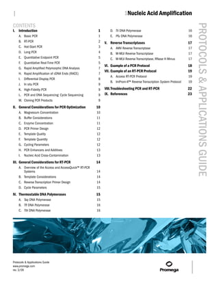
- 11. | 1Nucleic Acid Amplification PROTOCOLS & APPLICATIONS GUIDE Additional Resources for Cloning PCR Products Citations Technical Bulletins and Manuals Kurth, E.G. et al. (2008) Involvement of BmoR and BmoG TM042 in n-alkane metabolism in Pseudomonas butanovora. pGEM®-T and pGEM®-T Easy Vector Microbiology 154, 139–47. Systems Technical Manual (www.promega.com The authors characterized five open-reading frames /tbs/tm042/tm042.html) flanking the alcohol-inducible alkane monooxygenase (BMO) structural gene of Pseudomonas butanovora. Strains TM044 pTARGET™ Mammalian Expression Vector with mutated bmoR, encoding a putative transcriptional System Technical Manual regulator, or bmoG, encoding a putative chaperonin, were (www.promega.com created by gene inactivation. The bmoR gene was amplified /tbs/tm044/tm044.html) Promega Publications and cloned into the pGEM®-T Vector for disruption with a kanamycin cassette. The two termini of the bmoG gene PN082 Technically speaking: T-vector cloning were amplified separately, ligated to the kanamycin cassette (www.promega.com and cloned into the pGEM®-T Easy Vector. Plasmids /pnotes/82/10203_24/10203_24.html) encoding the disrupted genes were transformed into PN071 Rapid ligation for the pGEM®-T and Pseudomonas butanovora by electroporation. pGEM®-T Easy Vector Systems PubMed Number: 18174133 (www.promega.com /pnotes/71/7807_08/7807_08.html) Bröker, D. et al. (2008) The genomes of the non-clearing-zone-forming and natural-rubber-degrading PN071 Cloning blunt-end Pfu DNA species Gordonia polyisoprenivorans and Gordonia westfalica polymerase-generated PCR fragments into harbor genes expressing Lcp activity in Streptomyces strains. pGEM®-T Vector Systems Appl. Environ. Microbiol. 74, 2288–97. (www.promega.com /pnotes/71/7807_10/7807_10.html) Natural rubber-degrading bacteria fall into two categories: those forming clearing zones on latex overlay plates and PN068 Technically speaking: Optimized cloning those that do not. To investigate this degradation process, with T vectors the authors amplified latex-clearing protein (lcp) homologs (www.promega.com from non-clearing-zone-forming bacteria using degenerate /pnotes/68/7381_31/7381_31.html) PCR primers based on lcp sequences from clearing-zone PN060 Digestion of PCR and RT-PCR products forming species. The 3′ region of the lcp gene in G. westfalica with restriction endonucleases without was amplified by nested PCR using biotinylated primers, prior purification or precipitation and the amplified products were cloned in the pGEM®-T (www.promega.com Easy Vector and sequenced using universal M13 forward /pnotes/60/6079_23/promega.html) and reverse primers. More (www.promega.com PubMed Number: 18296529 publications /pnotes/apps/cloning/) Vector Maps pGEM®-T and pGEM®-T Easy Vectors (www.promega.com II. General Considerations for PCR Optimization /vectors/t_vectors.htm#b01) This discussion focuses on the use of Taq DNA polymerase pTARGET™ Mammalian Expression Vector in PCR, since this is the enzyme most commonly used in (www.promega.com/vectors/t_vectors.htm#b02) PCR. Many of these suggestions also apply when using other DNA polymerases. A. Magnesium Concentration Magnesium is a required cofactor for thermostable DNA polymerases, and magnesium concentration is a crucial factor that can affect amplification success. Template DNA concentration, chelating agents present in the sample (e.g., EDTA or citrate), dNTP concentration and the presence of proteins can all affect the amount of free magnesium in the reaction. In the absence of adequate free magnesium, Taq DNA polymerase is inactive (Figure 1.6). Excess free magnesium reduces enzyme fidelity (Eckert and Kunkel, 1990) and may increase the level of nonspecific amplification (Williams, 1989; Ellsworth et al. 1993). For these reasons, researchers should empirically determine the optimal magnesium concentration for each target. To do so, set up a series of reactions containing 1.0–4.0mM Protocols & Applications Guide www.promega.com 1-10 rev. 3/09
- 12. | 1Nucleic Acid Amplification PROTOCOLS & APPLICATIONS GUIDE Mg2+ in 0.5–1mM increments and visualize the results to B. Buffer Considerations determine which magnesium concentration produced the Most reaction buffers consist of a buffering agent, most highest yield of product and the minimal amount of often a Tris-based buffer, and salt, commonly KCl. The nonspecific product. The effect of magnesium concentration buffer regulates the pH of the reaction, which affects the and the optimal concentration range can vary with the DNA polymerase activity and fidelity. Modest particular DNA polymerase. For example, the performance concentrations of KCl will increase DNA polymerase of Pfu DNA polymerase seems to be less dependent on activity by 50–60% over activities in the absence of KCl; magnesium concentration, but when optimization is 50mM KCl is considered optimal (Gelfand, 1989). required, the optimal concentration is usually in the range of 2–6mM. GoTaq® DNA Polymerase contains native Taq DNA polymerase in a proprietary formulation. It is supplied with Many DNA polymerases are supplied with a 5X Green GoTaq® Reaction Buffer and 5X Colorless GoTaq® magnesium-free reaction buffer and a tube of 25mM MgCl2 Reaction Buffer. The 5X Green GoTaq® Reaction Buffer so that you can adjust the Mg2+ concentration to the level contains two dyes (blue and yellow) that separate during that is optimal for each reaction. Before assembling the electrophoresis to monitor migration progress. The buffer reactions, be sure to thaw the magnesium solution also contains a compound that increases the density of the completely prior to use and vortex the magnesium solution sample so that it will sink into the well of the agarose gel, for several seconds before pipetting. Magnesium chloride allowing reactions to be directly loaded onto an agarose solutions can form concentration gradients as a result of gel without the need for loading dye. The blue dye multiple freeze-thaw cycles, and vortex mixing is required comigrates at the same rate as a 3–5kb DNA fragment in a to obtain a uniform solution. These two steps, though 1% agarose gel. The yellow dye migrates at a rate faster seemingly simple, eliminate the cause of many failed than primers (<50bp) in a 1% agarose gel. The 5X Colorless experiments. GoTaq® Reaction Buffer and the 5X Green GoTaq® Reaction Some scientists prefer to use reaction buffers that already Buffer have the same formulation, except for the dyes. The contain MgCl2 at a final concentration of 1.5mM. It should 5X Colorless GoTaq® Reaction Buffer is recommended for be noted, however, that Hu et al. reported performance any applications where absorbance or fluorescence variability of reaction buffer solutions containing measurements of the PCR amplimer will be taken without magnesium (Hu et al. 1992). The free magnesium changes prior cleanup. Both buffers are supplied at pH 8.5 and of 0.6mM observed in their experiments dramatically contain MgCl2 at a concentration of 7.5mM for a final affected amplification yields in an allele-specific manner. concentration of 1.5mM. The authors found that heating the buffer at 90°C for 10 GoTaq® Flexi DNA Polymerase is supplied with 5X Green minutes restored the homogeneity of the solution. They GoTaq® Flexi Reaction Buffer and 5X Colorless GoTaq® postulated that magnesium chloride precipitates as a result Flexi Reaction Buffer. The compositions are identical to the of multiple freeze-thaw cycles. 5X Green GoTaq® Reaction Buffer and 5X Colorless GoTaq® M 1 2 3 4 5 6 7 8 Reaction Buffer, except that the GoTaq® Flexi reaction buffers do not contain MgCl2. Instead, the GoTaq® Flexi DNA Polymerase is supplied with a tube of 25mM MgCl2 2,645 – 1,605 – so that reactions can be supplemented with varying 1,198 – concentrations of magnesium. C. Enzyme Concentration We recommend using 1–1.25 units of Taq DNA polymerase in a 50μl amplification reaction. In most cases, this is an 1348TB01_6A excess of enzyme, and adding more enzyme will not significantly increase product yield. In fact, increased Mg2+ (mM) 0 0.5 1.0 1.5 2.0 2.5 3.0 3.5 amounts of enzyme increase the likelihood of generating Figure 1.6. Effects of magnesium concentration on amplification. artifacts associated with the intrinsic 5′→3′ exonuclease Amplifications were performed using various Mg2+ concentrations activity of Taq DNA polymerase, resulting in smeared bands to demonstrate the effect on the amplification of a 1.8kb target in an agarose gel (Longley et al. 1990; Bell and DeMarini, luciferase gene. The reaction products were analyzed by agarose 1991). gel electrophoresis followed by ethidium bromide staining. Lane M, Promega pGEM® DNA Markers (Cat.# G1741); lane 1, 0mM Pipetting errors are a frequent cause of excessive enzyme Mg2+; lane 2, 0.5mM Mg2+; lane 3, 1mM Mg2+; lane 4, 1.5mM Mg2+; levels. Accurate dispensing of small volumes of enzyme lane 5, 2mM Mg2+; lane 6, 2.5mM Mg2+; lane 7, 3mM Mg2+ and solutions in 50% glycerol is difficult, so we strongly lLane 8, 3.5mM Mg2+. recommend preparing a reaction master mix, which requires a larger volume of each reagent, to reduce pipetting errors. Protocols & Applications Guide www.promega.com 1-11 rev. 3/09