Top Rated Hyderabad Call Girls Erragadda ⟟ 9332606886 ⟟ Call Me For Genuine ...
Airwaysandappliances
1. Print this page
Airways and Appliances March 1989, Volume 82, Number 2
by Allen J. Moses, DDS The official publication of the Chicago
Dental Society
Editor's Note:
Human being are obligate nasal breathers. Should the nasal respiratory
mechanism malfunction, the human organism is capable of employing a
compensatory oral respiratory mechanism. This month, Allen J. Moses reviews
obstructive breathing conditions, both nasal and oral, which pose important
diagnostic criteria upon interceptive and corrective treatment judgments for the
dentist.
Moses describes an appliance termed a "passivator" which supports the
mandible in an anterior and slightly open bite yet neither allows nor causes
undesirable orthodontic tooth movements. This appliance is reported to support
the patient’s mandible during sleep, prevent collapse of the pharyngeal
musculature, allow greater volume of space for tongue activity and prevent
incorrect swallowing and snoring.
Moses feels that dentists must share a common interest with physicians of the
nose and throat while developing an expertise in measurement, diagnosis and
treatment of neuromuscular disorders which may affect the stomatognathic
system. By understanding the role of breathing problems in oral, facial and dental
development, dentists can play an equally important role in preventing many of
these problems.
Moses completed his BS degree in 1964 at the University of Illinois and in 1968
received his DDS degree from the University of Illinois College of Dentistry. He
has a special interest in temporomandibular joint problems and their effects on
the rest of the body. He has taught at the U of I College of Dentistry and lectured
at the National College of Chiropractic Medicine. He has published in various
journals including the CDS Review. Moses is a Fellow in the International
College of Craniomandibular Orthopedics. He currently maintains a private
practice of general dentistry in Arlington Heights, Illinois.
In a previous article, "Thumb Sucking or Thumb Propping?" (CDS Review,
December 1987), the differentiation was made that thumb suckers are nose
breathers and thumb proppers are mouth breathers. In proppers, the thumb is an
orthopedic appliance to keep the lips apart, mouth open and facilitate mouth
breathing in these nasally obstructed children.
The deleterious consequences of propping may be narrow V-shaped upper arch,
low tongue position, abnormal swallow, distal jaw position and forward head
2. posture. It is no coincidence that these signs characterize many TMJ patients.
Understanding the distinction between sucking and propping and timely referral
presents an ideal interceptive situation. The purpose here is to elaborate on the
subject of breathing and its effect on the types of neuromuscular problems
dentists commonly treat and what dentists can do to affect their patients’
breathing.
Nasal function/dysfunction
The nose is an incredible organ. In addition to its olfactory function, it is the
carburetor of the body. The nose provides for proper humidification, filtration and
warming of inspired air.
The nasal pulmonary reflex controls the volume of inspired air. Additionally the
nose has a role in immunity, voice resonance and certain hormonal responses.
Human beings are normally nasal breathers.1
There are numerous pathological conditions and abnormalities which can
obstruct nasal breathing the role of the backup breathing organ, but the air is not
properly filtered, humidified and warmed. This results in construction of bronchi
and depressed elasticity of the lungs. With less tidal volume there follows
decreased oxygenation and increased partial pressure of carbon dioxide in the
blood in mouth breathers.2
Some of the effects of impaired nasal respiration reported in the literature are:
Abnormal facial development, headaches, malocclusion, poor sleep, sore
throats, excessive daytime sleepiness, dry mouth, frequent infections, earaches
and sinus problems.3
At this point, one might think of the breathing possibilities as nose and/or mouth,
but one other possibility exists - no breathing. The condition known as apnea is
very serious. The causes and consequences will be discussed.
Animal experiments
Egil Harvold did some interesting experiments on rhesus monkeys to examine
the consequences of mouth breathing. The monkey, like humans and most
animals, is a nose breather. Harvold asked the question, "What changes would
occur if the primate were forced to utilize 100 percent oral respiration?"" To
achieve this, he obstructed the nasal passages with silastic button plugs
permanently sewn in place. No nasal inspiration was possible and only a small
amount of nasal expiration could occur.4
The experimental animals were kept in this condition for three years. The jaws
gradually lowered in rest positions and the tongue position also lowered to
establish an oral airway. The shape of the tongue changed and became long and
3. narrow. The total volume of the tongue, however, did not change. The palate
collapsed laterally and the canines went into crossbite. The size of the mandible
remained the same, but its habitual position relative to the skull changed.
Harvold clearly shows that oral respiration in monkeys alters neuromuscular
activity and causes changes in bone resulting in malocclusion.5
In another experiment, Harvold reduced the oral space by displacing the tongue
downward against the mandible. He did this by sewing objects in the posterior
palatal vault of the monkeys which simulated the effect of tonsils. These objects
did not interfere with the occlusion or mastication.
In time, changes in the shape and position of the tongue were noted as the
tongue tried to avoid the sensory interference. Indentations formed in the back of
the tongue which were not sores. Sensory input altered the shape of the tongue
and the following skeletal adaptation occurred: Change in mandibular positions
and shape, extrusion of teeth, open bites, increased face height, opening of the
gonial angle and steep mandibular plane angle.6
Harvold further points out that there are two types of movement involved –
rhythmic, such as respiration and tonic, which involves changes in posture.
Changes in rhythmic movement cause few skeletal variations but tonic changes
cause significant alterations in skeletal morphology.7 The muscles with which the
dental clinician is primarily concerned, are those involved in tonic changes, such
as the diagastrics, pterygoids, masseters and temporalis.
Harvold’s controlled experiments confirm and clarify many of our clinical
observations. Sten Linder-Aronsen’s study of human children establishes
convincing statistical correlation that conditions which can obstruct nasal air flow
and cause oral respiration may cause deviations in craniofacial growth,
particularly increased face height.8
Human breathing problems
Several of the more common causes of nasal obstructive breathing in human
beings are: Allergy, small external nares, enlarged tonsils and adenoids,
vasomotor rhinitis, deviated septum, trauma, enlarged turbinates, congenital
abnormalities and nasal polyps.9
As a referring doctor, one has to decide between allergist, ENT, pediatrician or
sleep lab. Referrals should always be for an evaluation by the specialist of
choice, accompanied by a note of one’s findings. Thomas Weimert, professor of
Otorhinolaryngology at the University of Michigan, found in a five-year study of
1,360 patients referred to him by orthodontists for suspicion of airway
compromise that 28 percent had no evidence of significant obstruction.10
4. Dentists are important as both referrers and treating doctors. As treating doctors,
early is best. At age 2-3, when one sees the finger habit, the allergic shiners, the
large tonsils, the mouth breathing, etc., that is the time to begin. At age 4, 60
percent of the facial growth is complete.11 At age 7, 70 percent of facial growth is
complete. At age 12, 90 percent of craniofacial growth is complete.
Both structural and functional normalizations must be achieved to get ideal
growth patterns. This is not always possible if the growth potential is lost.
Children do not begin to complain of nasal obstructive problems until
approximately age 16.12 It seems children have no frame of reference from which
to complain as this is the only way they know how to live. At about age 16, they
start of realize that this kind of condition is not normal.
Allergy is by far the most common and important cause of nasal obstruction. The
clinician signs of allergy which one may note are nasal salute, nasal crease
(turned up nose), allergic shiners, upper lip incompetence, marginal eyelid
eczema, nonfebrile pharyngitis (from mouth breathing), lymphoid nodules in the
throat and blue-gray color to nasal mucosa.13
Allergy must be screened out first. If one parent or sibling is allergic, the child has
a 40 percent chance of being allergic. If two parents are allergic, the child has a
70 percent chance of being allergic.14 Allergy is an example of a normal
protective mechanism gone awry – an exaggerated response to proteins and
large polypeptide molecules which ordinarily do not warrant such an extreme
response. The conventional treatment of allergies includes antihistamines,
desensitization, avoidance and prevention.
Probably the most important thing a concerned dentist can do to help prevent
allergy is to counsel and educate mothers on the importance of breast-feeding.16
If mothers must bottle-feed, the dental profession can begin the education
process by recommending use of soy formulas which are not as allergenic.
Human infants do not produce immunoglobulins of their own for the first three
months of life. Transplacental transfer from mother and breast milk are the two
sources. Children not breast fed do not get this immunity and, as such, are more
susceptible to childhood illnesses. According to a six-year study be Glaser, bottle
fed children have four times as many respiratory conditions as breast fed
children.17
Measurement and diagnosis
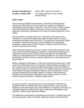
Dentists have some very important tools to aid in the diagnosis of nasal breathing
problems. The top two photographs (page at right) are of panoramic radiographs.
The patient in Figure 1 has a patent nasal airway and the one in Figure 2 has an
obstructed nasal area. The bottom two photos, Figure 3 and Figure 4, are
5. cephalometric radiographs in which the presence of enlarged adenoids can be
noted in the nasopharynx. (Radiographs courtesy of Dr. Dan Schwarb, Mt.
Clemens, Michigan)
It is enlightening and gratifying to deal with ENTs in today’s technological
environment. Dentistry prides itself on the refinement of diagnostic skills by the
newer electronic means, such as the electromyograph and computerized
mandibular scanning. Otorhinolaryngologists have equally sophisticated
measuring tools available to them, such as rhinomanometry and
polysomnography to determine the validity of their treatment and provide
documentation.
Rhinomanometry is a reproducible means of measuring nasal airway resistance.
The University of North Carolina has developed SNORT, an acronym which
stands for Simultaneous Nasal Oral Respirometric Technique. Using a custom
fitted face mask with separate valves attached to the nose and mouth and
attached to a flow meter, air pressure transducer, recorder and computer, it can
give the nasal versus the oral inspiration, expiration and their ratios.18
Jerold Principato has established age-related norms of nasal resistance using an
anterior rhinomanometric technique. Normal nasal resistance for an adult is 2.5
to 3.5 cm of water per liter per second. At readings above 7.0 at rest, an
individual must go to mouth breathing to get adequate air. Between 4.0 and 7.0,
orthodontic intervention is the treatment of choice. Above 7.0, surgery is usually
considered.19
After allergy, the second most common cause of nasal obstruction is adenoid
hypertrophy.20 Swollen adenoids block the distal part of the nasal airway, thus
decreasing nasal patency by increasing the resistance to airflow. Mouth
breathing is the result, often accompanied by a lower tongue position, extended
head position, lower mandibular posture and narrowed upper arch. This used to
be called adenoid facies but the more popular term today is long face syndrome
(LFS).
Accompanying hypertrophied adenoids often are enlarged lateral pharyngeal
tonsils. These tissue enlargements situated in the airway also alter air flow
resistance. Enlarged turbinates is another common cause of nasal airflow
obstruction. Chronic rhinitis is the result of thickening of the tissues lining the
inferior turbinates, particularly at the posterior end. The increased mucus
production causes increased pharyngeal symptoms and the need to constantly
clear the throat, better known as postnasal drip.21 Children who have
hypertrophied adenoids, tonsils and inferior turbinates develop long face
syndrome 30 percent of the time. In contrast, children with normal respiratory
airways develop long face syndrome 2 percent of the time.22
6. Harry Richter, professor of otorhinolaryngology at University of Michigan, feels
that the safety of nasal surgery has been well established, and that a clear nasal
airway is a prerequisite for developing normal facial form. He writes, that in
Current Therapy in Otolaryngology and Head and Neck Surgery, the authors
reviewing indications for nasal surgery only allude to a relationship between
adenoid hypertrophy and dentofacial development, but they do not yet feel that it
is a proper indication for surgery. Quality of life considerations allow for surgical
treatment of these patients on a selected basis.23
It is particularly the quality of life criteria that is making the ENTs and dentists
increasingly aware of the existence and importance of sleep apnea.
Apnea literally means want of breath. It can be of central nervous system origin
or obstructive. Obstructive sleep apnea is a collapse of the upper airway causing
total obstruction of breathing. Episodes usually over 10 seconds long with over
30 per night and over seven per hour cause chronic oxygen deprivation resulting
in symptoms such as morning headaches, intellectual deterioration, irritability,
impotence, hypertension, cerebral edema and cardiac arrhythmias.24
Snoring is obstructive sleep breathing. Not all snorers have apnea, but 90
percent of apnea patients snore. Human beings are the only animals to sleep in a
ventral positions. The sound of snoring originates in the collapsible part of the
airway from the epiglottis to the carinium. This area has no rigid support – only
the muscles keep it open. During sleep, human get some muscle flaccidity and in
combination with nasal obstructions, such as hypertrophied adenoids and tonsils,
the pharyngeal space can be occluded. The combination of long soft palate and
uvula can also occlude the airway by collapsing when the subject lies in the
ventral position.25
Polysomnography can help diagnose and document this condition so that it can
be appropriately treated. This is noninvasive testing done while the patient is
asleep. It is usually done in a sleep lab of a hospital. Patients are measured for
nasal air flow, arterial flow, heart, lungs, blood oxygen saturation, air flow
turbulence and brain wave patterns during a 5-6 hour sleeping period.
Treatment
Depending on the objective findings, proper steps can be taken ranging from
uvulopalatopharyngeoplasty (UPPP) to tonsillectomy and adenoidectomy to
turbinectomy or medication such are antihistamine sprays, proper sleep posture
or dental orthopedic appliances.
Dentists are an integral part of the health care team. As referrers, one has to
know when and whom to refer. Allergies generally must be treated first, before
consideration of any surgery. The greatest number of surgical failures are in
7. allergic patients.26 The greatest number of orthodontic relapses are in patients
who have not had their breathing problems successfully treated.27
What can a dentist do to treat? Normalization of face form following
adenoidectomy in a child can take five years. Donald Timms, in a study of 26
patients treated with Rapid Maxillary Expansion, reported a 37 percent mean
drop in nasal resistance with 7 mm of expansion. In another larger study of
children with a previous history respiratory disease treated with Rapid Maxillary
Expansion, 82 percent reported improvement in number of upper respiratory tract
infections and 60 percent reported improvement of allergic rhinitis.28
Functional orthopedic appliances can be designed which support patients
vertically during sleep and prevent collapse of pharyngeal musculature. It may be
that dentists can do as much to clear the airway for the snorer/apnea patient by
fabricating the appropriate appliance, as the physician can do with surgery.
Any appliance which is placed in the mouth has an effect on the volume of the
tongue box. This is a term coined by Dr. Henri Petit. The tongue box has the
pharynx as the posterior wall. The lateral and anterior walls are the teeth. The
palate is the superior wall and the inferior wall is the floor of the mouth. Petit
emphasizes that normal oral function cannot be achieved with inadequate tongue
space. It is important that intraoral appliances do not reduce the volume of space
for the tongue or they will set up the deleterious neuromuscular effect of pushing
the tongue back into the nasopharynx and adversely affect nose breathing.29
If an obstructive nasal breathing problem exists, dentists must direct themselves
toward constructing an appliance which does not compromise tongue space.
Appliances which increase tongue space may, in certain cases, be helpful in
alleviating nasal obstructive breathing problems. The mandibular orthotic which
corrects for deficient vertical dimension and brings the jaw anteriorly may also
have a favorable effect on the tongue box.
This may account for why maxillary orthotics, which compromise the swallow and
decrease tongue space by their palatal bulk, sometimes do not achieve success
when mandibular appliances do.
Activity levels are at their highest or close to it bilaterally for temporalis,
masseter, medial pterygoid and superior head of the lateral pterygoid. In an edge
to edge bite there are much lower EMG levels in these muscles and almost a cut-
off of activity in temporalis.30
By supporting the patients both vertically and anteroposteriorly during sleep and
allowing adequate room for the tongue, it has the ability of preventing collapse of
pharyngeal musculature. And because it supports the teeth in the exact
relationship that they are to each other at minimal EMG activity, it allows no
undesirable orthodontic movement to occur.
8. Passivator
• Prevents "orthodontic" movement
• Does not interfere with correct swallow
• Prevents incorrect swallow
• Allows adequate tongue space
• Helps prevent collapse of pharyngeal musculature
• May improve nose breathing
• May improve snoring
References
1. Proetz A. Physiology of the Nose, Ed. 2, St. Louis, Annals Pub. Co., pp 119-169, 153.
2. Rubin RM. Mode of respiration and facial growth. Am J Orthod, 1980, 78:504-510.
3. Timms DJ. Lecture. RME for medical reasons. Upper Airway Compromise Dentrofacial
Development Symposium III, Virginia Beach, VA. 1986.
4. Harvold EP, Chierici EG, Vergervik K. Experiments on the development of dental
malocclusions. Am J Orthodont. 1972. 61:38-4.
5. Harvold EP. Lecture. Experiments on bone remodeling. Upper Airway Compromise Dentofacial
Development Symposium III, Virginia Beach, VA. 1986.
6. Harvold EP, Vergervik K, Chierici G. Primate experiments on oral sensation and dental
malocclusion. Am J Orthodont. 1973. 63:494-508.
7. Vargevik H, Harvold EP. Experiments on interaction between orofacial function and
morphology. Monograph of Upper Airway Compromise Dentrofacial Development Symposium III,
Virginia Beach, VA. 1986.
8. Linder-Aronsen, Sten Lecture. Vertical dysplasia update. Amer. Assoc. Orthodontists Annual
Meeting, 1985.
9. Rubin RM. Lecture. Allergy, adenoids and airway: early recognition for optimal facial growth.
Amer. Assoc. Orthod. Annual Meeting. 1982.
10. Weimert T. Airway obstruction in orthodontic practice. Jour. Clin. Orthod.1986. 20:96-104.
11. Nelson WE, Vaughan VC and McKay RJ. Textbook of Pediatrics. WB
Saunders, pp. 23-42, 1969.
9. 12. Principato J. Lecture. Nasal airway assessment and management in the orthodontic patient.
Amer. Assoc. Orthodontists Annual Meeting, 1982.
13. Rubin, Lecture. Controversies regarding etiology of the Long Face Syndrome. Upper Airway
Compromise Dentrofacial Development Symposium III, Virginia Beach, VA. 1986.
14. Ibid.
16. Rubin, Lecture. Controversies regarding etiology of the Long Face Syndrome. Upper Airway
Compromise Dentrofacial Development Symposium III, Virginia Beach, VA. 1986.
17. Glaser J. The prophylaxis of allergic disease in infancy. Pediatrics. 1962. 29:835-841.
18. Vig P. Lecture. Respiration: a critique of conventional wisdom and an approach towards a
rational orthodontic perspective. Amer. Assc Orthodontists Annual Meeting, 1982.
19. Principato J. Lecture. Nasal airway assessment and management in the orthodontic patient.
Amer. Assoc. Orthodontists Annual Meeting, 1982.
20. Ibid
21. Ibid.
22. Shapiro GC and Shapiro PA. Nasal airway obstruction and facial development. Clin. Rev.
Allergy, 1984. 2:225-235.
23. Gates GA, Current therapy in otolaryngology: Head and Neck Surgery. C.V. Mosby, 1982-
1983.
24. Fairbanks DNF, Lecture. Snoring and obstructive sleep apnea and children. Upper Airway
Compromise Dentrofacial Development Symposium III, Virginia Beach, VA. 1986.
26. Rubin. Lecture. Controversies regarding etiology of the Long Face Syndrome. Upper Airway
Compromise Dentrofacial Development Symposium III, Virginia Beach, VA. 1986.
25. Ibid.
27. Principato J. Lecture. Nasal airway assessment and management in the orthodontic patient.
Amer. Assoc. Orthodontists Annual Meeting, 1982.
28. Timmons DJ, Rapid Maxillary Expansion. Chicago Quintessence Pub Co., pp. 41-45, 1981.
29. Petit, Henri Monograph, Upper airway problems and pre-orthodontic
30. Miller A, Lecture. Muscle Recruitment and the Chewing System. American Academy of
Craniomandibular Disorders Annual Meeting, March 1987.