
Recommended
More Related Content
What's hot
What's hot (16)
Similar to Thyroid gland anatomy part1
Similar to Thyroid gland anatomy part1 (20)
Recently uploaded
Recently uploaded (20)
Thyroid gland anatomy part1
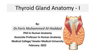
- 1. Thyroid Gland Anatomy - I By: Dr.Faris Muhammed Al-Haddad PhD In Human Anatomy Associate Professor In Human Anatomy Medical College/ Hawler Medical University February. 2022
- 2. Definition A bi-lobed endocrine gland located at the midline of the neck, it secrets hormones that regulate metabolism, growth & development T H Y R O I D
- 5. Location and Description of The thyroid gland: The Rt and Lt lobes are connected by an isthmus: 1. Each lobe • Pear shaped, • Its apex: being directed upward to oblique line of the thyroid cartilage; • Its base: lies below at the level of 4th or 5th tracheal ring.
- 6. 2. Isthmus • Connects between Rt & Lt lobes • Extends across the midline in front of the: 2nd 3rd 4th tracheal rings Location and Description of The thyroid gland Cont.
- 7. Location and Description of The thyroid gland 3. Pyramidal lobe • is often present, • it projects upward from the isthmus, usually to the left of the midline • Its connected to the hyoid bone by either: • Fibrous band • Muscular band, it is referred to as the levator glandulae thyroideae Pyramidal lobe levator glandulae thyroideae Cont.
- 8. surrounded by a sheath derived from the: The sheath attaches the gland to the larynx & the trachea. Fasciae Pretracheal layer of deep fascia.
- 9. Capsules of Thyroid Gland 1. True capsule: is its own connective tissue 2. False capsule: • from the pretracheal layer of the deep cervical fascia • It is thin along the posterior border of the lobes • It is thick on the inner surface of the gland where it forms a suspensory ligament (of Berry), which connects the lobe to the cricoid cartilage
- 11. End of Part 1