Best Rate (Hyderabad) Call Girls Jahanuma ⟟ 8250192130 ⟟ High Class Call Girl...
Caso ciego 8 5-12
1. CASO CIEGO
PRESENTA: ELISABET LOPEZ GONZALEZ
PRESENTATION OF CASE
A 35-year-old woman was admitted to the hospital because of recurrent melena after treatment for a
gastric ulcer with positive tests for Helicobacter pylori.
The patient had been well until 21 months earlier, when abdominal pain developed. Two months later
melena appeared, and she was admitted to another hospital, where endoscopic examination showed
a bleeding gastric ulcer. Four transfusions of packed red cells were given, and she was treated with
several histamine H2-receptor antagonists. At the time of discharge she was receiving ranitidine
therapy. The following month a repeated endoscopic examination revealed a healed gastric ulcer and
the presence of H. pylori. During the next 17 months she received three or four courses of
medications intended to eradicate the microorganism, including bismuth, tetracycline, metronidazole,
and amoxicillin in various combinations, with H2-receptor blockers or omeprazole. Each course
resulted in the disappearance of the abdominal pain, followed by its eventual return after the
withdrawal of all medications. The most recent course of treatment ended 25 days before admission,
and the patient was asymptomatic until two days before admission, when she felt warm, with
chilliness and weakness. On the day of admission melena recurred, and she came to this hospital.
The patient was married and employed in a stressful administrative position. She was of Greek
ancestry and had thalassemia minor. There was no history of hematemesis, hematochezia, diarrhea,
constipation, sweats, weight loss, or use of tobacco, alcohol, or nonsteroidal antiinflammatory drugs.
The temperature was 36.9°C, the pulse was 90, and the respirations were 18. The blood pressure
was 150/80 mm Hg, without orthostatic change.
On physical examination the patient was pale and mildly overweight. No lymphadenopathy was
found. The lungs, heart, and breasts were normal. Abdominal examination disclosed active bowel
sounds; the liver and spleen were not felt, and no tenderness or mass was detected. The extremities
were normal. Rectal examination was negative except for a tarry stool that was ++++ positive for
occult blood.
The hematologic laboratory findings are presented in Table 1. The conjugated bilirubin level was 0.3
mg per deciliter (5.1 μmol per liter), and the total bilirubin level was 1.1 mg per deciliter (19 μmol per
liter); the serum aspartate aminotransferase level was 46 U per liter. The values for urea nitrogen,
creatinine, glucose, calcium, phosphorus, protein, albumin, globulin, electrolytes, magnesium, lactate
dehydrogenase, amylase, and alkaline phosphatase were normal.
Oral intake was withheld. The patient was given cimetidine, fluid, and electrolytes intravenously.
Examination with a fiberoptic endoscope showed a normal esophagus. Fresh blood was present in
the stomach, with a large ulcer on the posterosuperior wall in the distal part of the body of the
stomach, covered by a large clot; epinephrine was injected around the base of the ulcer. The
duodenum was normal. No active bleeding was observed at the end of the procedure. Soon after the
examination the patient vomited 200 ml of material resembling coffee grounds. She continued to
pass dark stools that were positive for occult blood, and there was evidence of mild orthostatic
hypotension on several occasions. The hematocrit on the second hospital day was 24.6 percent. Two
2. units of packed red cells were transfused; the hematocrit rose as high as 27.5 percent and stabilized
between 26 and 26.5 percent. The temperature rose on most days to 37.3 to 37.8°C.
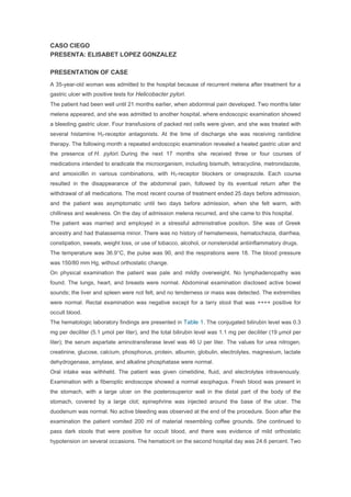
On the sixth hospital day a repeated esophagogastroduodenoscopic examination (Figure 1 and
figure 2) again revealed a normal esophagus and duodenum A large, slightly irregular gastric
ulcer was seen, with a deep crater that contained exudate; a red spot was observed on its distal
margin. The edge of the ulcer was slightly elevated above the surrounding mucosa. A white
submucosal plaque, 10 mm in diameter, was noted on the gastric wall opposite the ulcer.
A diagnostic procedure was performed.
3. Figure 1. Endoscopic View of the Ulcer in the Distal Body of the Stomach, with the Patient in
a Retroflexed Position.
The ulcer has an irregular shape, and its edge is slightly nodular.
Figure 2. White Submucosal Plaque, 10 mm in Diameter, on the Gastric Wall Opposite the
Ulcer.