SlideShare a Scribd company logo
1 of 11
Download to read offline
Human Molecular Genetics, 2004, Vol. 13, No. 10 1069–1079 
DOI: 10.1093/hmg/ddh115 
Advance Access published on March 25, 2004 
Evidence that unrestricted legumain activity is 
involved in disturbed epidermal cornification in 
cystatin M/E deficient mice 
Patrick L.J.M. Zeeuwen1,*, Ivonne M.J.J. van Vlijmen-Willems1, Diana Olthuis1, 
Harald T. Johansen2, Kiyotaka Hitomi3, Ikuko Hara-Nishimura4, James C. Powers5, 
Karen E. James5, Huub J. op den Camp6, Rob Lemmens1 and Joost Schalkwijk1 
1Department of Dermatology, Nijmegen Center for Molecular Life Sciences, University Medical Center Nijmegen, 
PO Box 9101, 6500 HB Nijmegen, The Netherlands, 2Department of Pharmacology, School of Pharmacy, Oslo, 
Norway, 3Department of Applied Molecular Bioscience, Graduate School of Bioagricultural Sciences, 
Nagoya University, Nagoya, Japan, 4Department of Botany, Graduate School of Science, Kyoto University, 
Kyoto, Japan, 5School of Chemistry and Biochemistry, Georgia Institute of Technology, Atlanta, Georgia, 
USA and 6Department of Microbiology, Faculty of Science, University of Nijmegen, The Netherlands 
Received January 28, 2004; Revised and Accepted March 11, 2004 
Homozygosity for Cst6 null alleles causes the phenotype of the ichq mouse, which is a model for human 
harlequin ichthyosis (OMIM 242500), a genetically heterogeneous group of keratinization disorders. Here we 
report evidence for the mechanism by which deficiency of the cysteine protease inhibitor cystatin M/E (the Cst6 
gene product) leads to disturbed cornification, impaired barrier function and dehydration. Absence of cystatin 
M/E causes unrestricted activity of its target protease legumain in hair follicles and epidermis, which is the 
exact location where cystatin M/E is normally expressed. Analysis of stratum corneum proteins revealed a 
strong decrease of soluble loricrin monomers in skin extracts of ichq mice, although normal levels of loricrin 
were present in the stratum granulosum and stratum corneum of ichq mice, as shown by immunohisto-chemistry. 
This suggested a premature or enhanced crosslinking of loricrin monomers in ichq mice by 
transglutaminase 3 (TGase 3). In these mice, we indeed found strongly increased levels of TGase 3 that was 
processed into its activated 30 and 47 kDa subunits, compared to wild-type mice. This study shows that cystatin 
M/E and legumain form a functional dyad in epidermis in vivo. Disturbance of this protease–antiprotease 
balance causes increased enzyme activity of TGase 3 that could explain the observed abnormal cornification. 
INTRODUCTION 
Over the past decade, significant progress has been made in our 
understanding of the molecular basis of disorders of epidermal 
differentiation (1– 4). A variety of genes have been identified 
that underlie inherited forms of ichthyosis, keratoderma and 
skin fragility. The encoded proteins have diverse cellular 
functions, ranging from structural properties (loricrin, keratins), 
cell–cell adhesion (desmoplakin, desmoglein-1, plakophilin, 
plakoglobin), lipid metabolism and/or trafficking (steroid 
sulfatase, fatty aldehyde dehydrogenase, lipoxygenases, 
ABCA12), regulation of post-translational protein proces-sing 
(transglutaminase 1, cathepsin C, LEKT1), intercellular 
communication (connexins) and calcium signaling (ATP2C1, 
ATP2A2). Despite these advances, a number of genodermatoses 
characterized by disturbed cornification remain unexplained at 
the molecular level. These include, for example, forms of 
lamellar ichthyosis that are not caused by known mutations, 
ichthyosis vulgaris (OMIM 146700) and harlequin ichthyosis 
(OMIM 242500). Harlequin ichthyosis (HI) is a severe 
congenital skin disorder usually leading to a stillborn fetus or 
early neonatal death. Its clinical features at birth include 
ectropion, eclabium, ear dysmorphology and a thickened 
fissured epidermis (5). We have recently demonstrated that 
the phenotype of the ichq mouse, a model for human HI, is 
caused by a null mutation in the Cst6 gene leading to 
deficiency for the epidermal cysteine protease inhibitor cystatin 
M/E (6). The ichq mice phenotype has morphological and 
*To whom correspondence should be addressed. Tel: þ31 243617245; Fax: þ31 243541184; Email: p.zeeuwen@derma.umcn.nl 
Human Molecular Genetics, Vol. 13, No. 10 # Oxford University Press 2004; all rights reserved 
Downloaded from http://hmg.oxfordjournals.org/ by guest on December 3, 2011
biochemical similarities to human HI, which includes excessive 
epidermal and follicular hyperkeratosis, abnormal large 
mitochondria, and a characteristic keratin expression pattern 
in the interfollicular epidermis. Although we have so far 
excluded CST6 mutations in humans as a major cause of 
harlequin ichthyosis type 2, CST6 and genes that have a role in 
biological pathways that are controlled by cystatin M/E, should 
be considered as candidate genes for human disorders of 
cornification of unknown etiology (7). The function of cystatin 
M/E in normal epidermal homeostasis is unknown and the 
mechanism that leads to the observed pathology in ichq mice 
remains unresolved. Clearly, the identification of its target 
protease(s) in vivo would be an essential step in answering 
these questions. 
A biochemical study on the novel asparaginyl endopeptidase 
legumain has indicated that cystatin M/E in vitro binds to this 
protease with high affinity and could possibly represent a 
physiological target enzyme, although no biological evidence 
has been provided so far (8). Legumain or asparaginyl 
endopeptidase (AEP, EC 3.4.22.34) belongs to clan CD of 
the cysteine proteases and has a strict specificity for hydrolysis 
of asparaginyl bonds. Initially, the enzyme was purified as a 
cysteine protease responsible for the maturation of seed storage 
proteins and designated vacuolar processing enzyme (VPE) (9). 
Subsequently, legumain has been described in mammals (10); it 
is found at low levels in many tissues, but is particularly 
abundant in kidney. Very recently, the generation of legumain 
deficient mice has been reported (11). Limited functional 
studies have shown disturbed biosynthetic processing of 
cathepsins and accumulation of macromolecules in the 
lysosomes in the proximal tubule cells of the kidney. 
In the present study, we have investigated the mechanism by 
which absence of cystatin M/E causes epidermal abnormalities 
in ichq mice. We report that legumain is a physiologically 
relevant target protease of cystatin M/E in vivo. Cystatin M/E 
deficiency in ichq mice leads to free cutaneous legumain 
activity at exactly the location where cystatin M/E is normally 
present in wild-type mice. These mice show increased 
transepidermal water loss and neonatal death, probably due to 
dehydration. We provide evidence that disturbed cornification 
is caused by abnormalities in loricrin processing, caused by 
uncontrolled legumain activity. 
RESULTS 
Skin barrier function is severely disturbed in cystatin 
M/E deficient mice 
Cystatin M/E deficient mice have defects in epidermal 
cornification and die between 5 and 12 days of age (6,12). 
Autopsy on neonatal ichq mice strongly suggested that these 
mice died of dehydration. This observation and the gross 
abnormalities in the stratum corneum prompted us to measure 
the rate of transepidermal water loss (TEWL) of ichq mice, 
compared to phenotypically normal littermates. As shown in 
Figure 1 (left hand y-axis), we found increased TEWL levels (up 
to 3-fold) from day 9 onwards. Figure 1 (right hand y-axis) 
also shows progressive weight loss in the cystatin M/E deficient 
mice starting at the time point that transepidermalwater loss was 
apparent. At day 11, body weights were 50% compared to 
normal littermates and most mice died shortly thereafter. 
Legumain is expressed in epidermis and hair follicles 
The antiprotease activity of cystatin M/E has been investigated 
previously (8,13–15). Cystatin M/E inhibits the asparaginyl 
endoprotease legumain with a high affinity (Fig. 2A). In 
addition to cathepsin B tested by others, we examined the 
inhibition of cathepsins C, H, L and S. None of these proteases 
was appreciably inhibited by cystatin M/E (Fig. 2B). These 
data indicate that legumain is a likely physiological target of 
cystatin M/E in skin in vivo, although nothing is known so far 
on the presence of legumain in cutaneous tissues. Using anti-legumain 
antibodies for immunohistochemical analysis of 
mouse tissues we found legumain expression in the entire 
epidermis of adult, wild-type mice (Fig. 3A) and in the inner 
root sheet of the hair follicle (Fig. 3B), most abundantly at the 
junction of dermis and subcutaneous fat (Fig. 3C). In addition 
to the known presence in kidney, legumain expression was 
further detected in the ciliated epithelium of the trachea and in 
bronchial epithelium (Fig. 3D and E). Immunohistochemical 
staining of dorsal skin in cystatin M/E deficient ichq mice 
revealed normal epidermal legumain expression (Fig. 3F). 
These results demonstrate that legumain is indeed expressed in 
skin and hair follicles, as well as in other mouse tissues, some 
of which were previously shown to express cystatin M/E (6). 
These findings were confirmed at the mRNA level in mouse 
and human skin by reverse transcriptase PCR analysis, and 
subsequent sequencing of the PCR product (data not shown). 
Free in situ legumain activity in skin of cystatin M/E 
deficient mice but not in wild-type mice 
Immunohistochemical localization of legumain cannot be 
equated with legumain activity because legumain can be 
present in an inactive zymogen form or complexed with 
endogenous inhibitors. In order to demonstrate legumain enzy-matic 
activity in tissue sections, we developed a cytochemical 
assay, based on hydrolysis of a specific fluorescent substrate 
that could be precipitated by nitrosalicylaldehyde in situ. As 
it was known from literature that kidney is a rich source of 
free legumain (as measured by biochemical assays) we used 
this tissue as a positive control. Enzymatic legumain activity 
is detected in situ as a green fluorescent precipitate in the 
proximal tubulus epithelial cells of pig kidney (Fig. 4A). 
Specificity of this reaction was confirmed using Cbz-Ala-Ala- 
Aasn-EPCOOEt, which completely blocks proteolysis of the 
substrate (Fig. 4B). This synthetic inhibitor is highly specific 
for legumain (16) and shows similar affinity and specificity as 
the natural inhibitor cystatin M/E. For comparison, immuno-histochemical 
staining of pig kidney for legumain shows the 
presence of legumain in epithelial cells of the proximal 
tubuli, whereas staining of the glomeruli was weak to absent 
(Fig. 4C); this is in accordance with the immunohistochemical 
localization of legumain in rat kidney reported by others (17), 
and it shows colocalization with enzymatically active legumain 
as demonstrated in Figure 4A. We subsequently used the 
cytochemical legumain assay to examine free legumain activity 
in the skin of wild-type and cystatin M/E deficient mice. 
1070 Human Molecular Genetics, 2004, Vol. 13, No. 10 
Downloaded from http://hmg.oxfordjournals.org/ by guest on December 3, 2011
Human Molecular Genetics, 2004, Vol. 13, No. 10 1071 
Figure 1. Disturbed barrier function in cystatin M/E deficient mice. TEWL of the ichq mutant mice is increased compared to phenotypically normal mice. 
Concomitantly with increased TEWL, body weights are decreasing until these mice die around day 11. Left hand y-axis: TEWL in g/m2/h. Right hand y-axis: 
decrease in body weight as a percentage of the control mice. 
Figure 4D visualizes free legumain activity in the stratum 
granulosum and in the hair follicles at the level of the infundi-bulum 
in the cystatin M/E deficient mice. No legumain activity 
could be demonstrated in the skin of wild type littermates 
(Fig. 4E), which indicates that the presence of cystatin M/E 
normally regulates legumain activity in skin in vivo. For 
comparison, immunohistochemical cystatin M/E staining of 
dorsal skin is shown in wild-type and cystatin M/E deficient 
mice. This confirms the absence of cystatin M/E expression at 
the protein level in the ichq mice (Fig. 4F), whereas the wild-type 
skin shows cystatin M/E expression in the infundibular 
epithelium of the hair follicle and the stratum granulosum of 
the interfollicular epidermis (Fig. 4G). The localization of free 
legumain activity in the infundibular part of the hair follicle 
in cystatin M/E deficient mice coincides with the reported 
localization of tissue pathology in these mice such as excessive 
cornification (6,12), which leads to plugging of the hair follicle 
and ichthyosis as shown before. 
Absence of loricrin monomers in cystatin M/E 
deficient mice 
Abnormal cornification in ichq mice could be the result of 
abnormal expression or processing of structural components of 
the cornified envelope. In previous studies, no abnormalities 
were found in structural proteins such as cytokeratins 1 and 14, 
involucrin and filaggrin (18). We now analyzed extracts of ichq 
and wild-type mice for the presence of loricrin and involucrin, 
two major components of the stratum corneum. We could 
confirm the reported normal presence of involucrin in ichq skin 
(data not shown), but remarkably, loricrin monomers and 
dimers were nearly absent in cystatin M/E deficient mice as 
revealed by western blot analysis (Fig. 5B). We took care to 
prepare the skin extracts in buffer with and without the specific 
synthetic legumain inhibitor Cbz-Ala-Ala-Aasn-EP-COOEt to 
preclude the action of excess free legumain in the cystatin M/E 
deficient skin during extraction. No low molecular weight 
degradation products of loricrin were detected in lanes 1 and 2 
of Figure 5B, nor did we observe high molecular weight 
loricrin complexes, although these probably cannot be 
extracted, due to their poor solubility. Immunohistochemical 
analysis, however, showed that loricrin was abundantly present 
in the upper layers of the interfollicular epidermis of dorsal skin 
of cystatin M/E deficient mice (Fig. 5C), arguing against 
loricrin degradation as a mechanism to explain the findings on 
western blot. Moreover, in the epidermis of the mutant mice, 
three of five cell layers were found positive for loricrin expres-sion, 
whereas in only one of three cell layers, positive staining 
was detected in the epidermis of wild-type mice (Fig. 5D). 
In Figure 5A, a Coomassie blue stained blot is shown to check 
for equal loading of the samples in Figure 5B. No gross 
differences were seen in most of the high molecular weight 
bands, which represent the major soluble structural proteins 
of mouse skin. Interestingly, two abundant protein bands of 
13 and 15kDa were observed in the extract of cystatin M/E 
deficient mice (boxed in Fig. 5A), which were less pronounced 
in wild-type skin. In order to exclude the possibility that these 
bands represented accumulated loricrin breakdown products 
not detected by the anti-mouse loricrin serum (directed against 
a single epitope), we subjected these bands to in-gel tryptic 
digestion followed by matrix-assisted laser desorption/ionization 
time-of-flight (MALDI-TOF) mass spectrometry. The lower 
13kDa band was identified as S100 calcium binding protein A9 
(or calgranulin B). This is a protein known to be induced in 
epidermis during inflammation (19), and it can also act as a 
modulator of intracellular calcium homeostasis during follicular 
differentiation (20). The upper 15kDa band was identified as 
epidermal fatty acid binding protein (E-FABP). Interestingly, 
E-FABP has been described as an epidermal protein that is 
strongly upregulated following skin barrier disruption (21), which 
is in accordance with the observed increase in transepidermal 
water loss described above. These findings also demonstrate 
that these low molecular weight bands are not derived from 
breakdown of loricrin monomers and dimers. 
Increased processing of TGase 3 in skin of ichq mice 
could explain abnormal cornification 
The strongly diminished presence of loricrin monomers and 
dimers in skin extracts of ichq mice, and the observation that 
Downloaded from http://hmg.oxfordjournals.org/ by guest on December 3, 2011
Figure 2. Protease inhibition by recombinant cystatin M/E. (A) Inhibition of free legumain activity in human kidney extract. Nanomolar concentrations of recom-binant 
cystatin M/E and the specific synthetic legumain inhibitor inhibited hydrolysis of the fluorogenic substrate. Representative curves of three experiments are 
shown. (B) Hydrolysis of the fluorogenic substrate by the plant protease papain was almost completely inhibited by nanomolar concentrations of recombinant 
cystatin M/E. Inhibition of a panel of lysosomal cathepsins was incomplete, even when 2 mM recombinant cystatin M/E was used. 
this phenomenon is not due to degradation, led us to consider 
misregulated crosslinking as an alternative explanation. It is 
known that loricrin and small proline-rich proteins (SPRs) are 
cross-linked by cytosolic TGase 3 enzyme primarily to form 
homodimers and heterodimers (22). Proteolytic cleavage of 
TGase 3 is required to achieve maximal specific activity of the 
enzyme (23). We now addressed the question of whether 
aberrant processing of the TGase 3 zymogen in the 
interfollicular epidermis of cystatin M/E deficient mice could 
be responsible for the observed defects in epidermal cornifica-tion. 
TGase 3 activation during keratinocyte differentiation 
involves cleavage of the 77 kDa zymogen by a hitherto 
unknown protease resulting in the release of 30 and 47 kDa 
fragments (Fig. 6A), which then associate non-covalently to 
form the active enzyme (24). To confirm the aberrant 
processing of the TGase 3 zymogen due to unrestricted 
legumain activity in the interfollicular epidermis of cystatin 
M/E deficient mice, we used a cleavage-site-specific antibody 
that was generated against a synthetic peptide (FGATS) that 
corresponds to the cleavage site of mouse TGase 3 (Fig. 6A). 
Western blot analysis revealed the abundant presence of 
proteolyzed (and hence activated) TGase 3 in skin extracts of 
cystatin M/E deficient mice at day 6, whereas no appreciable 
levels of this 30 kDA fragment could be detected in skin extract 
of wild-type littermates (Fig. 6B). This indicates that prema-ture, 
high levels of activated TGase 3 are present at the time 
when the skin lesions develop. 
The next step was to investigate if legumain could process 
human recombinant TGase 3 in vitro into its active form by 
proteolysis of the 77 kDa zymogen into the 30 and 47 kDa 
fragments. As we were unsuccessful in generating active 
recombinant legumain, and most tissues do not contain 
1072 Human Molecular Genetics, 2004, Vol. 13, No. 10 
Downloaded from http://hmg.oxfordjournals.org/ by guest on December 3, 2011
Human Molecular Genetics, 2004, Vol. 13, No. 10 1073 
Figure 3. Immunohistochemical localization of legumain in mouse tissues. (A) Expression of legumain in the epidermis of dorsal skin of an adult wild type mouse; 
(B) in the inner root sheet epithelium of a hair follicle; and (C) high expression around the hair follicle at the junction of dermis and subcutaneous fat. (D) 
Legumain expression in the ciliated epithelium of the trachea and the chondrocytes in the cartilage below and in (E) bronchial epithelial cells (the lung tissue 
is slightly positive). (F) Immunohistochemical staining of dorsal skin in an ichq mutant mouse (9 days of age) shows normal epidermal staining. Scale bars: B 
and F, 12.5 mm; C–E, 25 mm; A, 50 mm. 
measurable free legumain activity, we used human kidney 
extract that is reported by others as a tissue where free legumain 
is relatively abundant. As shown in Figure 6C (lanes 1 and 4), 
recombinant human TGase 3 was slowly processed to its 
active form by human kidney extract as detected by the 
monoclonal antibodies C2D and C9D that are directed against 
the human zymogen and the 30 and 47 kDa fragments, 
respectively. For comparison, we show cleavage of TGase 3 
by dispase, a neutral bacterial protease that is commonly 
used to activate TGase 3 in vitro. Dispase generated a fragment 
(Fig. 6C, lane 3) with the same electrophoretic properties 
as those generated using kidney extract (Fig. 6C, lane 4). 
Proteolytic processing of TGase 3 by kidney extract could 
be completely inhibited by the specific legumain inhibitor 
Cbz-Ala-Ala-Aasn-EP-COOEt (Fig. 6C, lanes 2 and 5). As it 
is known that legumain affects processing of lysosomal 
cathepsins (11), we considered the possibility that legumain 
mediates processing of TGase 3 in an indirect manner. Indeed, 
when we used E-64, a synthetic broad-spectrum inhibitor 
of lysosomal cysteine proteases that is not effective against 
legumain, a complete inhibition of TGase 3 processing 
was also found (Fig. 6C, lane 6). This finding was addressed 
in further detail by investigating the in vitro processing 
of TGase 3 by a panel of purified lysosomal cysteine proteases. 
Figure 6D shows that cathepsin S completely processed 
TGase 3 in 30 min (lane 7) similar to the positive control 
dispase (lane 2). Partial processing of TGase 3 was found using 
cathepsin B and cathepsin L (Fig. 6D, lanes 3 and 6), whereas 
cathepsins C and H were not able to do this (Fig. 6D, lanes 
4 and 5). The addition of E-64 to the reaction mixtures 
completely blocked the processing of the TGase 3 zymogen 
(data not shown). 
DISCUSSION 
In this report, we provide evidence that the asparaginyl 
endopeptidase legumain is a physiologically relevant target 
protease of cystatin M/E in skin. No detectable free legumain 
activity was found in the skin of wild-type mice whereas in 
ichq mice, legumain activity was found at exactly the same 
location where cystatin M/E is normally expressed. Absence of 
cystatin M/E and appearance of legumain activity colocalize 
with the observed pathology in ichq mice (i.e. excessive 
cornification in hair follicles and epidermis). 
Cystatin M/E is an unusual cystatin in its biochemical 
properties, chromosomal localization and restricted tissue 
distribution. Whereas most cystatins are extracellular inhibitors 
of cysteine proteases, cystatin M/E appears to function more 
locally and also intracellularly, as will be discussed below. 
Little is known on the specific biological functions of cystatin 
family members. Deficiency for cystatin B in humans causes 
myoclonic epilepsy (25); deficiency for cystatin C in mice 
causes no obvious spontaneous phenotype (26). In ichq mice, 
deficiency for cystatin M/E causes a dramatic, apparently 
tissue-specific phenotype, with pronounced morphological and 
ultrastructural abnormalities in epidermis and hair follicles. We 
considered a number of known lysosomal cysteine proteases as 
likely candidate target enzymes for cystatin M/E. Only the 
recently discovered asparaginyl endopeptidase legumain was 
found to be inhibited at physiologically relevant concentrations. 
Mammalian legumain is found in late endosomes and is 
postulated to have a regulatory role in the biosynthesis of 
lysosomal enzymes, as recently demonstrated using legumain 
deficient mice (11). The body weights of the legumain deficient 
mice were significantly decreased, however, they were normally 
Downloaded from http://hmg.oxfordjournals.org/ by guest on December 3, 2011
Figure 4. Cytochemical detection of free legumain activity. (A) In situ enzymatic legumain activity is shown as a green fluorescent precipitate (speckles) in the 
proximal tubulus epithelial cells (arrowheads) of pig kidney. Nuclei were counterstained with propidium iodide. (B) Legumain activity is blocked by Cbz-Ala-Ala- 
Aasn-EP-COOEt. (C) Immunohistochemical localization of legumain in pig kidney: epithelial cells of the proximal tubuli (arrowheads) showed positive staining; 
staining in the glomeruli (GL) was weak to absent. (D) Free in situ protease activity of legumain is visible as yellow speckles in the stratum granulosum in dorsal 
skin of ichq mutant mice, and in the hair follicles at the level of the infundibulum (arrows). Red staining is from the nuclei. A magnification of this signal is shown 
in the inset. (E) No legumain activity was found in the skin of wild type mice. (F) Immunohistochemical staining of dorsal skin in the ichq mutant mouse shows 
absence of cystatin M/E expression, whereas (G) the wild type mouse reveals cystatin M/E expression in the infundibular epithelium of the hair follicle and the 
stratum granulosum of the interfollicular epidermis. Scale bars: A–E 100 mm; F,G 50 mm. 
born and fertile. Disruption of the legumain gene led to the 
enlargement of lysosomes in the proximal tubule cells of the 
kidney, which suggests that materials to be degraded are being 
accumulated within the lysosomal compartments. It appeared 
that the processing of the lysosomal proteases, cathepsins B, H 
and L, from the single-chain forms into the two-chain forms 
was defective in the kidney cells of these legumain deficient 
mice. This observation confirms the earlier assumption that 
legumain could act as a protease responsible for processing of 
lysosomal cathepsins into their active forms (10). Interestingly, 
an increase of lysosomal cysteine protease activity has been 
observed in the terminal differentiation process of keratinocytes 
(27,28). Recent studies have reported increasing evidence that 
lysosomal proteases play important roles in physiological 
processes not restricted to lysosomes only (29). For example, 
protease activity and regulation outside lysosomes potentially 
contributes to propagation of apoptosis, a process that is 
distinct from terminal differentiation of the epidermis but 
nevertheless shares some molecular and cellular features. 
In epidermis there is a balanced regulation of protease 
activity that, when disturbed, could lead to faulty cornification 
processes in the epidermis and upper part of the hair follicle. 
Examples of a disturbed protease–antiprotease balance invol-ving 
lysosomal cathepsins, leading to abnormal cornification, 
include Papillon–Lefevre syndrome (cathepsin C deficiency) 
and the furless mouse (cathepsin L deficiency) (30,31). 
However, the exact role for these cathepsins in epidermal 
differentiation and desquamation is unknown. Our findings on 
the legumain dependent, cathepsin-mediated TGase 3 proces-sing 
fit very well with the proposed role of legumain in other 
cell types. Although it has to be verified experimentally, we 
assume that legumain regulates lysosomal cathepsin processing 
and TGase 3 processing in normal skin, a process that is 
apparently under tight control of cystatin M/E. Deficiency of 
cystatin M/E, as found in ichq mice, unleashes legumain 
activity thereby causing the observed phenotype. 
Much of the barrier function of human epidermis against the 
environment is provided by the cornified cell envelope (CE) 
1074 Human Molecular Genetics, 2004, Vol. 13, No. 10 
Downloaded from http://hmg.oxfordjournals.org/ by guest on December 3, 2011
Human Molecular Genetics, 2004, Vol. 13, No. 10 1075 
Figure 5. Western blot analysis of mouse skin extracts. Proteins were extracted in buffer with (þ) or without () the addition of Cbz-Ala-Ala-Aasn-EP-COOEt. 
(A) Coomassie blue staining, and (B) rabbit anti-mouse loricrin antiserum. Each lane contains 20 mg mouse skin extract. Note the diminished amounts of loricrin 
monomers and dimers in the skin extracts of ichq mice (d¼dimer, m¼monomer). Two abundant protein bands are found in ichq mouse skin extracts (boxed in A), 
which are identified as calgranulin B and E-FABP using MALDI-TOF mass spectrometry. (C) Immunohistochemical staining of loricrin in dorsal skin of an ichq 
mutant mouse, and (D) a wild-type littermate. Scale bars: 50 mm. 
(32–34), which is assembled by TGase mediated cross-linking 
of several structural proteins during the terminal stages of 
normal keratinocyte differentiation. Surprisingly, recent studies 
on mice in which major CE components were knocked out (e.g. 
loricrin, envoplakin and involucrin), have shown no discernable 
phenotype (35–37). This suggests that there are compensatory 
backup systems and additional unidentified components 
involved that maintain the skin barrier function. However, 
mutations or absence of desmosomal or cytoskeletal proteins in 
differentiated keratinocytes often leads to severe pathology and 
disturbance of barrier function (38–42). Deficiency for 
regulatory enzymes as TGase1 and steroid sulfatase also leads 
to disease in humans (43–45). The severe phenotype of cystatin 
M/E deficient mice provides an example of a disturbed 
protease–antiprotease balance that causes faulty differentiation 
processes in the epidermis and hair follicle. Another example is 
the recent identification of the serine protease inhibitor Kazal-type 
5 (SPINK5) as the defective gene in Netherton syndrome 
(NS, OMIM 256500) (46). It was shown that the proteolytic 
processing and distribution of the protein product of SPINK5, 
LEKT1, is disturbed in NS patients (46,47). NS is a congenital 
ichthyosis associated with erythroderma, a specific hair shaft 
defect and atopic features. It was hypothesized that defective 
inhibitory regulation by LEKT1 result in increased protease 
activity in the stratum corneum, accelerated degradation of 
desmoglein-1 and overdesquamation of corneocytes (48). 
Colocalization of LEKT1 transcripts with stratum corneum 
serine proteases (SCTE and SCCE) in hair follicles suggested 
that the regulation of the activity of these proteases by LEKT1 
might also affect hair growth and morphogenesis. Targeted 
epidermal overexpression of stratum corneum chymotryptic 
enzyme (SCCE) results in pathologic skin changes (49), which 
suggest that increased activity of proteases present in the skin 
may indeed play a significant part in skin pathophysiology. Our 
study underscores the importance of the regulation of 
proteolysis in the process of keratinocyte terminal differentia-tion. 
In addition, our study reveals that the function of cystatin 
M/E is to control keratinocyte legumain activity and thereby 
probably regulating correct TGase 3-dependent cross-linking of 
loricrin molecules, which is an important step in the formation 
of the cornified layer (22). In addition to excessive TGase 
activity, lack of TGase activity can also lead to disturbed 
cornification as witnessed by an autosomal recessive ichthyosis, 
termed lamellar ichthyosis (LI1, OMIM 242300) in which 
mutations in TGase 1 are responsible for the disease (43,44). 
Although this might seem paradoxical at first glance, both loss 
and inappropriate gain of TGase activity could explain 
ichthyotic changes, although the mechanisms could be 
different. Excessive TGase 3 activity could lead to retention 
of scales by hypercross-linked corneocytes, whereas deficiency 
for TGase 1 could lead to irregular scaling due to lack of 
attachment of involucrin to the o-hydroxyceramides (50). 
There are, however, other forms that are clinically similar to 
lamellar ichthyosis but are not linked to the locus of the TGase 
1 gene. The non-redundancy of TGase 3 is indicated by 
knockout mice that show an early embryonic-lethal phenotype 
(51), but no mutations in this gene have been found in humans 
thus far. New loci for autosomal recessive ichthyosis have 
recently been identified (reviewed in 51), but they do not link to 
the gene for TGase 3. This leaves open the possibility that 
mutations in genes that are involved in the processing of 
TGases into the active form might be causative for other forms 
Downloaded from http://hmg.oxfordjournals.org/ by guest on December 3, 2011
of ichthyosis. We suggest that cystatin M/E is a candidate gene 
for heritable human skin disorders that show faulty cornifica-tion 
and desquamation. 
In conclusion, the data presented in this study have identified 
cystatin M/E and legumain as a functional dyad in skin, and we 
provide evidence that legumain and lysosomal cysteine 
proteases are involved in the proteolytic activation of TGase 
3 during terminal epidermal differentiation. 
Figure 6. Processing of TGase 3. (A) The 77 kDa zymogen form of TGase 3 is 
proteolyzed into the activated form consisting of 30 and 47 kDa fragments. The 
boxed amino acid sequence corresponds to the N-terminus of the 30 kDa frag-ment 
that was used to generate the mouse-specific cleavage-site-directed anti-body 
(the arrow indicates the cleavage site). (B) Western blot analysis of 
mouse skin extracts. Processed TGase 3 is found in skin extract of 6-day-old 
cystatin M/E deficient mice (lane 2), whereas hardly any processed TGase 3 
is found in skin of wild type littermates (lane 1). Each lane contains 10 mg 
mouse skin extract. (C) Processing of recombinant human TGase 3 by human 
kidney extract (HKE), and detection by western blot analysis of the zymogen 
and proteolyzed fragments of 30 and 47 kDa, which could be recognized by 
the monoclonal anti-human TGase 3 antibodies C2D and C9D, respectively. 
60 ng recombinant TGase 3 was partially processed by HKE (lanes 1 and 4), 
however, no proteolyzed fragments were found in the presence of Cbz-Ala- 
Ala-Aasn-EP-COOEt (lanes 2 and 5) or E-64 (lane 6). Processing by dispase 
is shown using the C2D antibody (lane 3). (D) Processing of recombinant 
TGase 3 by cathepsins, and detection of the zymogen and proteolyzed frag-ments 
with the C2D monoclonal antibody. Recombinant TGase 3 (lane 1) is 
completely processed in 30 min by dispase (lane 2) and by cathepsin S (lane 
7). Partially processed TGase 3 was found using cathepsin B (lane 3) and cathe-psin 
L (lane 6). TGase 3 could not be processed by cathepsin C (lane 4) and 
cathepsin H (lane 5). 
MATERIALS AND METHODS 
Animal studies 
Mice were housed in specific pathogen-free facilities at the 
Central Animal Laboratory, University of Nijmegen, The 
Netherlands. Genotyping of the mice was performed as 
described previously (6). TEWL of cystatin M/E deficient 
mice (ichq/ichq) and phenotypically normal littermates (þ/þ 
and ichq/þ) was measured using a Tewameter TM 210, in 
accordance with current guidelines (52). 
Recombinant proteins 
Recombinant human cystatin M/E was produced both in a 
bacterial expression system as a GST fusion protein and as a 
fully processed protein in an eukaryotic system using the 
baculovirus in insect cells as described previously (15). 
Recombinant human TGase 3 was produced as a full length 
zymogen in a baculovirus system and as a bacterially expressed 
His6-tagged fusion protein as previously described in detail (53). 
Antibodies 
Purified GST-cystatin M/E fusion protein was used to 
immunize a New Zealand White rabbit. Antisera raised against 
the GST-cystatin M/E fusion protein were purified by affinity 
chromatography as described previously (15). Anti-mouse 
legumain antibodies were prepared by immunizing rabbits 
with a bacterially expressed His6-tagged fusion protein of 
legumain as previously described (11). Purified human 
recombinant TGase 3 from Escherichia coli was used to 
immunize mice for the establishment of monoclonal antibodies 
(C2D and C9D) as described in detail by Hitomi (53). Affinity 
purified polyclonal rabbit anti-mouse-FGATS antibodies were 
generated as described previously (54). Affinity purified 
polyclonal rabbit anti-mouse loricrin antibodies (AF-62) were 
purchased from BAbCO (BAbCO, Richmond, CA). 
1076 Human Molecular Genetics, 2004, Vol. 13, No. 10 
Downloaded from http://hmg.oxfordjournals.org/ by guest on December 3, 2011
Extraction of proteins and immunoblotting 
Human kidney and mouse skin biopsies were frozen in 
liquid nitrogen and subsequently grinded using a Micro 
Dismembrator U (B. Braun Biotech International, Melsungen, 
Germany). Proteins were extracted in buffer containing 50mM 
citrate (pH 5.8), 1mM EDTA, 0.1M NaCl and 2mM DTT, 
followed by mild sonification of the lysate for 1 min at 4C. 
Mouse skin proteins were extracted in the absence and presence 
of Cbz-Ala-Ala-AAsn-EP-COOEt. Samples were diluted with 
SDS sample buffer (containing a reducing agent) and boiled for 
3 min. These protein samples were separated by SDS–PAGE on 
a 12% Bis-Tris-Gel and blotted onto polyvinylidenedifluoride 
(PVDF) membrane using the NuPAGE system (Invitrogen, 
Carlsbad, CA), according to the protocol provided by the 
manufacturer. Membranes were stained with Coomassie blue, 
or incubated with polyclonal antibodies directed against 
mouse loricrin (1 : 5000), mouse anti-FGATS (1 : 100), and the 
C2D (1 : 1000) and C9D (1 : 4000) monoclonal antibodies 
against human TGase 3. Proteins were detected with the 
Phototope-HRP Western Blot Detection Kit (Cell Signaling 
Technology, Beverly, MA), according to the manufacturer’s 
protocol. 
MALDI-TOF mass spectrometry analysis 
In-gel digestion coupled with mass spectrometry analysis was 
used for the identification of proteins extracted from mouse 
skin. To perform digestions on Coomassie blue stained protein 
bands that were abundantly found in the skin extract of cystatin 
M/E deficient mice, the In-Gel Tryptic Digestion Kit (Pierce, 
Rockford, IL) was used in accordance to the protocol provided 
by the manufacturer. For MALDI-TOF mass spectrometry, 
0.3 ml of the tryptic digest was spotted on the target plate 
followed by 0.3 ml of matrix solution (a saturated solution of 
a-cyano-4-hydroxy-cinnamic acid in a 50 : 50 mixture of 
acetonitrile and 0.1% TFA, v/v). Positive mass ion spectra 
were collected in the reflectron mode, with pulsed ion 
extraction on a Biflex III instrument (Bruker-Franzen, 
Bremen, FRG). Mass calibration was performed externally 
using a mixture of peptide standards (Sigma, St Louis, MO). A 
total of 150 single laser shots were accumulated for each 
sample spot. For identification of proteins, peptide mass lists 
were used to search protein databases with the Mascot software 
system (Matrix Science, London, UK). 
TGase 3 processing 
Baculovirus expressed recombinant TGase 3 zymogen was 
processed by treatment with dispase, cathepsins and by a 
protein extract from human kidney. To prepare the proteolyzed 
form, 0.6 mg zymogen was treated with 5mU dispase (Roche 
Diagnostics, Mannheim, Germany) in the presence of 5mM 
CaCl2 at 37C for 30 min. The same reaction conditions were 
used to test proteolytic activity of human cathepsin B (Sigma), 
bovine cathepsin C (Sigma), human cathepsin H (Calbiochem, 
San Diego, CA), human cathepsin L (Sigma) and bovine 
cathepsin S (Calbiochem). To test whether the TGase 3 
zymogen could be processed by free legumain activity, 0.6 mg 
zymogen was treated with protein extracts from human kidney 
Human Molecular Genetics, 2004, Vol. 13, No. 10 1077 
in the presence of 5mM CaCl2 at 37C for 0.5 and 24 h. 
For controls the same reaction mixtures were prepared but with 
the addition of Cbz-Ala-Ala-Aasn-EPCOOEt, or compound E-64 
[trans-epoxysuccinyl-L-leucylamido-(4-guanidino)butane]. The 
reaction mixtures were subjected to SDS–PAGE and electro-blotted 
onto PVDF membrane. The membrane was incubated 
with the monoclonal mouse anti-human TGase 3 antibodies 
(C2D and C9D) and detection was established as described 
above. 
In situ assay for legumain activity 
This assay is based on data that have shown that 5- 
nitrosalicylaldehyde forms an insoluble fluorescent complex 
with NH2NapOMe, which has been used to visualize cathepsin 
B in fibroblasts (55). Unfixed cryostat sections (6 mm) of mouse 
skin and pig kidney were air-dried for 10 min. These tissue 
sections were subsequently incubated with 100 ml legumain in 
situ assay buffer under a coverslip. Enzyme activity was 
measured after an incubation period of 30 min at 37C with the 
selective fluorescent Suc-Ala-Ala-Asn-4-methoxy-2-naphthy-lamide 
(Suc-Ala-Ala-Asn-NHNapOME) substrate. This sub-strate 
was originally synthesized for the measurement of 
legumain in colorimetric and fluorimetric microplate assays as 
described previously (56). Legumain in situ assay buffer was 
prepared by the addition of 5-nitro-salicylaldehyde and Suc- 
Ala-Ala-Asn-NHNapOME substrate to a final concentration of 
1 and 2mM, respectively, in a buffer that contained 121mM 
phosphate (pH 5.8), 39.5mM citric acid, 1mM EDTA, 1mM 
DTT and 0.01% CHAPS. The specificity of the reaction was 
verified by incubation in the absence of substrate or in the 
presence of Cbz-Ala-Ala-Aasn-EP-COOEt. 
Fluorimetric enzyme assays 
Protease inhibitory activity of recombinant cystatin M/E 
against cysteine proteases was determined by measuring the 
inhibition of papain and lysosomal cathepsins (B, C, H, L and 
S) using fluorogenic synthetic substrates, essentially described 
by Abrahamson (53). Protease inhibitory activity of recombi-nant 
cystatin M/E and the synthetic aza-peptide epoxide 
inhibitor against legumain was determined by measuring the 
inhibition of free legumain activity in human kidney extract. 
Kidney extract was titrated in the absence and presence of 
increasing concentrations of recombinant cystatin M/E or the 
specific legumain inhibitor as a positive control. Preincubation 
time of kidney extract and inhibitor was 30 min at room 
temperature. Enzyme activity was measured after an incuba-tion 
period of 30 min at 37C with the selective fluorescent 
Z-Ala-Ala-Asn-MCA substrate (Peptides International, Louisville, 
KY). The buffer that was used contained 0.1M phosphate ( pH 
5.7), 2mM EDTA, 1mM DTT and 2.7mM L-cystein. 
ACKNOWLEDGEMENTS 
We thank Geert Poelen and Debby Smits for their assistance 
with the animal experiments. Wiljan Hendriks and Bruce Jenks 
are acknowledged for critical reading of this manuscript. This 
work was financially supported by Grant 902.11.092 from the 
Netherlands Organization for Scientific Research (NWO). 
Downloaded from http://hmg.oxfordjournals.org/ by guest on December 3, 2011
J.C.P. would like to acknowledge grants from the National 
Institute of General Medical Sciences (Grant GM54401 and 
GM61964). 
REFERENCES 
1. Irvine, A.D. and McLean, W.H. (2003) The molecular genetics 
of the genodermatoses: progress to date and future directions. 
Br. J. Dermatol., 148, 1–13. 
2. McGrath, J.A. and Eady, R.A. (2001) Recent advances in the molecular 
basis of inherited skin diseases. Adv. Genet., 43, 1–32. 
3. Hohl, D. (2000) Towards a better classification of erythrokeratodermias. 
Br. J. Dermatol., 143, 1133–1137. 
4. Griffiths, W.A.D., Judge, M.R. and Leigh, I.M. (1998) Disorders of 
keratinization. In Champion, R.H., Burton, J.L., Burns, D.A. and 
Breathnach, S.M. (eds), Textbook of Dermatology, Sixth Edition. Blackwell 
Science, Inc., Malden, pp. 1483–1588. 
5. Williams, M.L. and Elias, P.M. (1987) Genetically transmitted, generalized 
disorders of cornification. The ichthyoses. Dermatol. Clin., 5, 155–178. 
6. Zeeuwen, P.L., Vlijmen-Willems, I.M., Hendriks, W., Merkx, G.F. and 
Schalkwijk, J. (2002) A null mutation in the cystatin M/E gene of ichq 
mice causes juvenile lethality and defects in epidermal cornification. 
Hum. Mol. Genet., 11, 2867–2875. 
7. Zeeuwen, P.L., Dale, B.A., de Jongh, G.J., Vlijmen-Willems, I.M., 
Fleckman, P., Kimball, J.R., Stephens, K. and Schalkwijk, J. (2003) The 
human cystatin M/E gene (CST6): exclusion as a candidate gene for 
harlequin ichthyosis. J. Invest Dermatol., 121, 65–68. 
8. Alvarez-Fernandez, M., Barrett, A.J., Gerhartz, B., Dando, P.M., Ni, J. 
and Abrahamson, M. (1999) Inhibition of mammalian legumain 
by some cystatins is due to a novel second reactive site. J. Biol. 
Chem., 274, 19195–19203. 
9. Hara-Nishimura, I., Inoue, K. and Nishimura, M. (1991) A unique 
vacuolar processing enzyme responsible for conversion of several 
proprotein precursors into the mature forms. FEBS Lett., 294, 89–93. 
10. Chen, J.M., Dando, P.M., Rawlings, N.D., Brown, M.A., Young, N.E., 
Stevens, R.A., Hewitt, E., Watts, C. and Barrett, A.J. (1997) Cloning, 
isolation, and characterization of mammalian legumain, an asparaginyl 
endopeptidase. J. Biol. Chem., 272, 8090–8098. 
11. Shirahama-Noda, K., Yamamoto, A., Sugihara, K., Hashimoto, N., 
Asano, M., Nishimura, M.and Hara-Nishimura, I. (2003) Biosynthetic 
processing of cathepsins and lysosomal degradation are abolished in 
asparaginyl endopeptidase-deficient mice. J. Biol. Chem., 278, 
33194–33199. 
12. Sundberg, J.P., Boggess, D., Hogan, M.E., Sundberg, B.A., Rourk, M.H., 
Harris, B., Johnson, K., Dunstan, R.W. and Davisson, M.T. (1997) 
Harlequin ichthyosis (ichq): a juvenile lethal mouse mutation with 
ichthyosiform dermatitis. Am. J. Pathol., 151, 293–310. 
13. Ni, J., Abrahamson, M., Zhang, M., Fernandez, M.A., Grubb, A., 
Su, J., Yu, G.L., Li, Y., Parmelee, D., Xing, L. et al. (1997) Cystatin 
E is a novel human cysteine proteinase inhibitor with structural 
resemblance to family 2 cystatins. J. Biol. Chem., 272, 10853–10858. 
14. Sotiropoulou, G., Anisowicz, A. and Sager, R. (1997) Identification, 
cloning, and characterization of cystatin M, a novel cysteine 
proteinase inhibitor, down-regulated in breast cancer. J. Biol. Chem., 
272, 903–910. 
15. Zeeuwen, P.L., Vlijmen-Willems, I.M., Jansen, B.J., Sotiropoulou, G., 
Curfs, J.H., Meis, J.F., Janssen, J.J., van Ruissen, F. and Schalkwijk, J. 
(2001) Cystatin M/E expression is restricted to differentiated 
epidermal keratinocytes and sweat glands: a new skin-specific 
proteinase inhibitor that is a target for cross-linking by 
transglutaminase. J. Invest Dermatol., 116, 693–701. 
16. James, K.E., Gotz, M.G., Caffrey, C.R., Hansell, E., Carter,W., Barrett, A.J., 
McKerrow, J.H. and Powers, J.C. (2003) Aza-peptide epoxides: potent 
and selective inhibitors of Schistosoma mansoni and pig kidney 
legumains (asparaginyl endopeptidases). Biol. Chem., 384, 1613–1618. 
17. Yamane, T., Takeuchi, K., Yamamoto, Y., Li, Y.H., Fujiwara, M., Nishi, K., 
Takahashi, S. and Ohkubo, I. (2002) Legumain from bovine kidney: 
its purification, molecular cloning, immunohistochemical localization 
and degradation of annexin II and vitamin D-binding protein. 
Biochim. Biophys. Acta, 1596, 108–120. 
18. Dunnwald, M., Zuberi, A.R., Stephens, K., Le, R., Sundberg, J.P., 
Fleckman, P. and Dale, B.A. (2003) The ichq mutant mouse, a 
model for the human skin disorder harlequin ichthyosis: mapping, 
keratinocyte culture, and consideration of candidate genes involved in 
epidermal growth regulation. Exp. Dermatol., 12, 245–254. 
19. Broome, A.M., Ryan, D. and Eckert, R.L. (2003) S100 protein 
subcellular localization during epidermal differentiation and psoriasis. 
J. Histochem. Cytochem., 51, 675–685. 
20. Schmidt, M., Gillitzer, R., Toksoy, A., Brocker, E.B., Rapp, U.R., 
Paus, R., Roth, J., Ludwig, S. and Goebeler, M. (2001) Selective 
expression of calcium-binding proteins S100a8 and S100a9 at distinct 
sites of hair follicles. J. Invest Dermatol., 117, 748–750. 
21. Yamaguchi, H., Yamamoto, A., Watanabe, R., Uchiyama, N., Fujii, H., 
Ono, T. and Ito, M. (1998) High transepidermal water loss induces 
fatty acid synthesis and cutaneous fatty acidbinding protein 
expression in rat skin. J. Dermatol. Sci., 17, 205–213. 
22. Kalinin, A., Marekov, L.N. and Steinert, P.M. (2001) Assembly of the 
epidermal cornified cell envelope. J. Cell Sci., 114, 3069–3070. 
23. Kim, H.C., Nemes, Z., Idler, W.W., Hyde, C.C., Steinert, P.M. and 
Ahvazi, B. (2001) Crystallization and preliminary X-ray analysis 
of human transglutaminase 3 from zymogen to active form. 
J. Struct. Biol., 135, 73–77. 
24. Ahvazi, B., Kim, H.C., Kee, S.H., Nemes, Z. and Steinert, P.M. (2002) 
Three-dimensional structure of the human transglutaminase 3 enzyme: 
binding of calcium ions changes structure for activation. EMBO J., 21, 
2055–2067. 
25. Pennacchio, L.A., Lehesjoki, A.E., Stone, N.E.,Willour, V.L., Virtaneva, K., 
Miao, J., D’Amato, E., Ramirez, L., Faham, M., Koskiniemi, M. et al. 
(1996) Mutations in the gene encoding cystatin B in progressive myoclonus 
epilepsy (EPM1). Science, 271, 1731–1734. 
26. Huh, C.G., Hakansson, K., Nathanson, C.M., Thorgeirsson, U.P., 
Jonsson, N., Grubb, A., Abrahamson, M. and Karlsson, S. (1999) 
Decreased metastatic spread in mice homozygous for a null allele of the 
cystatin C protease inhibitor gene. Mol. Pathol., 52, 332–340. 
27. Kawada, A., Hara, K., Kominami, E., Hiruma, M., Noguchi, H. and 
Ishibashi, A. (1997) Processing of cathepsins L, B and D in psoriatic 
epidermis. Arch. Dermatol. Res., 289, 87–93. 
28. Tanabe, H., Kumagai, N., Tsukahara, T., Ishiura, S., Kominami, E., 
Nishina, H. and Sugita, H. (1991) Changes of lysosomal proteinase 
activities and their expression in rat cultured keratinocytes during 
differentiation. Biochim. Biophys. Acta, 1094, 281–287. 
29. Turk, B., Stoka, V., Rozman-Pungercar, J., Cirman, T., Droga-Mazovec, G., 
Oreic, K. and Turk, V. (2002) Apoptotic pathways: involvement of 
lysosomal proteases. Biol. Chem., 383, 1035–1044. 
30. Toomes, C., James, J., Wood, A.J., Wu, C.L., McCormick, D., Lench, N., 
Hewitt, C., Moynihan, L., Roberts, E., Woods, C.G. et al. (1999) 
Loss-of-function mutations in the cathepsin C gene result in periodontal 
disease and palmoplantar keratosis. Nat. Genet., 23, 421–424. 
31. Roth, W., Deussing, J., Botchkarev, V.A., Pauly-Evers, M., Saftig, P., 
Hafner, A., Schmidt, P., Schmahl,W., Scherer, J., Anton-Lamprecht, I. et al. 
(2000) Cathepsin L deficiency as molecular defect of furless: 
hyperproliferation of keratinocytes and pertubation of hair follicle 
cycling. FASEB J., 14, 2075–2086. 
32. Robinson, N.A., Lapic, S., Welter, J.F. and Eckert, R.L. (1997) S100A11, 
S100A10, annexin I, desmosomal proteins, small proline-rich proteins, 
plasminogen activator inhibitor-2, and involucrin are components 
of the cornified envelope of cultured human epidermal keratinocytes. 
J. Biol. Chem., 272, 12035–12046. 
33. Nemes, Z. and Steinert, P.M. (1999) Bricks and mortar of the epidermal 
barrier. Exp. Mol. Med., 31, 5–19. 
34. Presland, R.B. and Dale, B.A. (2000) Epithelial structural proteins 
of the skin and oral cavity: function in health and disease. Crit Rev. 
Oral Biol. Med., 11, 383–408. 
35. Koch, P.J., de Viragh, P.A., Scharer, E., Bundman, D., Longley, M.A., 
Bickenbach, J., Kawachi, Y., Suga, Y., Zhou, Z., Huber, M. et al. (2000) 
Lessons from loricrin-deficient mice: compensatory mechanisms 
maintaining skin barrier function in the absence of a major cornified 
envelope protein. J. Cell. Biol., 151, 389–400. 
36. Maatta, A., DiColandrea, T., Groot, K. and Watt, F.M. (2001) Gene 
targeting of envoplakin, a cytoskeletal linker protein and precursor 
of the epidermal cornified envelope. Mol. Cell Biol., 21, 7047–7053. 
37. Djian, P., Easley, K. and Green, H. (2000) Targeted ablation of the 
murine involucrin gene. J. Cell Biol., 151, 381–388. 
1078 Human Molecular Genetics, 2004, Vol. 13, No. 10 
Downloaded from http://hmg.oxfordjournals.org/ by guest on December 3, 2011
38. Porter, R.M. and Lane, E.B. (2003) Phenotypes, genotypes and their 
contribution to understanding keratin function. Trends Genet., 19, 278–285. 
39. Fuchs, E. and Cleveland, D.W. (1998) A structural scaffolding of 
intermediate filaments in health and disease. Science, 279, 514–519. 
40. McGrath, J.A., McMillan, J.R., Shemanko, C.S., Runswick, S.K., 
Leigh, I.M., Lane, E.B., Garrod, D.R. and Eady, R.A. (1997) 
Mutations in the plakophilin 1 gene result in ectodermal dysplasia/skin 
fragility syndrome. Nat. Genet., 17, 240–244. 
41. Vasioukhin, V., Bowers, E., Bauer, C., Degenstein, L. and Fuchs, E. 
(2001) Desmoplakin is essential in epidermal sheet formation. 
Nat. Cell Biol., 3, 1076–1085. 
42. McKoy, G., Protonotarios, N., Crosby, A., Tsatsopoulou, A., 
Anastasakis, A., Coonar, A., Norman, M., Baboonian, C., Jeffery, S. 
and McKenna, W.J. (2000) Identification of a deletion in plakoglobin in 
arrhythmogenic right ventricular cardiomyopathy with palmoplantar 
keratoderma and woolly hair (Naxos disease). Lancet, 355, 2119–2124. 
43. Huber, M., Rettler, I., Bernasconi, K., Frenk, E., Lavrijsen, S.P., Ponec, M., 
Bon, A., Lautenschlager, S., Schorderet, D.F. and Hohl, D. (1995) 
Mutations of keratinocyte transglutaminase in lamellar ichthyosis. 
Science, 267, 525–528. 
44. Russell, L.J., DiGiovanna, J.J., Rogers, G.R., Steinert, P.M., 
Hashem, N., Compton, J.G. and Bale, S.J. (1995) Mutations in the 
gene for transglutaminase 1 in autosomal recessive lamellar 
ichthyosis. Nat. Genet., 9, 279–283. 
45. Webster, D., France, J.T., Shapiro, L.J. and Weiss, R. (1978) X-linked 
ichthyosis due to steroidsulphatase deficiency. Lancet, 1, 70–72. 
46. Chavanas, S., Bodemer, C., Rochat, A., Hamel-Teillac, D., Ali, M., 
Irvine, A.D., Bonafe, J.L., Wilkinson, J., Taieb, A., Barrandon, Y. et al., 
(2000) Mutations in SPINK5, encoding a serine protease inhibitor, cause 
Netherton syndrome. Nat. Genet., 25, 141–142. 
47. Bitoun, E., Micheloni, A., Lamant, L., Bonnart, C., Tartaglia-Polcini, A., 
Cobbold, C., Al Saati, T., Mariotti, F., Mazereeuw-Hautier, J., 
Boralevi, F. et al. (2003) LEKTI proteolytic processing in human 
primary keratinocytes, tissue distribution and defective expression in 
Netherton syndrome. Hum. Mol. Genet., 12, 2417–2430. 
Human Molecular Genetics, 2004, Vol. 13, No. 10 1079 
48. Komatsu, N., Takata, M., Otsuki, N., Ohka, R., Amano, O., Takehara, K. 
and Saijoh, K. (2002) Elevated stratum corneum hydrolytic activity in 
Netherton syndrome suggests an inhibitory regulation of desquamation by 
SPINK5-derived peptides. J. Invest Dermatol., 118, 436–443. 
49. Hansson, L., Backman, A., Ny, A., Edlund, M., Ekholm, E., Ekstrand, H.B., 
Tornell, J., Wallbrandt, P., Wennbo, H. and Egelrud, T. (2002) Epidermal 
overexpression of stratum corneum chymotryptic enzyme in mice: 
a model for chronic itchy dermatitis. J. Invest Dermatol., 118, 444–449. 
50. Nemes, Z., Marekov, L.N., Fesus, L. and Steinert, P.M. (1999) 
A novel function for transglutaminase 1: attachment of long-chain 
omega- hydroxyceramides to involucrin by ester bond formation. 
Proc. Natl Acad. Sci. USA, 96, 8402–8407. 
51. Kim, S.Y., Jeitner, T.M. and Steinert, P.M. (2002) Transglutaminases in 
disease. Neurochem. Int., 40, 85–103. 
52. Pinnagoda, J., Tupker, R.A., Agner, T. and Serup, J. (1990) Guidelines 
for transepidermal water loss (TEWL) measurement. A report 
from the Standardization Group of the European Society of Contact 
Dermatitis. Contact Dermatitis, 22, 164–178. 
53. Hitomi, K., Presland, R.B., Nakayama, T., Fleckman, P., Dale, B.A. 
and Maki, M. (2003) Analysis of epidermal-type transglutaminase 
(transglutaminase 3) in human stratified epithelia and cultured 
keratinocytes using monoclonal antibodies. J. Dermatol. Sci., 
32, 95–103. 
54. Hitomi, K., Ikeda, N. and Maki, M. (2003) Immunological 
detection of proteolytically activated epidermal-type transglutaminase 
(TGase 3) using cleavage-site-specific antibody. Biosci. Biotechnol. 
Biochem., 67, 2492–2494. 
55. Van Noorden, C.J., Vogels, I.M., Everts, V. and Beertsen, W. (1987) 
Localization of cathepsin B activity in fibroblasts and chondrocytes by 
continuous monitoring of the formation of a final fluorescent reaction 
product using 5-nitrosalicylaldehyde. Histochem. J., 19, 483–487. 
56. Johansen, H.T., Knight, C.G. and Barrett, A.J. (1999) Colorimetric and 
fluorimetric microplate assays for legumain and a staining reaction for 
detection of the enzyme after electrophoresis. Anal. Biochem., 273, 
278–283. 
Downloaded from http://hmg.oxfordjournals.org/ by guest on December 3, 2011

More Related Content

What's hot

Ldb 145 Geni Mutanti_2014-11-19 Jamora - dalla ricerca al prodotto 5
Ldb 145 Geni Mutanti_2014-11-19 Jamora - dalla ricerca al prodotto 5Ldb 145 Geni Mutanti_2014-11-19 Jamora - dalla ricerca al prodotto 5
Ldb 145 Geni Mutanti_2014-11-19 Jamora - dalla ricerca al prodotto 5laboratoridalbasso
 
Ld b 145 geni mutanti_2014-11-18 jamora - ricerca scientifica 3
Ld b 145 geni mutanti_2014-11-18 jamora - ricerca scientifica 3Ld b 145 geni mutanti_2014-11-18 jamora - ricerca scientifica 3
Ld b 145 geni mutanti_2014-11-18 jamora - ricerca scientifica 3laboratoridalbasso
 
education_11-24
education_11-24education_11-24
education_11-24pharmdude
 
José Luis Gómez-Skarmeta-Lo último en obesidad
José Luis Gómez-Skarmeta-Lo último en obesidadJosé Luis Gómez-Skarmeta-Lo último en obesidad
José Luis Gómez-Skarmeta-Lo último en obesidadFundación Ramón Areces
 
Cold Spring Harb Symp Quant Biol-2015-Leseva-sqb.2015.80.027441
Cold Spring Harb Symp Quant Biol-2015-Leseva-sqb.2015.80.027441Cold Spring Harb Symp Quant Biol-2015-Leseva-sqb.2015.80.027441
Cold Spring Harb Symp Quant Biol-2015-Leseva-sqb.2015.80.027441Milena Leseva
 
Epigenetic regulation of rice flowering and reproduction
Epigenetic regulation of rice flowering and reproductionEpigenetic regulation of rice flowering and reproduction
Epigenetic regulation of rice flowering and reproductionRoshan Parihar
 
SHSARP paper final
SHSARP paper finalSHSARP paper final
SHSARP paper finalKaylee Racs
 
CHARACTERIZATION OF THE INTERACTOME OF DYNLT1 AND ITS BIOLOGICAL FUNCTIONS
CHARACTERIZATION OF THE INTERACTOME OF DYNLT1 AND ITS BIOLOGICAL FUNCTIONSCHARACTERIZATION OF THE INTERACTOME OF DYNLT1 AND ITS BIOLOGICAL FUNCTIONS
CHARACTERIZATION OF THE INTERACTOME OF DYNLT1 AND ITS BIOLOGICAL FUNCTIONSMeghnaSalil
 
Kumar-Ricker-Poster-mesa_2013_V2
Kumar-Ricker-Poster-mesa_2013_V2Kumar-Ricker-Poster-mesa_2013_V2
Kumar-Ricker-Poster-mesa_2013_V2shantanu kumar
 
TILLING- Eco tilling
TILLING- Eco tillingTILLING- Eco tilling
TILLING- Eco tillingDivya S
 
Luis Velasquez Cumplido (Differences in the Endometrial)
Luis Velasquez Cumplido (Differences in the Endometrial)Luis Velasquez Cumplido (Differences in the Endometrial)
Luis Velasquez Cumplido (Differences in the Endometrial)Luis Alberto Velasquez Cumplido
 
The Impact of Lysogenic and Tail Assembly Chaperone Proteins on the Life Cycl...
The Impact of Lysogenic and Tail Assembly Chaperone Proteins on the Life Cycl...The Impact of Lysogenic and Tail Assembly Chaperone Proteins on the Life Cycl...
The Impact of Lysogenic and Tail Assembly Chaperone Proteins on the Life Cycl...Wyatt Nelson
 
Epigenetic role in plant
Epigenetic role in plant Epigenetic role in plant
Epigenetic role in plant harshdeep josan
 
Sigma xi 2014 for slides
Sigma xi 2014 for slidesSigma xi 2014 for slides
Sigma xi 2014 for slidesalanjacobsacks
 
2011 - Cellular inhibitor of apoptosis protein-1 (cIAP1) can regulate E2F1 tr...
2011 - Cellular inhibitor of apoptosis protein-1 (cIAP1) can regulate E2F1 tr...2011 - Cellular inhibitor of apoptosis protein-1 (cIAP1) can regulate E2F1 tr...
2011 - Cellular inhibitor of apoptosis protein-1 (cIAP1) can regulate E2F1 tr...Simon Gemble
 
Nutrition Research - Decreased ZnT-2 transporter expression in offspring prod...
Nutrition Research - Decreased ZnT-2 transporter expression in offspring prod...Nutrition Research - Decreased ZnT-2 transporter expression in offspring prod...
Nutrition Research - Decreased ZnT-2 transporter expression in offspring prod...Aroldo Trejo
 

What's hot (20)

Ldb 145 Geni Mutanti_2014-11-19 Jamora - dalla ricerca al prodotto 5
Ldb 145 Geni Mutanti_2014-11-19 Jamora - dalla ricerca al prodotto 5Ldb 145 Geni Mutanti_2014-11-19 Jamora - dalla ricerca al prodotto 5
Ldb 145 Geni Mutanti_2014-11-19 Jamora - dalla ricerca al prodotto 5
 
Ld b 145 geni mutanti_2014-11-18 jamora - ricerca scientifica 3
Ld b 145 geni mutanti_2014-11-18 jamora - ricerca scientifica 3Ld b 145 geni mutanti_2014-11-18 jamora - ricerca scientifica 3
Ld b 145 geni mutanti_2014-11-18 jamora - ricerca scientifica 3
 
education_11-24
education_11-24education_11-24
education_11-24
 
José Luis Gómez-Skarmeta-Lo último en obesidad
José Luis Gómez-Skarmeta-Lo último en obesidadJosé Luis Gómez-Skarmeta-Lo último en obesidad
José Luis Gómez-Skarmeta-Lo último en obesidad
 
Cold Spring Harb Symp Quant Biol-2015-Leseva-sqb.2015.80.027441
Cold Spring Harb Symp Quant Biol-2015-Leseva-sqb.2015.80.027441Cold Spring Harb Symp Quant Biol-2015-Leseva-sqb.2015.80.027441
Cold Spring Harb Symp Quant Biol-2015-Leseva-sqb.2015.80.027441
 
Epigenomics gyanika
Epigenomics   gyanikaEpigenomics   gyanika
Epigenomics gyanika
 
Epigenetic regulation of rice flowering and reproduction
Epigenetic regulation of rice flowering and reproductionEpigenetic regulation of rice flowering and reproduction
Epigenetic regulation of rice flowering and reproduction
 
SHSARP paper final
SHSARP paper finalSHSARP paper final
SHSARP paper final
 
CHARACTERIZATION OF THE INTERACTOME OF DYNLT1 AND ITS BIOLOGICAL FUNCTIONS
CHARACTERIZATION OF THE INTERACTOME OF DYNLT1 AND ITS BIOLOGICAL FUNCTIONSCHARACTERIZATION OF THE INTERACTOME OF DYNLT1 AND ITS BIOLOGICAL FUNCTIONS
CHARACTERIZATION OF THE INTERACTOME OF DYNLT1 AND ITS BIOLOGICAL FUNCTIONS
 
Kumar-Ricker-Poster-mesa_2013_V2
Kumar-Ricker-Poster-mesa_2013_V2Kumar-Ricker-Poster-mesa_2013_V2
Kumar-Ricker-Poster-mesa_2013_V2
 
Mutants2
Mutants2Mutants2
Mutants2
 
TILLING- Eco tilling
TILLING- Eco tillingTILLING- Eco tilling
TILLING- Eco tilling
 
BIOL3200 PROPOSAL
BIOL3200 PROPOSALBIOL3200 PROPOSAL
BIOL3200 PROPOSAL
 
Luis Velasquez Cumplido (Differences in the Endometrial)
Luis Velasquez Cumplido (Differences in the Endometrial)Luis Velasquez Cumplido (Differences in the Endometrial)
Luis Velasquez Cumplido (Differences in the Endometrial)
 
The Impact of Lysogenic and Tail Assembly Chaperone Proteins on the Life Cycl...
The Impact of Lysogenic and Tail Assembly Chaperone Proteins on the Life Cycl...The Impact of Lysogenic and Tail Assembly Chaperone Proteins on the Life Cycl...
The Impact of Lysogenic and Tail Assembly Chaperone Proteins on the Life Cycl...
 
Epigenetic role in plant
Epigenetic role in plant Epigenetic role in plant
Epigenetic role in plant
 
Sigma xi 2014 for slides
Sigma xi 2014 for slidesSigma xi 2014 for slides
Sigma xi 2014 for slides
 
NSMB_2008
NSMB_2008NSMB_2008
NSMB_2008
 
2011 - Cellular inhibitor of apoptosis protein-1 (cIAP1) can regulate E2F1 tr...
2011 - Cellular inhibitor of apoptosis protein-1 (cIAP1) can regulate E2F1 tr...2011 - Cellular inhibitor of apoptosis protein-1 (cIAP1) can regulate E2F1 tr...
2011 - Cellular inhibitor of apoptosis protein-1 (cIAP1) can regulate E2F1 tr...
 
Nutrition Research - Decreased ZnT-2 transporter expression in offspring prod...
Nutrition Research - Decreased ZnT-2 transporter expression in offspring prod...Nutrition Research - Decreased ZnT-2 transporter expression in offspring prod...
Nutrition Research - Decreased ZnT-2 transporter expression in offspring prod...
 

Similar to Zeeuwen 2004-evidence that unrest

Zeeuwen 2003-the human cystatin m
Zeeuwen 2003-the human cystatin mZeeuwen 2003-the human cystatin m
Zeeuwen 2003-the human cystatin mdrntcanh
 
Generation of transgenic non human primates with germline transmission
Generation of transgenic non human primates with germline transmissionGeneration of transgenic non human primates with germline transmission
Generation of transgenic non human primates with germline transmissionUniversity Of Wuerzburg,Germany
 
(Reproductive biology and endocrinology) Luis Alberto Velasquez Cumplido
(Reproductive biology and endocrinology) Luis Alberto Velasquez Cumplido(Reproductive biology and endocrinology) Luis Alberto Velasquez Cumplido
(Reproductive biology and endocrinology) Luis Alberto Velasquez CumplidoLuis Alberto Velasquez Cumplido
 
INTERACTION BETWEEN PROLACTIN AND GONADOTROPINS
INTERACTION BETWEEN PROLACTIN AND GONADOTROPINSINTERACTION BETWEEN PROLACTIN AND GONADOTROPINS
INTERACTION BETWEEN PROLACTIN AND GONADOTROPINSRohit Aggarwal
 
Monotreme alchemy : mystical milk and lethal elixir
Monotreme alchemy : mystical milk and lethal elixirMonotreme alchemy : mystical milk and lethal elixir
Monotreme alchemy : mystical milk and lethal elixirSimransingh934900
 
2011 repeated restraint stress reduces the ig a producing cells in peyers pat...
2011 repeated restraint stress reduces the ig a producing cells in peyers pat...2011 repeated restraint stress reduces the ig a producing cells in peyers pat...
2011 repeated restraint stress reduces the ig a producing cells in peyers pat...LUVIA ENID SANCHEZ TORRES
 
The mammalian-specific Tex19.1 gene plays an essential role in spermatogenesi...
The mammalian-specific Tex19.1 gene plays an essential role in spermatogenesi...The mammalian-specific Tex19.1 gene plays an essential role in spermatogenesi...
The mammalian-specific Tex19.1 gene plays an essential role in spermatogenesi...Yara Tarabay
 
Transgenic animals by parth surana
Transgenic animals by parth suranaTransgenic animals by parth surana
Transgenic animals by parth suranaParthSurana1
 
Wong J et al. - PLoS ONE - 2013
Wong J et al. - PLoS ONE - 2013Wong J et al. - PLoS ONE - 2013
Wong J et al. - PLoS ONE - 2013Jacob Wong
 
SIR_final_paper.docx
SIR_final_paper.docxSIR_final_paper.docx
SIR_final_paper.docxSung Yeo
 
Troy University Tissue Culture Biology Discussion.pdf
Troy University Tissue Culture Biology Discussion.pdfTroy University Tissue Culture Biology Discussion.pdf
Troy University Tissue Culture Biology Discussion.pdfsdfghj21
 
OECD Guidlines By Genotoxicity
OECD Guidlines By GenotoxicityOECD Guidlines By Genotoxicity
OECD Guidlines By GenotoxicityShital Magar
 
Abstract piis0022202 x1831114x
Abstract piis0022202 x1831114xAbstract piis0022202 x1831114x
Abstract piis0022202 x1831114xWaddah Moghram
 
4. effect of-hydroxyprogesterone-17ohpc-on-placenta-in-a-rat-model-ofpreeclam...
4. effect of-hydroxyprogesterone-17ohpc-on-placenta-in-a-rat-model-ofpreeclam...4. effect of-hydroxyprogesterone-17ohpc-on-placenta-in-a-rat-model-ofpreeclam...
4. effect of-hydroxyprogesterone-17ohpc-on-placenta-in-a-rat-model-ofpreeclam...Minia university, Faculty of Medicine
 
1-s2.0-S0014480015000970-main
1-s2.0-S0014480015000970-main1-s2.0-S0014480015000970-main
1-s2.0-S0014480015000970-mainHelene Schulz
 
Mastócitos
MastócitosMastócitos
Mastócitosgumaaa
 
Abstract conference mbsmb 2009
Abstract conference mbsmb 2009Abstract conference mbsmb 2009
Abstract conference mbsmb 2009Norhafilda Ismail
 

Similar to Zeeuwen 2004-evidence that unrest (20)

Zeeuwen 2003-the human cystatin m
Zeeuwen 2003-the human cystatin mZeeuwen 2003-the human cystatin m
Zeeuwen 2003-the human cystatin m
 
Generation of transgenic non human primates with germline transmission
Generation of transgenic non human primates with germline transmissionGeneration of transgenic non human primates with germline transmission
Generation of transgenic non human primates with germline transmission
 
(Reproductive biology and endocrinology) Luis Alberto Velasquez Cumplido
(Reproductive biology and endocrinology) Luis Alberto Velasquez Cumplido(Reproductive biology and endocrinology) Luis Alberto Velasquez Cumplido
(Reproductive biology and endocrinology) Luis Alberto Velasquez Cumplido
 
INTERACTION BETWEEN PROLACTIN AND GONADOTROPINS
INTERACTION BETWEEN PROLACTIN AND GONADOTROPINSINTERACTION BETWEEN PROLACTIN AND GONADOTROPINS
INTERACTION BETWEEN PROLACTIN AND GONADOTROPINS
 
Editing example 1
Editing example 1Editing example 1
Editing example 1
 
Monotreme alchemy : mystical milk and lethal elixir
Monotreme alchemy : mystical milk and lethal elixirMonotreme alchemy : mystical milk and lethal elixir
Monotreme alchemy : mystical milk and lethal elixir
 
Austin Andrology
Austin AndrologyAustin Andrology
Austin Andrology
 
CTR 2016 april ALW & H
CTR 2016 april ALW & HCTR 2016 april ALW & H
CTR 2016 april ALW & H
 
2011 repeated restraint stress reduces the ig a producing cells in peyers pat...
2011 repeated restraint stress reduces the ig a producing cells in peyers pat...2011 repeated restraint stress reduces the ig a producing cells in peyers pat...
2011 repeated restraint stress reduces the ig a producing cells in peyers pat...
 
The mammalian-specific Tex19.1 gene plays an essential role in spermatogenesi...
The mammalian-specific Tex19.1 gene plays an essential role in spermatogenesi...The mammalian-specific Tex19.1 gene plays an essential role in spermatogenesi...
The mammalian-specific Tex19.1 gene plays an essential role in spermatogenesi...
 
Transgenic animals by parth surana
Transgenic animals by parth suranaTransgenic animals by parth surana
Transgenic animals by parth surana
 
Wong J et al. - PLoS ONE - 2013
Wong J et al. - PLoS ONE - 2013Wong J et al. - PLoS ONE - 2013
Wong J et al. - PLoS ONE - 2013
 
SIR_final_paper.docx
SIR_final_paper.docxSIR_final_paper.docx
SIR_final_paper.docx
 
Troy University Tissue Culture Biology Discussion.pdf
Troy University Tissue Culture Biology Discussion.pdfTroy University Tissue Culture Biology Discussion.pdf
Troy University Tissue Culture Biology Discussion.pdf
 
OECD Guidlines By Genotoxicity
OECD Guidlines By GenotoxicityOECD Guidlines By Genotoxicity
OECD Guidlines By Genotoxicity
 
Abstract piis0022202 x1831114x
Abstract piis0022202 x1831114xAbstract piis0022202 x1831114x
Abstract piis0022202 x1831114x
 
4. effect of-hydroxyprogesterone-17ohpc-on-placenta-in-a-rat-model-ofpreeclam...
4. effect of-hydroxyprogesterone-17ohpc-on-placenta-in-a-rat-model-ofpreeclam...4. effect of-hydroxyprogesterone-17ohpc-on-placenta-in-a-rat-model-ofpreeclam...
4. effect of-hydroxyprogesterone-17ohpc-on-placenta-in-a-rat-model-ofpreeclam...
 
1-s2.0-S0014480015000970-main
1-s2.0-S0014480015000970-main1-s2.0-S0014480015000970-main
1-s2.0-S0014480015000970-main
 
Mastócitos
MastócitosMastócitos
Mastócitos
 
Abstract conference mbsmb 2009
Abstract conference mbsmb 2009Abstract conference mbsmb 2009
Abstract conference mbsmb 2009
 

Zeeuwen 2004-evidence that unrest

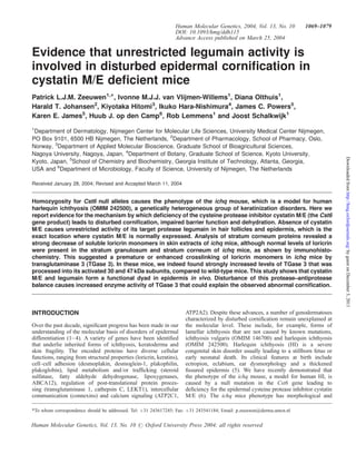
  • 1. Human Molecular Genetics, 2004, Vol. 13, No. 10 1069–1079 DOI: 10.1093/hmg/ddh115 Advance Access published on March 25, 2004 Evidence that unrestricted legumain activity is involved in disturbed epidermal cornification in cystatin M/E deficient mice Patrick L.J.M. Zeeuwen1,*, Ivonne M.J.J. van Vlijmen-Willems1, Diana Olthuis1, Harald T. Johansen2, Kiyotaka Hitomi3, Ikuko Hara-Nishimura4, James C. Powers5, Karen E. James5, Huub J. op den Camp6, Rob Lemmens1 and Joost Schalkwijk1 1Department of Dermatology, Nijmegen Center for Molecular Life Sciences, University Medical Center Nijmegen, PO Box 9101, 6500 HB Nijmegen, The Netherlands, 2Department of Pharmacology, School of Pharmacy, Oslo, Norway, 3Department of Applied Molecular Bioscience, Graduate School of Bioagricultural Sciences, Nagoya University, Nagoya, Japan, 4Department of Botany, Graduate School of Science, Kyoto University, Kyoto, Japan, 5School of Chemistry and Biochemistry, Georgia Institute of Technology, Atlanta, Georgia, USA and 6Department of Microbiology, Faculty of Science, University of Nijmegen, The Netherlands Received January 28, 2004; Revised and Accepted March 11, 2004 Homozygosity for Cst6 null alleles causes the phenotype of the ichq mouse, which is a model for human harlequin ichthyosis (OMIM 242500), a genetically heterogeneous group of keratinization disorders. Here we report evidence for the mechanism by which deficiency of the cysteine protease inhibitor cystatin M/E (the Cst6 gene product) leads to disturbed cornification, impaired barrier function and dehydration. Absence of cystatin M/E causes unrestricted activity of its target protease legumain in hair follicles and epidermis, which is the exact location where cystatin M/E is normally expressed. Analysis of stratum corneum proteins revealed a strong decrease of soluble loricrin monomers in skin extracts of ichq mice, although normal levels of loricrin were present in the stratum granulosum and stratum corneum of ichq mice, as shown by immunohisto-chemistry. This suggested a premature or enhanced crosslinking of loricrin monomers in ichq mice by transglutaminase 3 (TGase 3). In these mice, we indeed found strongly increased levels of TGase 3 that was processed into its activated 30 and 47 kDa subunits, compared to wild-type mice. This study shows that cystatin M/E and legumain form a functional dyad in epidermis in vivo. Disturbance of this protease–antiprotease balance causes increased enzyme activity of TGase 3 that could explain the observed abnormal cornification. INTRODUCTION Over the past decade, significant progress has been made in our understanding of the molecular basis of disorders of epidermal differentiation (1– 4). A variety of genes have been identified that underlie inherited forms of ichthyosis, keratoderma and skin fragility. The encoded proteins have diverse cellular functions, ranging from structural properties (loricrin, keratins), cell–cell adhesion (desmoplakin, desmoglein-1, plakophilin, plakoglobin), lipid metabolism and/or trafficking (steroid sulfatase, fatty aldehyde dehydrogenase, lipoxygenases, ABCA12), regulation of post-translational protein proces-sing (transglutaminase 1, cathepsin C, LEKT1), intercellular communication (connexins) and calcium signaling (ATP2C1, ATP2A2). Despite these advances, a number of genodermatoses characterized by disturbed cornification remain unexplained at the molecular level. These include, for example, forms of lamellar ichthyosis that are not caused by known mutations, ichthyosis vulgaris (OMIM 146700) and harlequin ichthyosis (OMIM 242500). Harlequin ichthyosis (HI) is a severe congenital skin disorder usually leading to a stillborn fetus or early neonatal death. Its clinical features at birth include ectropion, eclabium, ear dysmorphology and a thickened fissured epidermis (5). We have recently demonstrated that the phenotype of the ichq mouse, a model for human HI, is caused by a null mutation in the Cst6 gene leading to deficiency for the epidermal cysteine protease inhibitor cystatin M/E (6). The ichq mice phenotype has morphological and *To whom correspondence should be addressed. Tel: þ31 243617245; Fax: þ31 243541184; Email: p.zeeuwen@derma.umcn.nl Human Molecular Genetics, Vol. 13, No. 10 # Oxford University Press 2004; all rights reserved Downloaded from http://hmg.oxfordjournals.org/ by guest on December 3, 2011
  • 2. biochemical similarities to human HI, which includes excessive epidermal and follicular hyperkeratosis, abnormal large mitochondria, and a characteristic keratin expression pattern in the interfollicular epidermis. Although we have so far excluded CST6 mutations in humans as a major cause of harlequin ichthyosis type 2, CST6 and genes that have a role in biological pathways that are controlled by cystatin M/E, should be considered as candidate genes for human disorders of cornification of unknown etiology (7). The function of cystatin M/E in normal epidermal homeostasis is unknown and the mechanism that leads to the observed pathology in ichq mice remains unresolved. Clearly, the identification of its target protease(s) in vivo would be an essential step in answering these questions. A biochemical study on the novel asparaginyl endopeptidase legumain has indicated that cystatin M/E in vitro binds to this protease with high affinity and could possibly represent a physiological target enzyme, although no biological evidence has been provided so far (8). Legumain or asparaginyl endopeptidase (AEP, EC 3.4.22.34) belongs to clan CD of the cysteine proteases and has a strict specificity for hydrolysis of asparaginyl bonds. Initially, the enzyme was purified as a cysteine protease responsible for the maturation of seed storage proteins and designated vacuolar processing enzyme (VPE) (9). Subsequently, legumain has been described in mammals (10); it is found at low levels in many tissues, but is particularly abundant in kidney. Very recently, the generation of legumain deficient mice has been reported (11). Limited functional studies have shown disturbed biosynthetic processing of cathepsins and accumulation of macromolecules in the lysosomes in the proximal tubule cells of the kidney. In the present study, we have investigated the mechanism by which absence of cystatin M/E causes epidermal abnormalities in ichq mice. We report that legumain is a physiologically relevant target protease of cystatin M/E in vivo. Cystatin M/E deficiency in ichq mice leads to free cutaneous legumain activity at exactly the location where cystatin M/E is normally present in wild-type mice. These mice show increased transepidermal water loss and neonatal death, probably due to dehydration. We provide evidence that disturbed cornification is caused by abnormalities in loricrin processing, caused by uncontrolled legumain activity. RESULTS Skin barrier function is severely disturbed in cystatin M/E deficient mice Cystatin M/E deficient mice have defects in epidermal cornification and die between 5 and 12 days of age (6,12). Autopsy on neonatal ichq mice strongly suggested that these mice died of dehydration. This observation and the gross abnormalities in the stratum corneum prompted us to measure the rate of transepidermal water loss (TEWL) of ichq mice, compared to phenotypically normal littermates. As shown in Figure 1 (left hand y-axis), we found increased TEWL levels (up to 3-fold) from day 9 onwards. Figure 1 (right hand y-axis) also shows progressive weight loss in the cystatin M/E deficient mice starting at the time point that transepidermalwater loss was apparent. At day 11, body weights were 50% compared to normal littermates and most mice died shortly thereafter. Legumain is expressed in epidermis and hair follicles The antiprotease activity of cystatin M/E has been investigated previously (8,13–15). Cystatin M/E inhibits the asparaginyl endoprotease legumain with a high affinity (Fig. 2A). In addition to cathepsin B tested by others, we examined the inhibition of cathepsins C, H, L and S. None of these proteases was appreciably inhibited by cystatin M/E (Fig. 2B). These data indicate that legumain is a likely physiological target of cystatin M/E in skin in vivo, although nothing is known so far on the presence of legumain in cutaneous tissues. Using anti-legumain antibodies for immunohistochemical analysis of mouse tissues we found legumain expression in the entire epidermis of adult, wild-type mice (Fig. 3A) and in the inner root sheet of the hair follicle (Fig. 3B), most abundantly at the junction of dermis and subcutaneous fat (Fig. 3C). In addition to the known presence in kidney, legumain expression was further detected in the ciliated epithelium of the trachea and in bronchial epithelium (Fig. 3D and E). Immunohistochemical staining of dorsal skin in cystatin M/E deficient ichq mice revealed normal epidermal legumain expression (Fig. 3F). These results demonstrate that legumain is indeed expressed in skin and hair follicles, as well as in other mouse tissues, some of which were previously shown to express cystatin M/E (6). These findings were confirmed at the mRNA level in mouse and human skin by reverse transcriptase PCR analysis, and subsequent sequencing of the PCR product (data not shown). Free in situ legumain activity in skin of cystatin M/E deficient mice but not in wild-type mice Immunohistochemical localization of legumain cannot be equated with legumain activity because legumain can be present in an inactive zymogen form or complexed with endogenous inhibitors. In order to demonstrate legumain enzy-matic activity in tissue sections, we developed a cytochemical assay, based on hydrolysis of a specific fluorescent substrate that could be precipitated by nitrosalicylaldehyde in situ. As it was known from literature that kidney is a rich source of free legumain (as measured by biochemical assays) we used this tissue as a positive control. Enzymatic legumain activity is detected in situ as a green fluorescent precipitate in the proximal tubulus epithelial cells of pig kidney (Fig. 4A). Specificity of this reaction was confirmed using Cbz-Ala-Ala- Aasn-EPCOOEt, which completely blocks proteolysis of the substrate (Fig. 4B). This synthetic inhibitor is highly specific for legumain (16) and shows similar affinity and specificity as the natural inhibitor cystatin M/E. For comparison, immuno-histochemical staining of pig kidney for legumain shows the presence of legumain in epithelial cells of the proximal tubuli, whereas staining of the glomeruli was weak to absent (Fig. 4C); this is in accordance with the immunohistochemical localization of legumain in rat kidney reported by others (17), and it shows colocalization with enzymatically active legumain as demonstrated in Figure 4A. We subsequently used the cytochemical legumain assay to examine free legumain activity in the skin of wild-type and cystatin M/E deficient mice. 1070 Human Molecular Genetics, 2004, Vol. 13, No. 10 Downloaded from http://hmg.oxfordjournals.org/ by guest on December 3, 2011
  • 3. Human Molecular Genetics, 2004, Vol. 13, No. 10 1071 Figure 1. Disturbed barrier function in cystatin M/E deficient mice. TEWL of the ichq mutant mice is increased compared to phenotypically normal mice. Concomitantly with increased TEWL, body weights are decreasing until these mice die around day 11. Left hand y-axis: TEWL in g/m2/h. Right hand y-axis: decrease in body weight as a percentage of the control mice. Figure 4D visualizes free legumain activity in the stratum granulosum and in the hair follicles at the level of the infundi-bulum in the cystatin M/E deficient mice. No legumain activity could be demonstrated in the skin of wild type littermates (Fig. 4E), which indicates that the presence of cystatin M/E normally regulates legumain activity in skin in vivo. For comparison, immunohistochemical cystatin M/E staining of dorsal skin is shown in wild-type and cystatin M/E deficient mice. This confirms the absence of cystatin M/E expression at the protein level in the ichq mice (Fig. 4F), whereas the wild-type skin shows cystatin M/E expression in the infundibular epithelium of the hair follicle and the stratum granulosum of the interfollicular epidermis (Fig. 4G). The localization of free legumain activity in the infundibular part of the hair follicle in cystatin M/E deficient mice coincides with the reported localization of tissue pathology in these mice such as excessive cornification (6,12), which leads to plugging of the hair follicle and ichthyosis as shown before. Absence of loricrin monomers in cystatin M/E deficient mice Abnormal cornification in ichq mice could be the result of abnormal expression or processing of structural components of the cornified envelope. In previous studies, no abnormalities were found in structural proteins such as cytokeratins 1 and 14, involucrin and filaggrin (18). We now analyzed extracts of ichq and wild-type mice for the presence of loricrin and involucrin, two major components of the stratum corneum. We could confirm the reported normal presence of involucrin in ichq skin (data not shown), but remarkably, loricrin monomers and dimers were nearly absent in cystatin M/E deficient mice as revealed by western blot analysis (Fig. 5B). We took care to prepare the skin extracts in buffer with and without the specific synthetic legumain inhibitor Cbz-Ala-Ala-Aasn-EP-COOEt to preclude the action of excess free legumain in the cystatin M/E deficient skin during extraction. No low molecular weight degradation products of loricrin were detected in lanes 1 and 2 of Figure 5B, nor did we observe high molecular weight loricrin complexes, although these probably cannot be extracted, due to their poor solubility. Immunohistochemical analysis, however, showed that loricrin was abundantly present in the upper layers of the interfollicular epidermis of dorsal skin of cystatin M/E deficient mice (Fig. 5C), arguing against loricrin degradation as a mechanism to explain the findings on western blot. Moreover, in the epidermis of the mutant mice, three of five cell layers were found positive for loricrin expres-sion, whereas in only one of three cell layers, positive staining was detected in the epidermis of wild-type mice (Fig. 5D). In Figure 5A, a Coomassie blue stained blot is shown to check for equal loading of the samples in Figure 5B. No gross differences were seen in most of the high molecular weight bands, which represent the major soluble structural proteins of mouse skin. Interestingly, two abundant protein bands of 13 and 15kDa were observed in the extract of cystatin M/E deficient mice (boxed in Fig. 5A), which were less pronounced in wild-type skin. In order to exclude the possibility that these bands represented accumulated loricrin breakdown products not detected by the anti-mouse loricrin serum (directed against a single epitope), we subjected these bands to in-gel tryptic digestion followed by matrix-assisted laser desorption/ionization time-of-flight (MALDI-TOF) mass spectrometry. The lower 13kDa band was identified as S100 calcium binding protein A9 (or calgranulin B). This is a protein known to be induced in epidermis during inflammation (19), and it can also act as a modulator of intracellular calcium homeostasis during follicular differentiation (20). The upper 15kDa band was identified as epidermal fatty acid binding protein (E-FABP). Interestingly, E-FABP has been described as an epidermal protein that is strongly upregulated following skin barrier disruption (21), which is in accordance with the observed increase in transepidermal water loss described above. These findings also demonstrate that these low molecular weight bands are not derived from breakdown of loricrin monomers and dimers. Increased processing of TGase 3 in skin of ichq mice could explain abnormal cornification The strongly diminished presence of loricrin monomers and dimers in skin extracts of ichq mice, and the observation that Downloaded from http://hmg.oxfordjournals.org/ by guest on December 3, 2011
  • 4. Figure 2. Protease inhibition by recombinant cystatin M/E. (A) Inhibition of free legumain activity in human kidney extract. Nanomolar concentrations of recom-binant cystatin M/E and the specific synthetic legumain inhibitor inhibited hydrolysis of the fluorogenic substrate. Representative curves of three experiments are shown. (B) Hydrolysis of the fluorogenic substrate by the plant protease papain was almost completely inhibited by nanomolar concentrations of recombinant cystatin M/E. Inhibition of a panel of lysosomal cathepsins was incomplete, even when 2 mM recombinant cystatin M/E was used. this phenomenon is not due to degradation, led us to consider misregulated crosslinking as an alternative explanation. It is known that loricrin and small proline-rich proteins (SPRs) are cross-linked by cytosolic TGase 3 enzyme primarily to form homodimers and heterodimers (22). Proteolytic cleavage of TGase 3 is required to achieve maximal specific activity of the enzyme (23). We now addressed the question of whether aberrant processing of the TGase 3 zymogen in the interfollicular epidermis of cystatin M/E deficient mice could be responsible for the observed defects in epidermal cornifica-tion. TGase 3 activation during keratinocyte differentiation involves cleavage of the 77 kDa zymogen by a hitherto unknown protease resulting in the release of 30 and 47 kDa fragments (Fig. 6A), which then associate non-covalently to form the active enzyme (24). To confirm the aberrant processing of the TGase 3 zymogen due to unrestricted legumain activity in the interfollicular epidermis of cystatin M/E deficient mice, we used a cleavage-site-specific antibody that was generated against a synthetic peptide (FGATS) that corresponds to the cleavage site of mouse TGase 3 (Fig. 6A). Western blot analysis revealed the abundant presence of proteolyzed (and hence activated) TGase 3 in skin extracts of cystatin M/E deficient mice at day 6, whereas no appreciable levels of this 30 kDA fragment could be detected in skin extract of wild-type littermates (Fig. 6B). This indicates that prema-ture, high levels of activated TGase 3 are present at the time when the skin lesions develop. The next step was to investigate if legumain could process human recombinant TGase 3 in vitro into its active form by proteolysis of the 77 kDa zymogen into the 30 and 47 kDa fragments. As we were unsuccessful in generating active recombinant legumain, and most tissues do not contain 1072 Human Molecular Genetics, 2004, Vol. 13, No. 10 Downloaded from http://hmg.oxfordjournals.org/ by guest on December 3, 2011
  • 5. Human Molecular Genetics, 2004, Vol. 13, No. 10 1073 Figure 3. Immunohistochemical localization of legumain in mouse tissues. (A) Expression of legumain in the epidermis of dorsal skin of an adult wild type mouse; (B) in the inner root sheet epithelium of a hair follicle; and (C) high expression around the hair follicle at the junction of dermis and subcutaneous fat. (D) Legumain expression in the ciliated epithelium of the trachea and the chondrocytes in the cartilage below and in (E) bronchial epithelial cells (the lung tissue is slightly positive). (F) Immunohistochemical staining of dorsal skin in an ichq mutant mouse (9 days of age) shows normal epidermal staining. Scale bars: B and F, 12.5 mm; C–E, 25 mm; A, 50 mm. measurable free legumain activity, we used human kidney extract that is reported by others as a tissue where free legumain is relatively abundant. As shown in Figure 6C (lanes 1 and 4), recombinant human TGase 3 was slowly processed to its active form by human kidney extract as detected by the monoclonal antibodies C2D and C9D that are directed against the human zymogen and the 30 and 47 kDa fragments, respectively. For comparison, we show cleavage of TGase 3 by dispase, a neutral bacterial protease that is commonly used to activate TGase 3 in vitro. Dispase generated a fragment (Fig. 6C, lane 3) with the same electrophoretic properties as those generated using kidney extract (Fig. 6C, lane 4). Proteolytic processing of TGase 3 by kidney extract could be completely inhibited by the specific legumain inhibitor Cbz-Ala-Ala-Aasn-EP-COOEt (Fig. 6C, lanes 2 and 5). As it is known that legumain affects processing of lysosomal cathepsins (11), we considered the possibility that legumain mediates processing of TGase 3 in an indirect manner. Indeed, when we used E-64, a synthetic broad-spectrum inhibitor of lysosomal cysteine proteases that is not effective against legumain, a complete inhibition of TGase 3 processing was also found (Fig. 6C, lane 6). This finding was addressed in further detail by investigating the in vitro processing of TGase 3 by a panel of purified lysosomal cysteine proteases. Figure 6D shows that cathepsin S completely processed TGase 3 in 30 min (lane 7) similar to the positive control dispase (lane 2). Partial processing of TGase 3 was found using cathepsin B and cathepsin L (Fig. 6D, lanes 3 and 6), whereas cathepsins C and H were not able to do this (Fig. 6D, lanes 4 and 5). The addition of E-64 to the reaction mixtures completely blocked the processing of the TGase 3 zymogen (data not shown). DISCUSSION In this report, we provide evidence that the asparaginyl endopeptidase legumain is a physiologically relevant target protease of cystatin M/E in skin. No detectable free legumain activity was found in the skin of wild-type mice whereas in ichq mice, legumain activity was found at exactly the same location where cystatin M/E is normally expressed. Absence of cystatin M/E and appearance of legumain activity colocalize with the observed pathology in ichq mice (i.e. excessive cornification in hair follicles and epidermis). Cystatin M/E is an unusual cystatin in its biochemical properties, chromosomal localization and restricted tissue distribution. Whereas most cystatins are extracellular inhibitors of cysteine proteases, cystatin M/E appears to function more locally and also intracellularly, as will be discussed below. Little is known on the specific biological functions of cystatin family members. Deficiency for cystatin B in humans causes myoclonic epilepsy (25); deficiency for cystatin C in mice causes no obvious spontaneous phenotype (26). In ichq mice, deficiency for cystatin M/E causes a dramatic, apparently tissue-specific phenotype, with pronounced morphological and ultrastructural abnormalities in epidermis and hair follicles. We considered a number of known lysosomal cysteine proteases as likely candidate target enzymes for cystatin M/E. Only the recently discovered asparaginyl endopeptidase legumain was found to be inhibited at physiologically relevant concentrations. Mammalian legumain is found in late endosomes and is postulated to have a regulatory role in the biosynthesis of lysosomal enzymes, as recently demonstrated using legumain deficient mice (11). The body weights of the legumain deficient mice were significantly decreased, however, they were normally Downloaded from http://hmg.oxfordjournals.org/ by guest on December 3, 2011
  • 6. Figure 4. Cytochemical detection of free legumain activity. (A) In situ enzymatic legumain activity is shown as a green fluorescent precipitate (speckles) in the proximal tubulus epithelial cells (arrowheads) of pig kidney. Nuclei were counterstained with propidium iodide. (B) Legumain activity is blocked by Cbz-Ala-Ala- Aasn-EP-COOEt. (C) Immunohistochemical localization of legumain in pig kidney: epithelial cells of the proximal tubuli (arrowheads) showed positive staining; staining in the glomeruli (GL) was weak to absent. (D) Free in situ protease activity of legumain is visible as yellow speckles in the stratum granulosum in dorsal skin of ichq mutant mice, and in the hair follicles at the level of the infundibulum (arrows). Red staining is from the nuclei. A magnification of this signal is shown in the inset. (E) No legumain activity was found in the skin of wild type mice. (F) Immunohistochemical staining of dorsal skin in the ichq mutant mouse shows absence of cystatin M/E expression, whereas (G) the wild type mouse reveals cystatin M/E expression in the infundibular epithelium of the hair follicle and the stratum granulosum of the interfollicular epidermis. Scale bars: A–E 100 mm; F,G 50 mm. born and fertile. Disruption of the legumain gene led to the enlargement of lysosomes in the proximal tubule cells of the kidney, which suggests that materials to be degraded are being accumulated within the lysosomal compartments. It appeared that the processing of the lysosomal proteases, cathepsins B, H and L, from the single-chain forms into the two-chain forms was defective in the kidney cells of these legumain deficient mice. This observation confirms the earlier assumption that legumain could act as a protease responsible for processing of lysosomal cathepsins into their active forms (10). Interestingly, an increase of lysosomal cysteine protease activity has been observed in the terminal differentiation process of keratinocytes (27,28). Recent studies have reported increasing evidence that lysosomal proteases play important roles in physiological processes not restricted to lysosomes only (29). For example, protease activity and regulation outside lysosomes potentially contributes to propagation of apoptosis, a process that is distinct from terminal differentiation of the epidermis but nevertheless shares some molecular and cellular features. In epidermis there is a balanced regulation of protease activity that, when disturbed, could lead to faulty cornification processes in the epidermis and upper part of the hair follicle. Examples of a disturbed protease–antiprotease balance invol-ving lysosomal cathepsins, leading to abnormal cornification, include Papillon–Lefevre syndrome (cathepsin C deficiency) and the furless mouse (cathepsin L deficiency) (30,31). However, the exact role for these cathepsins in epidermal differentiation and desquamation is unknown. Our findings on the legumain dependent, cathepsin-mediated TGase 3 proces-sing fit very well with the proposed role of legumain in other cell types. Although it has to be verified experimentally, we assume that legumain regulates lysosomal cathepsin processing and TGase 3 processing in normal skin, a process that is apparently under tight control of cystatin M/E. Deficiency of cystatin M/E, as found in ichq mice, unleashes legumain activity thereby causing the observed phenotype. Much of the barrier function of human epidermis against the environment is provided by the cornified cell envelope (CE) 1074 Human Molecular Genetics, 2004, Vol. 13, No. 10 Downloaded from http://hmg.oxfordjournals.org/ by guest on December 3, 2011
  • 7. Human Molecular Genetics, 2004, Vol. 13, No. 10 1075 Figure 5. Western blot analysis of mouse skin extracts. Proteins were extracted in buffer with (þ) or without () the addition of Cbz-Ala-Ala-Aasn-EP-COOEt. (A) Coomassie blue staining, and (B) rabbit anti-mouse loricrin antiserum. Each lane contains 20 mg mouse skin extract. Note the diminished amounts of loricrin monomers and dimers in the skin extracts of ichq mice (d¼dimer, m¼monomer). Two abundant protein bands are found in ichq mouse skin extracts (boxed in A), which are identified as calgranulin B and E-FABP using MALDI-TOF mass spectrometry. (C) Immunohistochemical staining of loricrin in dorsal skin of an ichq mutant mouse, and (D) a wild-type littermate. Scale bars: 50 mm. (32–34), which is assembled by TGase mediated cross-linking of several structural proteins during the terminal stages of normal keratinocyte differentiation. Surprisingly, recent studies on mice in which major CE components were knocked out (e.g. loricrin, envoplakin and involucrin), have shown no discernable phenotype (35–37). This suggests that there are compensatory backup systems and additional unidentified components involved that maintain the skin barrier function. However, mutations or absence of desmosomal or cytoskeletal proteins in differentiated keratinocytes often leads to severe pathology and disturbance of barrier function (38–42). Deficiency for regulatory enzymes as TGase1 and steroid sulfatase also leads to disease in humans (43–45). The severe phenotype of cystatin M/E deficient mice provides an example of a disturbed protease–antiprotease balance that causes faulty differentiation processes in the epidermis and hair follicle. Another example is the recent identification of the serine protease inhibitor Kazal-type 5 (SPINK5) as the defective gene in Netherton syndrome (NS, OMIM 256500) (46). It was shown that the proteolytic processing and distribution of the protein product of SPINK5, LEKT1, is disturbed in NS patients (46,47). NS is a congenital ichthyosis associated with erythroderma, a specific hair shaft defect and atopic features. It was hypothesized that defective inhibitory regulation by LEKT1 result in increased protease activity in the stratum corneum, accelerated degradation of desmoglein-1 and overdesquamation of corneocytes (48). Colocalization of LEKT1 transcripts with stratum corneum serine proteases (SCTE and SCCE) in hair follicles suggested that the regulation of the activity of these proteases by LEKT1 might also affect hair growth and morphogenesis. Targeted epidermal overexpression of stratum corneum chymotryptic enzyme (SCCE) results in pathologic skin changes (49), which suggest that increased activity of proteases present in the skin may indeed play a significant part in skin pathophysiology. Our study underscores the importance of the regulation of proteolysis in the process of keratinocyte terminal differentia-tion. In addition, our study reveals that the function of cystatin M/E is to control keratinocyte legumain activity and thereby probably regulating correct TGase 3-dependent cross-linking of loricrin molecules, which is an important step in the formation of the cornified layer (22). In addition to excessive TGase activity, lack of TGase activity can also lead to disturbed cornification as witnessed by an autosomal recessive ichthyosis, termed lamellar ichthyosis (LI1, OMIM 242300) in which mutations in TGase 1 are responsible for the disease (43,44). Although this might seem paradoxical at first glance, both loss and inappropriate gain of TGase activity could explain ichthyotic changes, although the mechanisms could be different. Excessive TGase 3 activity could lead to retention of scales by hypercross-linked corneocytes, whereas deficiency for TGase 1 could lead to irregular scaling due to lack of attachment of involucrin to the o-hydroxyceramides (50). There are, however, other forms that are clinically similar to lamellar ichthyosis but are not linked to the locus of the TGase 1 gene. The non-redundancy of TGase 3 is indicated by knockout mice that show an early embryonic-lethal phenotype (51), but no mutations in this gene have been found in humans thus far. New loci for autosomal recessive ichthyosis have recently been identified (reviewed in 51), but they do not link to the gene for TGase 3. This leaves open the possibility that mutations in genes that are involved in the processing of TGases into the active form might be causative for other forms Downloaded from http://hmg.oxfordjournals.org/ by guest on December 3, 2011
  • 8. of ichthyosis. We suggest that cystatin M/E is a candidate gene for heritable human skin disorders that show faulty cornifica-tion and desquamation. In conclusion, the data presented in this study have identified cystatin M/E and legumain as a functional dyad in skin, and we provide evidence that legumain and lysosomal cysteine proteases are involved in the proteolytic activation of TGase 3 during terminal epidermal differentiation. Figure 6. Processing of TGase 3. (A) The 77 kDa zymogen form of TGase 3 is proteolyzed into the activated form consisting of 30 and 47 kDa fragments. The boxed amino acid sequence corresponds to the N-terminus of the 30 kDa frag-ment that was used to generate the mouse-specific cleavage-site-directed anti-body (the arrow indicates the cleavage site). (B) Western blot analysis of mouse skin extracts. Processed TGase 3 is found in skin extract of 6-day-old cystatin M/E deficient mice (lane 2), whereas hardly any processed TGase 3 is found in skin of wild type littermates (lane 1). Each lane contains 10 mg mouse skin extract. (C) Processing of recombinant human TGase 3 by human kidney extract (HKE), and detection by western blot analysis of the zymogen and proteolyzed fragments of 30 and 47 kDa, which could be recognized by the monoclonal anti-human TGase 3 antibodies C2D and C9D, respectively. 60 ng recombinant TGase 3 was partially processed by HKE (lanes 1 and 4), however, no proteolyzed fragments were found in the presence of Cbz-Ala- Ala-Aasn-EP-COOEt (lanes 2 and 5) or E-64 (lane 6). Processing by dispase is shown using the C2D antibody (lane 3). (D) Processing of recombinant TGase 3 by cathepsins, and detection of the zymogen and proteolyzed frag-ments with the C2D monoclonal antibody. Recombinant TGase 3 (lane 1) is completely processed in 30 min by dispase (lane 2) and by cathepsin S (lane 7). Partially processed TGase 3 was found using cathepsin B (lane 3) and cathe-psin L (lane 6). TGase 3 could not be processed by cathepsin C (lane 4) and cathepsin H (lane 5). MATERIALS AND METHODS Animal studies Mice were housed in specific pathogen-free facilities at the Central Animal Laboratory, University of Nijmegen, The Netherlands. Genotyping of the mice was performed as described previously (6). TEWL of cystatin M/E deficient mice (ichq/ichq) and phenotypically normal littermates (þ/þ and ichq/þ) was measured using a Tewameter TM 210, in accordance with current guidelines (52). Recombinant proteins Recombinant human cystatin M/E was produced both in a bacterial expression system as a GST fusion protein and as a fully processed protein in an eukaryotic system using the baculovirus in insect cells as described previously (15). Recombinant human TGase 3 was produced as a full length zymogen in a baculovirus system and as a bacterially expressed His6-tagged fusion protein as previously described in detail (53). Antibodies Purified GST-cystatin M/E fusion protein was used to immunize a New Zealand White rabbit. Antisera raised against the GST-cystatin M/E fusion protein were purified by affinity chromatography as described previously (15). Anti-mouse legumain antibodies were prepared by immunizing rabbits with a bacterially expressed His6-tagged fusion protein of legumain as previously described (11). Purified human recombinant TGase 3 from Escherichia coli was used to immunize mice for the establishment of monoclonal antibodies (C2D and C9D) as described in detail by Hitomi (53). Affinity purified polyclonal rabbit anti-mouse-FGATS antibodies were generated as described previously (54). Affinity purified polyclonal rabbit anti-mouse loricrin antibodies (AF-62) were purchased from BAbCO (BAbCO, Richmond, CA). 1076 Human Molecular Genetics, 2004, Vol. 13, No. 10 Downloaded from http://hmg.oxfordjournals.org/ by guest on December 3, 2011
  • 9. Extraction of proteins and immunoblotting Human kidney and mouse skin biopsies were frozen in liquid nitrogen and subsequently grinded using a Micro Dismembrator U (B. Braun Biotech International, Melsungen, Germany). Proteins were extracted in buffer containing 50mM citrate (pH 5.8), 1mM EDTA, 0.1M NaCl and 2mM DTT, followed by mild sonification of the lysate for 1 min at 4C. Mouse skin proteins were extracted in the absence and presence of Cbz-Ala-Ala-AAsn-EP-COOEt. Samples were diluted with SDS sample buffer (containing a reducing agent) and boiled for 3 min. These protein samples were separated by SDS–PAGE on a 12% Bis-Tris-Gel and blotted onto polyvinylidenedifluoride (PVDF) membrane using the NuPAGE system (Invitrogen, Carlsbad, CA), according to the protocol provided by the manufacturer. Membranes were stained with Coomassie blue, or incubated with polyclonal antibodies directed against mouse loricrin (1 : 5000), mouse anti-FGATS (1 : 100), and the C2D (1 : 1000) and C9D (1 : 4000) monoclonal antibodies against human TGase 3. Proteins were detected with the Phototope-HRP Western Blot Detection Kit (Cell Signaling Technology, Beverly, MA), according to the manufacturer’s protocol. MALDI-TOF mass spectrometry analysis In-gel digestion coupled with mass spectrometry analysis was used for the identification of proteins extracted from mouse skin. To perform digestions on Coomassie blue stained protein bands that were abundantly found in the skin extract of cystatin M/E deficient mice, the In-Gel Tryptic Digestion Kit (Pierce, Rockford, IL) was used in accordance to the protocol provided by the manufacturer. For MALDI-TOF mass spectrometry, 0.3 ml of the tryptic digest was spotted on the target plate followed by 0.3 ml of matrix solution (a saturated solution of a-cyano-4-hydroxy-cinnamic acid in a 50 : 50 mixture of acetonitrile and 0.1% TFA, v/v). Positive mass ion spectra were collected in the reflectron mode, with pulsed ion extraction on a Biflex III instrument (Bruker-Franzen, Bremen, FRG). Mass calibration was performed externally using a mixture of peptide standards (Sigma, St Louis, MO). A total of 150 single laser shots were accumulated for each sample spot. For identification of proteins, peptide mass lists were used to search protein databases with the Mascot software system (Matrix Science, London, UK). TGase 3 processing Baculovirus expressed recombinant TGase 3 zymogen was processed by treatment with dispase, cathepsins and by a protein extract from human kidney. To prepare the proteolyzed form, 0.6 mg zymogen was treated with 5mU dispase (Roche Diagnostics, Mannheim, Germany) in the presence of 5mM CaCl2 at 37C for 30 min. The same reaction conditions were used to test proteolytic activity of human cathepsin B (Sigma), bovine cathepsin C (Sigma), human cathepsin H (Calbiochem, San Diego, CA), human cathepsin L (Sigma) and bovine cathepsin S (Calbiochem). To test whether the TGase 3 zymogen could be processed by free legumain activity, 0.6 mg zymogen was treated with protein extracts from human kidney Human Molecular Genetics, 2004, Vol. 13, No. 10 1077 in the presence of 5mM CaCl2 at 37C for 0.5 and 24 h. For controls the same reaction mixtures were prepared but with the addition of Cbz-Ala-Ala-Aasn-EPCOOEt, or compound E-64 [trans-epoxysuccinyl-L-leucylamido-(4-guanidino)butane]. The reaction mixtures were subjected to SDS–PAGE and electro-blotted onto PVDF membrane. The membrane was incubated with the monoclonal mouse anti-human TGase 3 antibodies (C2D and C9D) and detection was established as described above. In situ assay for legumain activity This assay is based on data that have shown that 5- nitrosalicylaldehyde forms an insoluble fluorescent complex with NH2NapOMe, which has been used to visualize cathepsin B in fibroblasts (55). Unfixed cryostat sections (6 mm) of mouse skin and pig kidney were air-dried for 10 min. These tissue sections were subsequently incubated with 100 ml legumain in situ assay buffer under a coverslip. Enzyme activity was measured after an incubation period of 30 min at 37C with the selective fluorescent Suc-Ala-Ala-Asn-4-methoxy-2-naphthy-lamide (Suc-Ala-Ala-Asn-NHNapOME) substrate. This sub-strate was originally synthesized for the measurement of legumain in colorimetric and fluorimetric microplate assays as described previously (56). Legumain in situ assay buffer was prepared by the addition of 5-nitro-salicylaldehyde and Suc- Ala-Ala-Asn-NHNapOME substrate to a final concentration of 1 and 2mM, respectively, in a buffer that contained 121mM phosphate (pH 5.8), 39.5mM citric acid, 1mM EDTA, 1mM DTT and 0.01% CHAPS. The specificity of the reaction was verified by incubation in the absence of substrate or in the presence of Cbz-Ala-Ala-Aasn-EP-COOEt. Fluorimetric enzyme assays Protease inhibitory activity of recombinant cystatin M/E against cysteine proteases was determined by measuring the inhibition of papain and lysosomal cathepsins (B, C, H, L and S) using fluorogenic synthetic substrates, essentially described by Abrahamson (53). Protease inhibitory activity of recombi-nant cystatin M/E and the synthetic aza-peptide epoxide inhibitor against legumain was determined by measuring the inhibition of free legumain activity in human kidney extract. Kidney extract was titrated in the absence and presence of increasing concentrations of recombinant cystatin M/E or the specific legumain inhibitor as a positive control. Preincubation time of kidney extract and inhibitor was 30 min at room temperature. Enzyme activity was measured after an incuba-tion period of 30 min at 37C with the selective fluorescent Z-Ala-Ala-Asn-MCA substrate (Peptides International, Louisville, KY). The buffer that was used contained 0.1M phosphate ( pH 5.7), 2mM EDTA, 1mM DTT and 2.7mM L-cystein. ACKNOWLEDGEMENTS We thank Geert Poelen and Debby Smits for their assistance with the animal experiments. Wiljan Hendriks and Bruce Jenks are acknowledged for critical reading of this manuscript. This work was financially supported by Grant 902.11.092 from the Netherlands Organization for Scientific Research (NWO). Downloaded from http://hmg.oxfordjournals.org/ by guest on December 3, 2011
  • 10. J.C.P. would like to acknowledge grants from the National Institute of General Medical Sciences (Grant GM54401 and GM61964). REFERENCES 1. Irvine, A.D. and McLean, W.H. (2003) The molecular genetics of the genodermatoses: progress to date and future directions. Br. J. Dermatol., 148, 1–13. 2. McGrath, J.A. and Eady, R.A. (2001) Recent advances in the molecular basis of inherited skin diseases. Adv. Genet., 43, 1–32. 3. Hohl, D. (2000) Towards a better classification of erythrokeratodermias. Br. J. Dermatol., 143, 1133–1137. 4. Griffiths, W.A.D., Judge, M.R. and Leigh, I.M. (1998) Disorders of keratinization. In Champion, R.H., Burton, J.L., Burns, D.A. and Breathnach, S.M. (eds), Textbook of Dermatology, Sixth Edition. Blackwell Science, Inc., Malden, pp. 1483–1588. 5. Williams, M.L. and Elias, P.M. (1987) Genetically transmitted, generalized disorders of cornification. The ichthyoses. Dermatol. Clin., 5, 155–178. 6. Zeeuwen, P.L., Vlijmen-Willems, I.M., Hendriks, W., Merkx, G.F. and Schalkwijk, J. (2002) A null mutation in the cystatin M/E gene of ichq mice causes juvenile lethality and defects in epidermal cornification. Hum. Mol. Genet., 11, 2867–2875. 7. Zeeuwen, P.L., Dale, B.A., de Jongh, G.J., Vlijmen-Willems, I.M., Fleckman, P., Kimball, J.R., Stephens, K. and Schalkwijk, J. (2003) The human cystatin M/E gene (CST6): exclusion as a candidate gene for harlequin ichthyosis. J. Invest Dermatol., 121, 65–68. 8. Alvarez-Fernandez, M., Barrett, A.J., Gerhartz, B., Dando, P.M., Ni, J. and Abrahamson, M. (1999) Inhibition of mammalian legumain by some cystatins is due to a novel second reactive site. J. Biol. Chem., 274, 19195–19203. 9. Hara-Nishimura, I., Inoue, K. and Nishimura, M. (1991) A unique vacuolar processing enzyme responsible for conversion of several proprotein precursors into the mature forms. FEBS Lett., 294, 89–93. 10. Chen, J.M., Dando, P.M., Rawlings, N.D., Brown, M.A., Young, N.E., Stevens, R.A., Hewitt, E., Watts, C. and Barrett, A.J. (1997) Cloning, isolation, and characterization of mammalian legumain, an asparaginyl endopeptidase. J. Biol. Chem., 272, 8090–8098. 11. Shirahama-Noda, K., Yamamoto, A., Sugihara, K., Hashimoto, N., Asano, M., Nishimura, M.and Hara-Nishimura, I. (2003) Biosynthetic processing of cathepsins and lysosomal degradation are abolished in asparaginyl endopeptidase-deficient mice. J. Biol. Chem., 278, 33194–33199. 12. Sundberg, J.P., Boggess, D., Hogan, M.E., Sundberg, B.A., Rourk, M.H., Harris, B., Johnson, K., Dunstan, R.W. and Davisson, M.T. (1997) Harlequin ichthyosis (ichq): a juvenile lethal mouse mutation with ichthyosiform dermatitis. Am. J. Pathol., 151, 293–310. 13. Ni, J., Abrahamson, M., Zhang, M., Fernandez, M.A., Grubb, A., Su, J., Yu, G.L., Li, Y., Parmelee, D., Xing, L. et al. (1997) Cystatin E is a novel human cysteine proteinase inhibitor with structural resemblance to family 2 cystatins. J. Biol. Chem., 272, 10853–10858. 14. Sotiropoulou, G., Anisowicz, A. and Sager, R. (1997) Identification, cloning, and characterization of cystatin M, a novel cysteine proteinase inhibitor, down-regulated in breast cancer. J. Biol. Chem., 272, 903–910. 15. Zeeuwen, P.L., Vlijmen-Willems, I.M., Jansen, B.J., Sotiropoulou, G., Curfs, J.H., Meis, J.F., Janssen, J.J., van Ruissen, F. and Schalkwijk, J. (2001) Cystatin M/E expression is restricted to differentiated epidermal keratinocytes and sweat glands: a new skin-specific proteinase inhibitor that is a target for cross-linking by transglutaminase. J. Invest Dermatol., 116, 693–701. 16. James, K.E., Gotz, M.G., Caffrey, C.R., Hansell, E., Carter,W., Barrett, A.J., McKerrow, J.H. and Powers, J.C. (2003) Aza-peptide epoxides: potent and selective inhibitors of Schistosoma mansoni and pig kidney legumains (asparaginyl endopeptidases). Biol. Chem., 384, 1613–1618. 17. Yamane, T., Takeuchi, K., Yamamoto, Y., Li, Y.H., Fujiwara, M., Nishi, K., Takahashi, S. and Ohkubo, I. (2002) Legumain from bovine kidney: its purification, molecular cloning, immunohistochemical localization and degradation of annexin II and vitamin D-binding protein. Biochim. Biophys. Acta, 1596, 108–120. 18. Dunnwald, M., Zuberi, A.R., Stephens, K., Le, R., Sundberg, J.P., Fleckman, P. and Dale, B.A. (2003) The ichq mutant mouse, a model for the human skin disorder harlequin ichthyosis: mapping, keratinocyte culture, and consideration of candidate genes involved in epidermal growth regulation. Exp. Dermatol., 12, 245–254. 19. Broome, A.M., Ryan, D. and Eckert, R.L. (2003) S100 protein subcellular localization during epidermal differentiation and psoriasis. J. Histochem. Cytochem., 51, 675–685. 20. Schmidt, M., Gillitzer, R., Toksoy, A., Brocker, E.B., Rapp, U.R., Paus, R., Roth, J., Ludwig, S. and Goebeler, M. (2001) Selective expression of calcium-binding proteins S100a8 and S100a9 at distinct sites of hair follicles. J. Invest Dermatol., 117, 748–750. 21. Yamaguchi, H., Yamamoto, A., Watanabe, R., Uchiyama, N., Fujii, H., Ono, T. and Ito, M. (1998) High transepidermal water loss induces fatty acid synthesis and cutaneous fatty acidbinding protein expression in rat skin. J. Dermatol. Sci., 17, 205–213. 22. Kalinin, A., Marekov, L.N. and Steinert, P.M. (2001) Assembly of the epidermal cornified cell envelope. J. Cell Sci., 114, 3069–3070. 23. Kim, H.C., Nemes, Z., Idler, W.W., Hyde, C.C., Steinert, P.M. and Ahvazi, B. (2001) Crystallization and preliminary X-ray analysis of human transglutaminase 3 from zymogen to active form. J. Struct. Biol., 135, 73–77. 24. Ahvazi, B., Kim, H.C., Kee, S.H., Nemes, Z. and Steinert, P.M. (2002) Three-dimensional structure of the human transglutaminase 3 enzyme: binding of calcium ions changes structure for activation. EMBO J., 21, 2055–2067. 25. Pennacchio, L.A., Lehesjoki, A.E., Stone, N.E.,Willour, V.L., Virtaneva, K., Miao, J., D’Amato, E., Ramirez, L., Faham, M., Koskiniemi, M. et al. (1996) Mutations in the gene encoding cystatin B in progressive myoclonus epilepsy (EPM1). Science, 271, 1731–1734. 26. Huh, C.G., Hakansson, K., Nathanson, C.M., Thorgeirsson, U.P., Jonsson, N., Grubb, A., Abrahamson, M. and Karlsson, S. (1999) Decreased metastatic spread in mice homozygous for a null allele of the cystatin C protease inhibitor gene. Mol. Pathol., 52, 332–340. 27. Kawada, A., Hara, K., Kominami, E., Hiruma, M., Noguchi, H. and Ishibashi, A. (1997) Processing of cathepsins L, B and D in psoriatic epidermis. Arch. Dermatol. Res., 289, 87–93. 28. Tanabe, H., Kumagai, N., Tsukahara, T., Ishiura, S., Kominami, E., Nishina, H. and Sugita, H. (1991) Changes of lysosomal proteinase activities and their expression in rat cultured keratinocytes during differentiation. Biochim. Biophys. Acta, 1094, 281–287. 29. Turk, B., Stoka, V., Rozman-Pungercar, J., Cirman, T., Droga-Mazovec, G., Oreic, K. and Turk, V. (2002) Apoptotic pathways: involvement of lysosomal proteases. Biol. Chem., 383, 1035–1044. 30. Toomes, C., James, J., Wood, A.J., Wu, C.L., McCormick, D., Lench, N., Hewitt, C., Moynihan, L., Roberts, E., Woods, C.G. et al. (1999) Loss-of-function mutations in the cathepsin C gene result in periodontal disease and palmoplantar keratosis. Nat. Genet., 23, 421–424. 31. Roth, W., Deussing, J., Botchkarev, V.A., Pauly-Evers, M., Saftig, P., Hafner, A., Schmidt, P., Schmahl,W., Scherer, J., Anton-Lamprecht, I. et al. (2000) Cathepsin L deficiency as molecular defect of furless: hyperproliferation of keratinocytes and pertubation of hair follicle cycling. FASEB J., 14, 2075–2086. 32. Robinson, N.A., Lapic, S., Welter, J.F. and Eckert, R.L. (1997) S100A11, S100A10, annexin I, desmosomal proteins, small proline-rich proteins, plasminogen activator inhibitor-2, and involucrin are components of the cornified envelope of cultured human epidermal keratinocytes. J. Biol. Chem., 272, 12035–12046. 33. Nemes, Z. and Steinert, P.M. (1999) Bricks and mortar of the epidermal barrier. Exp. Mol. Med., 31, 5–19. 34. Presland, R.B. and Dale, B.A. (2000) Epithelial structural proteins of the skin and oral cavity: function in health and disease. Crit Rev. Oral Biol. Med., 11, 383–408. 35. Koch, P.J., de Viragh, P.A., Scharer, E., Bundman, D., Longley, M.A., Bickenbach, J., Kawachi, Y., Suga, Y., Zhou, Z., Huber, M. et al. (2000) Lessons from loricrin-deficient mice: compensatory mechanisms maintaining skin barrier function in the absence of a major cornified envelope protein. J. Cell. Biol., 151, 389–400. 36. Maatta, A., DiColandrea, T., Groot, K. and Watt, F.M. (2001) Gene targeting of envoplakin, a cytoskeletal linker protein and precursor of the epidermal cornified envelope. Mol. Cell Biol., 21, 7047–7053. 37. Djian, P., Easley, K. and Green, H. (2000) Targeted ablation of the murine involucrin gene. J. Cell Biol., 151, 381–388. 1078 Human Molecular Genetics, 2004, Vol. 13, No. 10 Downloaded from http://hmg.oxfordjournals.org/ by guest on December 3, 2011
  • 11. 38. Porter, R.M. and Lane, E.B. (2003) Phenotypes, genotypes and their contribution to understanding keratin function. Trends Genet., 19, 278–285. 39. Fuchs, E. and Cleveland, D.W. (1998) A structural scaffolding of intermediate filaments in health and disease. Science, 279, 514–519. 40. McGrath, J.A., McMillan, J.R., Shemanko, C.S., Runswick, S.K., Leigh, I.M., Lane, E.B., Garrod, D.R. and Eady, R.A. (1997) Mutations in the plakophilin 1 gene result in ectodermal dysplasia/skin fragility syndrome. Nat. Genet., 17, 240–244. 41. Vasioukhin, V., Bowers, E., Bauer, C., Degenstein, L. and Fuchs, E. (2001) Desmoplakin is essential in epidermal sheet formation. Nat. Cell Biol., 3, 1076–1085. 42. McKoy, G., Protonotarios, N., Crosby, A., Tsatsopoulou, A., Anastasakis, A., Coonar, A., Norman, M., Baboonian, C., Jeffery, S. and McKenna, W.J. (2000) Identification of a deletion in plakoglobin in arrhythmogenic right ventricular cardiomyopathy with palmoplantar keratoderma and woolly hair (Naxos disease). Lancet, 355, 2119–2124. 43. Huber, M., Rettler, I., Bernasconi, K., Frenk, E., Lavrijsen, S.P., Ponec, M., Bon, A., Lautenschlager, S., Schorderet, D.F. and Hohl, D. (1995) Mutations of keratinocyte transglutaminase in lamellar ichthyosis. Science, 267, 525–528. 44. Russell, L.J., DiGiovanna, J.J., Rogers, G.R., Steinert, P.M., Hashem, N., Compton, J.G. and Bale, S.J. (1995) Mutations in the gene for transglutaminase 1 in autosomal recessive lamellar ichthyosis. Nat. Genet., 9, 279–283. 45. Webster, D., France, J.T., Shapiro, L.J. and Weiss, R. (1978) X-linked ichthyosis due to steroidsulphatase deficiency. Lancet, 1, 70–72. 46. Chavanas, S., Bodemer, C., Rochat, A., Hamel-Teillac, D., Ali, M., Irvine, A.D., Bonafe, J.L., Wilkinson, J., Taieb, A., Barrandon, Y. et al., (2000) Mutations in SPINK5, encoding a serine protease inhibitor, cause Netherton syndrome. Nat. Genet., 25, 141–142. 47. Bitoun, E., Micheloni, A., Lamant, L., Bonnart, C., Tartaglia-Polcini, A., Cobbold, C., Al Saati, T., Mariotti, F., Mazereeuw-Hautier, J., Boralevi, F. et al. (2003) LEKTI proteolytic processing in human primary keratinocytes, tissue distribution and defective expression in Netherton syndrome. Hum. Mol. Genet., 12, 2417–2430. Human Molecular Genetics, 2004, Vol. 13, No. 10 1079 48. Komatsu, N., Takata, M., Otsuki, N., Ohka, R., Amano, O., Takehara, K. and Saijoh, K. (2002) Elevated stratum corneum hydrolytic activity in Netherton syndrome suggests an inhibitory regulation of desquamation by SPINK5-derived peptides. J. Invest Dermatol., 118, 436–443. 49. Hansson, L., Backman, A., Ny, A., Edlund, M., Ekholm, E., Ekstrand, H.B., Tornell, J., Wallbrandt, P., Wennbo, H. and Egelrud, T. (2002) Epidermal overexpression of stratum corneum chymotryptic enzyme in mice: a model for chronic itchy dermatitis. J. Invest Dermatol., 118, 444–449. 50. Nemes, Z., Marekov, L.N., Fesus, L. and Steinert, P.M. (1999) A novel function for transglutaminase 1: attachment of long-chain omega- hydroxyceramides to involucrin by ester bond formation. Proc. Natl Acad. Sci. USA, 96, 8402–8407. 51. Kim, S.Y., Jeitner, T.M. and Steinert, P.M. (2002) Transglutaminases in disease. Neurochem. Int., 40, 85–103. 52. Pinnagoda, J., Tupker, R.A., Agner, T. and Serup, J. (1990) Guidelines for transepidermal water loss (TEWL) measurement. A report from the Standardization Group of the European Society of Contact Dermatitis. Contact Dermatitis, 22, 164–178. 53. Hitomi, K., Presland, R.B., Nakayama, T., Fleckman, P., Dale, B.A. and Maki, M. (2003) Analysis of epidermal-type transglutaminase (transglutaminase 3) in human stratified epithelia and cultured keratinocytes using monoclonal antibodies. J. Dermatol. Sci., 32, 95–103. 54. Hitomi, K., Ikeda, N. and Maki, M. (2003) Immunological detection of proteolytically activated epidermal-type transglutaminase (TGase 3) using cleavage-site-specific antibody. Biosci. Biotechnol. Biochem., 67, 2492–2494. 55. Van Noorden, C.J., Vogels, I.M., Everts, V. and Beertsen, W. (1987) Localization of cathepsin B activity in fibroblasts and chondrocytes by continuous monitoring of the formation of a final fluorescent reaction product using 5-nitrosalicylaldehyde. Histochem. J., 19, 483–487. 56. Johansen, H.T., Knight, C.G. and Barrett, A.J. (1999) Colorimetric and fluorimetric microplate assays for legumain and a staining reaction for detection of the enzyme after electrophoresis. Anal. Biochem., 273, 278–283. Downloaded from http://hmg.oxfordjournals.org/ by guest on December 3, 2011