
Recommended
More Related Content
What's hot
What's hot (20)
Similar to lacrimal apparatus
Similar to lacrimal apparatus (20)
Recently uploaded
Recently uploaded (20)
lacrimal apparatus
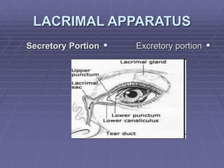
- 1. LACRIMAL APPARATUS Secretory Portion Excretory portion
- 2. Secretory Portion Accessory Lacrimal glands (see before) => Basic secretion Main lacrimai gland => Reflex secretion.
- 3. Gross Anatomy o Size: 20 X 12 X 5 mm o Weight: 75 gm o Portions: the gland is devided by the lateral part of levator aponeurosis into:
- 4. orbital portion Size : Small Site: Lacrimal gland fossa (in the anterior lateral part of the orbital roof) Shape Almond shaped Superior surface —> convex lies in the fossa connected to it by weak trabeculae Inferior surface —> concave lies on the levator aponeurosis & LR Anterior border —> Sharp in contact with the orbital septum (reached by division of skin. Orbicularis oculi & orbital septum) Posterior border —> Rounded in contact with orbital fat Medial extremity —» rests on levator Lateral extremity -> rests on LR
- 5. Palpepral portion Smaller (1/3 of the orbital portion) In the lateral part of upper eyelid (can be seen through the conjunctiva when the upper lid is everted Flattened from above downwards Anterior border —» lies just above the lateral part of the superior fornix. Posterior border —> continuous with the rest of the gland around the lateral border of the levator aponeurosis.
- 6. Ducts: 3-5 ducts pass from the orbital portion through the palpebral portion to open into the superior fornix. 5-7 ducts from the palpebral portion open separately into the superior fornix -» So, surgical removal of the palpebral part will destroy the whole gland.
- 7. Structure: Tubulo-alveolar (tubulo-aciner) gland Consist of lobules (each about the size of pin head) formed of: numerous Acini (composed of a double layers of cells resting on B.M) 1. outer myoepithelial contractile cells 2. inner secretory cylindrical cells 3. surrounding a central canal 4. open into larger ducts (intralobular then interlobular ducts) 5. open into excretory ducts (lined by columnar epithelium & have a fibrous coat)
- 8. Intralobular (between acini) & interlobular (between lobules) C.T reticulum is present containing Blood vessels, Nerve fibers, Plasma cells & lymphocytes (aggregate into follicles)
- 9. Ligaments & support: • Superior —> to the lacrimal fossa = suspensory ligament • Inferior pole —> to zygomatic bone • Posterior —> where lacrimal nerve & vessels enter the periorbita • Internally accompanying the ducts.
- 10. Blood supply: o Arterial -> Lacrimal artery (enters the posterior border of the gland) o Venous —> Corresponding vein join the ophthalmic vein o Lymphatic —> Conjunctival lymphatics —» Pre- auricular LNs
- 11. Function: Tears Secretion (mainly the reflex secretion) (Moisting the eye - Prevent friction between globe & lids - Keep the cornea transparent)
- 12. Nerve supply: 1. Parasympathatic 2. Sympathatic 3. Sensory
- 13. A. Parasympathetic lacrimal (Superior salivalory nucleus of the facial n.) (N VII) in the floor of the 4th ventricle) Preganglionic fibers extends with facial n.(nervus intermedius part) geniculate ganglion (no relay) greater superficial petrosal n. nerve of pterygoid canal sphenopalatine ganglion (synapse) postganglionic parasvmpatheic fibers Join maxilary n. (of trigeminal) Zygomatico-temporal branch Lacrimal n. Lacrimal Gland. (Vasomotor)
- 14. B. Sympathetic Superior cervical sympathetic ganglion Postganglionic fibers accompany I.C.A. Deep petrosal n. nerve of pterygoid canal‘ sphenopalatine ganglion(no relay) postganglionic sympatheic fibers Join maxilary n. (of trigeminal) Zygomatico-temporal branch Lacrimal n. Lacrimal Gland.
- 15. C. Sensory:! -> Lacrimal nerve.
- 16. Excretory Portion Puncta: • Shape: slightly elevated, rounded or slightly oval openings • Diameter: About 0.3 mm • Site: on the upper & lower eyelid margins (at the junction of the ciliary & lacrimal portions), on the papilla lacrimalis, about 6mm (upper) — 6.5mm (lower) from the medial canthus
- 17. • The puncta openings of the lacrimal canaliculi are inverted into the lacrimal lake when the eyelids are closed. • The puncta are surrounded by: - Dense, Fibrous, relatively avascular C.T. —> Prevents their collapse + gives the pale colour - Orbicularis oculi muscle —> Helps tear drainage How to know its site ? => 6 mm from the medial canthus + 0.5 mm from the tarsal gland + elevated + paler (avascular)
- 18. Canaliculi: • The upper and lower canaliculi consist of: o vertical portion about 2- 3.5 mm o horizontal portion about 8 mm (directed medially) => two join to Form, the common canaliculus —> opens in the lacrimal sinus of Maier • Diameter: About 0.5 mm. • Site:Lacrimal portion of the lid margin. • Structure & surroundings: o Lined by stratified squamous epithelium. o Surrounded by elastic tissue then orbicularis fibers. (Homer's muscle)
- 19. Lacrimal sac: • Site: lacrimal fossa (in the anterior part of the medial orbital wall), between: 1. the anterior lacrimal crest of the frontal process of the maxilla. 2. the posterior lacrimal crest of the lacrimal bone. Dimentions; 15 X 5 X 5 (When distended) • Parts: Fundus (above M.P.L) Body (behind M.P.L) Neck (continuous with NLD) • Lateral sinus of Maier: is a diverticulum that arise just behind the middle of the lateral surface (2.5 mm from the apex) —» in which open the canaliculi.
- 20. Structure: A. Sac: Lined by 2 layers of columnar cells with goblet cells, surrounded by fibro-elastic CT B. Lacrimal fascia: o Formed by splitting of the periorbita into 2 layers at the posterior lacrimal crest —> enclose the sac —> meet again at the anterior lacrimal crest. o Venous plexus is present between the lacrimal sac & lacrimal fascia (continuous with that around the duct)
- 21. Nasolacrirmal duct: • Definition: A downward extension of the neck of lacrimal sac till the inferior nasal meatus (at the anterior portion of the lateral wall) (lateral to & below the inferior turbinate) • Length: 15-18 mm • Site: o In the bone of the nasolacrimal canal => formed by a groove on the maxilla & completed by lacrimal bone & lacrimal process of inferior concha => The duct is loosely adherent to the canal above, but strongly adherent below —> mucoperiosteum (infection is easily transmitted from bone to duct & vice versa) o The duct may pass for millimeters in the nasal mucous membrane before its opening • Direction: Downwards, Laterally & posteriorly (Massage -> stretch & press down laterally & little back)
- 22. Valves: numerous mucous membrane folds, not valvular in function Hasner's valve (guards the opening of the NLD) —> prevent sudden blast of air from nose to the sac as during blowing the nose into a handkerchief)
- 23. • Structure: o Epithelium: 2 layers • Superficial columnar (reach the BM through deep cells) with goblet cells & mucous glands. . Deep flattened. o Subepithelial layer: contains lymphocytes o Fibroelastic membranous wall o Dense vascular erectile venous plexus surrounding the duct (engorgement -» duct obstruction)
- 24. • Relations; o Laterally => Maxillary antrum o Medially => Middle meatus of the nose • Blood supply: o Arteries —> Medial palpebral a. Angular a. Infraorbital a. Nasal br. of sphenopalatine o Veins —> Angular v. & Infraorbital v. (above) Nasal veins (below) • Lymphatic supply; -> Submandibular LNs • Nerve supply: o Infratrochlear n. (branch of ophthalmic) _ o Anterior superior alveolar n. (branch of maxillary)