
Recommended
More Related Content
What's hot
What's hot (20)
Similar to AMELOBLASTOMA [op].pptx
Similar to AMELOBLASTOMA [op].pptx (20)
Recently uploaded
Recently uploaded (20)
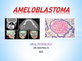
AMELOBLASTOMA [op].pptx
- 2. CONTENT 1. Introduction 2. Types of ameloblastoma 3. Pathogenesis 4. Clinical features 5. Radiographic features 6. Histopathological feature a) Multicystic ameloblastoma 1) follicular ameloblastoma 2) plexiform ameloblastoma b) unicystic ameloblastoma 7. Treatment
- 3. Introduction SYNONYMS: adamantinoma , adamantoblastoma, multilocular cyst DEFINATION:Ameloblastoma is a true neoplasm of enamel organ type of tissue which does not undergo differentiation to the point of enamel formation. 2ND most common odontogenic neoplasm worldwide. By ROBINSON: 1) unicentric 2) non functional { UNIAC } 3) intermittent 4) anatomically benign 5) clinically persistent
- 4. TYPES OF AMELOBLASTOMA Clinico-radiographically [due to differing therapeutic prognosis]: *Conventional , solid or multicystic (about 86% of all cases) *Unicystic (13% of all cases) *Peripheral (extraosseous)(1% of all cases)
- 5. PATHOGENESIS Varied origin • Cell rest of enamel organ (rest of serre’s/ malassez) • Epithelium of odontogenic cyst ( dentigerous cyst) • Disturbances in developing tooth • Basal cells of surface epithielium of oral cavity.
- 7. CLINICAL FEATURES *AGE: 3rd to 7th decade ,rare in children with no gender predilection. *High recurrence rate even after treatment. *Slow growing with no noticeable swelling in early stages. *EARLY STAGE: swelling is evident (hard boney in consistency) *LATER STAGE: tumor can perforate the bone cause egg shell cracking and spreads into surrounding soft tissues. * displacment ,mobility and resorption of teeth occur. *Pain and paresthesia are uncommon.
- 8. *Most common site is MANDIBLE MOLAR – ASCENDING RAMUS REGION *MAXILLA POSTERIOR REGION
- 9. RADIOGRAPHIC FEATURES *Most commonly multilocular radiolucent lesion. *Scalloped margins “soap bubble” or “honey comb”like pattern. *Resorption of root and bone adjacent to tumor. *Buccal and lingual cortical expansion is present. *It can also be associated with unerupted tooth as unilocular radioleucency.
- 11. HISTOPATHOLOGIC FEATURES AMELOBLASTOMA MULTICYSTIC UNICYSTIC Multicystic type have 6 histopathologic subtypes of ameloblastoma are recognized: 1. Follicular ameloblastoma 2. Plexiform ameloblastoma 3. Acanthomatous ameloblastoma 4. Granular ameloblastoma 5. Basal cell ameloblastoma 6. Desmoplastic ameloblatoma
- 12. *The tumor features includes following: Peripheral layer of tall columnar cells Hyperchromasia Reverse polarityof nuclei Subnuclear vacoule formation *Thus during the growth stage of tumor it mimic the normal embrylogic development of tooth bud at the stage of enamel matrix formation.
- 13. 1) Follicular type *Most common variant. *Composed of small descrete islands of tumor ( composed of peripheral tall columnar layer of cells with central mass of polyhedral resembles stellate reticullum) *The stellate reticulum like tissue undergoes breakdown or cystic degeneration, thus cyst formation is common in this type.
- 15. 2) Plexiform type *The tumor like cells are arranged as a network of interconnecting strands of cells . *Stellate reticulum like cells are less common . *Ares of cystic degeneration of stroma (connective tissue is common)
- 16. B) UNICYSTIC AMELOBLASTOMA *It is a cystic tumor ie , morphology as cyst by behavior is like tumor. *Tend to occur in younger patient with no sex predilection. *Recurrence rate is less than multicystic lesion. *There is high chances of these lesions that are associated with an impacted tooth and mostly have provisional diagnosis with dentigerous cyst. *It can be misdiagnosed as cyst thus confirmed microscopically. *Ameloblastomatous lining epithelium proliferating into connective tissue wall ( mural growth)
- 18. TREATMENT Preferable method of treatment of ameloblastoma includes : • Radical and conservative surgical exicision. • Curettage • Local resection • Chemical and electrocautery
- 20. THANKYOU