orthodontic dental casts the case against
•
1 like•80 views

Artículo referente ala ATM

Recommended
Recommended
More Related Content
What's hot
What's hot (20)
Similar to orthodontic dental casts the case against
Similar to orthodontic dental casts the case against (20)
More from Dr. Carlos Joel Sequeira.
More from Dr. Carlos Joel Sequeira. (19)
Recently uploaded
Recently uploaded (20)
orthodontic dental casts the case against
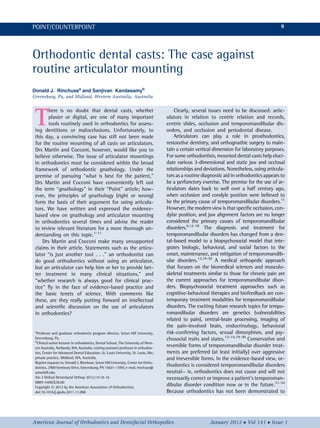
- 1. Orthodontic dental casts: The case against routine articulator mounting Donald J. Rinchusea and Sanjivan Kandasamyb Greensburg, Pa, and Midland, Western Australia, Australia T here is no doubt that dental casts, whether plaster or digital, are one of many important tools routinely used in orthodontics for assess- ing dentitions or malocclusions. Unfortunately, to this day, a convincing case has still not been made for the routine mounting of all casts on articulators. Drs Martin and Cocconi, however, would like you to believe otherwise. The issue of articulator mountings in orthodontics must be considered within the broad framework of orthodontic gnathology. Under the premise of pursuing “what is best for the patient,” Drs Martin and Cocconi have conveniently left out the term “gnathology” in their “Point” article; how- ever, the principles of gnathology (right or wrong) form the basis of their argument for using articula- tors. We have written and expressed the evidence- based view on gnathology and articulator mounting in orthodontics several times and advise the reader to review relevant literature for a more thorough un- derstanding on this topic.1-11 Drs Martin and Cocconi make many unsupported claims in their article. Statements such as the articu- lator “is just another tool . . .” an orthodontist can do good orthodontics without using an articulator, but an articulator can help him or her to provide bet- ter treatment in many clinical situations,” and “whether research is always good for clinical prac- tice” fly in the face of evidence-based practice and the basic tenets of science. With comments like these, are they really putting forward an intellectual and scientific discussion on the use of articulators in orthodontics? Clearly, several issues need to be discussed: artic- ulators in relation to centric relation and records, centric slides, occlusion and temporomandibular dis- orders, and occlusion and periodontal disease. Articulators can play a role in prosthodontics, restorative dentistry, and orthognathic surgery to main- tain a certain vertical dimension for laboratory purposes. For some orthodontists, mounted dental casts help eluci- date various 3-dimensional and static jaw and occlusal relationships and deviations. Nonetheless, using articula- tors as a routine diagnostic aid in orthodontics appears to be a perfunctory exercise. The premise for the use of ar- ticulators dates back to well over a half century ago, when occlusion and condyle position were believed to be the primary cause of temporomandibular disorders.11 However, the modern view is that specific occlusion, con- dylar position, and jaw alignment factors are no longer considered the primary causes of temporomandibular disorders.6,12-18 The diagnosis and treatment for temporomandibular disorders has changed from a den- tal-based model to a biopsychosocial model that inte- grates biologic, behavioral, and social factors to the onset, maintenance, and mitigation of temporomandib- ular disorders.12,19-30 A medical orthopedic approach that focuses on the biomedical sciences and musculo- skeletal treatments similar to those for chronic pain are the current approaches for temporomandibular disor- ders. Biopsychosocial treatment approaches such as cognitive-behavioral therapies and biofeedback are con- temporary treatment modalities for temporomandibular disorders. The exciting future research topics for tempo- romandibular disorders are genetics (vulnerabilities related to pain), central-brain processing, imaging of the pain-involved brain, endocrinology, behavioral risk-conferring factors, sexual dimorphism, and psy- chosocial traits and states.12-15,19-30 Conservative and reversible forms of temporomandibular disorder treat- ments are preferred (at least initially) over aggressive and irreversible forms. In the evidence-based view, or- thodontics is considered temporomandibular disorders neutral— ie, orthodontics does not cause and will not necessarily correct or improve a patient’s temporoman- dibular disorder condition now or in the future.31-34 Because orthodontics has not been demonstrated to a Professor and graduate orthodontic program director, Seton Hill University, Greensburg, Pa. b Clinical senior lecturer in orthodontics, Dental School, The University of West- ern Australia, Nedlands, WA, Australia; visiting assistant professor in orthodon- tics, Center for Advanced Dental Education, St. Louis University, St. Louis, Mo; private practice, Midland, WA, Australia. Reprint requests to: Donald J. Rinchuse, Seton Hill University, Center for Ortho- dontics, 2900 Seminary Drive, Greensburg, PA 15601-1599; e-mail, rinchuse@ setonhill.edu. Am J Orthod Dentofacial Orthop 2012;141:8-16 0889-5406/$36.00 Copyright Ó 2012 by the American Association of Orthodontists. doi:10.1016/j.ajodo.2011.11.008 American Journal of Orthodontics and Dentofacial Orthopedics January 2012 Vol 141 Issue 1 POINT/COUNTERPOINT 9
- 2. cause temporomandibular disorders, this further sup- ports the notion that occlusion and condyle position do not cause temporomandibular disorders: ie, the premise that orthodontics could cause temporoman- dibular disorders is grounded in the dental-based model. Of note, the evidence-based view does not argue that occlusion and condyle position have no relation- ship to temporomandibular disorders and that ortho- dontists should ignore them. The gross evaluation of the patient’s occlusion still are useful: “assessment of occlusion is necessary as part of the initial oral exami- nation to identify and eliminate gross occlusal discrep- ancies.”35 The amelioration of gross occlusal interferences that cause tooth mobility, fremitus, and deviation or deflection on mandibular closure and movement are within the scope of the evidence-based paradigm. Because there has been a paradigm shift regarding the etiology, diagnosis, and treatment of temporoman- dibular disorders away from occlusion and condyle posi- tion, occlusionists and gna- thologists should reconsider and abandon age-old views and techniques that are not supported by science and ev- idence. Certainly, the evidence-based view on the role of occlusion in relation to temporomandibular dis- orders should have had a negative impact on the rou- tine use of articulators in orthodontics.4,12 Greene16,17 wrote: “I would encourage orthodontists to be somewhat flexible in applying the standards of ideal jaw relationships as well as ideal occlusion rela- tionships to their finishing of patients . . . we should not have conversations about tenths of millimeters when discussing where the condyle should be. . . . The failure to produce some expert’s version of a so- called good/ideal occlusion or CR position is not a risk for developing TMD.” Does it make sense to focus and study occlusion and condyle position the same way that we did more than 50 years ago? The belief that centric occlusion or (or maximum in- tercuspation or intercuspal position) should be coinci- dent with an arbitrary centric relation position is not evidence-based. The preponderance of evidence sug- gests that for the population at large there is a range of acceptable positions and not 1 centric relation posi- tion that is optimal for every patient.5 That is, a partic- ular 3-dimensional position of the temporomandibular joint condyles in the glenoid fossa has not been found to predict temporomandibular disorders.5 Johnston36 wrote: “I know of no convincing evidence that condyles of patients with intact dentitions should be placed in CR or that once placed or that once having been placed there, the resulting improvement on nature will be sta- ble.” Interestingly, centric slides greater than 4 mm that have been found to be associated with temporoman- dibular joint arthropathies are most likely the result of the temporomandibular disorders rather than the cause, contrary to the beliefs of Drs Martin and Coc- coni.32 Many of the problems we have with articulators as diagnostic aids in orthodontics start before the articu- lator is actually used (Fig 1). Centric relation records can be demonstrated to be reliable, but there is no ev- idence to support their validity.5 Nonetheless, proper attention to an orthodontic patient’s centric records is an important consideration in orthodontics and all of dentistry. We do know that doctor-generated patient cen- tric records are more reliable than patient-generated re- cords, but they are also less valid (physiologic).5 In addi- tion, the same bite registra- tions used to make dental casts should be consistently used throughout all the pa- tient records—dental models, photographs, cephalometric radiographs, and so on. And, to merely ask orthodontic patients to bite on their back teeth will not always provide an accurate centric bite registration, and one might miss diagnosing a “Sunday bite.” Gnathologic centric records are static and not func- tional (Fig 1). Patients are not asked to exercise any rel- evant physiologic mandibular movements to generate centric relation records. Chewing kinematics are not evaluated, such as chewing pattern dynamics, which might determine whether a patient is more or less a ver- tical or horizontal chewer.4,37 Furthermore, the harsh- est occlusal forces are those generated via parafunctional mandibular movements, such as brux- ing and clenching. These movements and forces are not evaluated by centric relation records and articulator mountings.4 Based on information from the study of Alexander et al,38 clinicians, in general, cannot predict where con- dyles are positioned and recorded based on their partic- ular type of centric bite registration. This makes it imperative that orthodontic gnathologists provide The premise for the use of articula- tors dates back to well over a half century ago, when occlusion and condyle position were believed to be the primary cause of temporo- mandibular disorders. Counterpoint 11 American Journal of Orthodontics and Dentofacial Orthopedics January 2012 Vol 141 Issue 1
- 3. magnetic resonance imaging research data of the tem- poromandibular joints that substantiate that the sub- jects’ condyles are actually placed and recorded in positions determined by such registrations. In addition, the difference between gnathologic and nongnatho- logic diagnostics is on average as little as 1 mm or less, and this is mostly in the vertical dimension.39 Are such minor differences really a health concern? Also, in growing children, the temporomandibular joint condyle-glenoid fossa complex changes location with growth; the fossae on average are displaced posteriorly and inferiorly.40 One would therefore need to perform new mountings regularly throughout treatment to di- agnose and maintain their so-called ideal centric rela- tion position. This however does not occur. The shortcomings of articulators in orthodontics were discussed in detail in our article and are listed in Figure 2.4 Diseases of the temporomandibular joint such as disc displacement and osteoarthrosis are best di- agnosed with magnetic resonance imaging of the tem- poromandibular joints and a clinical examination, and not with articulators. The major premise for the use of articulators is based on the incorrect concept of a “ter- minal hinge axis” of Posselt41 dating back more than a half century. Posselt conjectured that, in the initial 20 mm or so of opening and closing, the mandible (con- dyles) only rotates similar to a door hinge (and does not simultaneously translate). However, Posselt’s theory was made in the era when centric relation was consid- ered a retruded, posterior position of the condyles in the glenoid fossa. During this time period, retruded cen- tric relation was recorded with distal guided pressure applied to the chin, a probable reason for Posselt’s find- ing of a “terminal hinge axis.” In 1995, Lindauer et al42 demonstrated that, during opening and closing, the condyles not only rotate, but also simultaneously Fig 1. Limitations of centric relation, condyle position records, and measurements. Fig 2. Limitations of articulators. Counterpoint 13 American Journal of Orthodontics and Dentofacial Orthopedics January 2012 Vol 141 Issue 1
- 4. translate (move downward and forward). They demon- strated that the terminal hinge axis does not exist, and their findings support an “instantaneous center of rota- tion” that varies in every patient and cannot be simu- lated via an articulator (Fig 2).4 Drs Martin and Cocconi failed to point out that an association between “occlusal discrepancies and the progression of periodontal disease” is controversial. In this respect, they referenced an article by Harrel et al43 entitled “Is there an association between occlu- sion and periodontal destruction? Yes—occlusal forces can contribute to periodontal destruction.” Unfortu- nately, this was only 1 side of a debate published in the Journal of the American Dental Association in October 2006; they neglected to mention the accom- panying counterpoint article in which Deas and Mealey44 discussed several clinical studies with results contradicting those of Harrel and Nunn,45 Nunn,46 Nunn and Harrel.47 As Deas and Mealey44 pointed out, “the determining factor of whether an occlusal contact produces occlusal trauma is the presence of periodontal injury, not the physical manifestation of the teeth, temporomandibular joints or muscles of mastication.” Even Harrel et al43 acknowledged the limitations of their research: “Our study should be viewed in the context of its design. It does not meet the level of what is considered the gold standard of clinical research. . . . Our study was retrospective in na- ture, a single practitioner performed all evaluations and data gathering. Furthermore, the patients’ oral hygiene and maintenance compliance was not standardized. All these are significant concerns regarding our research design.” Interestingly, according to the research by Nunn44 and Deas and Mealey,43 the recording of sub- jects’ occlusal discrepancies was not determined by the use articulated mounted dental casts in this research. So we ask Drs Martin and Cocconi, if they think it so important to use articulators to deduce occlusal dis- crepancies, why did they cite and focus on research that did not use articulators as the clinical tool of choice for recording such discrepancies? CONCLUSIONS If we know that specific condylar positions, slides, and the minute details of occlusion are not predictive of temporomandibular disorders, that articulators do not simulate condylar movements accurately, and that centric relation records do not really place condyles in their deemed positions, then what are we actually trying to achieve by routinely mounting dental casts on artic- ulators? Such a mechanistic and instrument-oriented approach to patient diagnosis and treatment planning is only a disservice to patients. The burden of proof is and should always lie on those who are advocating a controversial tool or procedure. So we ask the readers, did Drs Martin and Cocconi present a convincing argu- ment for the routine use of articulators in orthodontics based on scientific and evidence-based considerations rather than on anecdotes and blind rhetoric? The con- vincing evidence is against the routine mounting of casts on articulators. What would William Shakespeare say, as Hamlet’s famous quote is placed in the context of this debate on articulators? Perhaps it is whether one chooses “to believe or not to believe.” REFERENCES 1. Rinchuse DJ, Rinchuse DJ, Kandasamy S. Evidence-based versus experience-based views on occlusion and TMD. Am J Orthod Dentofacial Orthop 2005;127:249-54. 2. Rinchuse DJ, Sweitzer EM, Rinchuse DJ, Rinchuse DL. Under- standing science and evidence-based decision making in or- thodontics. Am J Orthod Dentofacial Orthop 2005;127: 618-24. 3. Rinchuse DJ, Rinchuse DJ. Developmental occlusion, orthodontic interventions, and orthognathic surgery for adolescents. Dent Clin North Am 2006;50:69-86. 4. Rinchuse DJ, Kandasamy S. Articulators in orthodontics: an evidence-based perspective. Am J Orthod Dentofacial Orthop 2006;129:299-308. 5. Rinchuse DJ, Kandasamy S. Centric relation: a historical and con- temporary orthodontic perspective. J Am Dent Assoc 2006;137: 494-501. 6. Rinchuse DJ, McMinn J. Summary of evidence-based systematic reviews of temporomandibular disorders. Am J Orthod Dentofa- cial Orthop 2006;130:715-20. 7. Rinchuse DJ, Kandasamy S, Sciote J. A contemporary and evidence-based view of canine protected occlusion. Am J Orthod Dentofacial Orthop 2007;132:90-102. 8. Rinchuse DJ, Rinchuse DJ, Kandasamy S, Ackerman MB. Decon- structing evidence in orthodontics: making sense of systematic reviews, randomized clinical trials (RCTs) and meta-analyses. World J Orthod 2008;9:167-76. 9. Rinchuse DJ, Kandasamy S. Implications of the inclination of the mandibular first molars in the extractionist versus expansionist debate. World J Orthod 2008;9:383-90. 10. Rinchuse DJ, Kandasamy S. The myths of orthodontic gnathol- ogy. Am J Orthod Dentofacial Orthop 2009;136:322-30. 11. Rinchuse DJ, Kandasamy S. Orthodontics and TMD management. In: Manfredini D, editor. Current concepts on temporomandibu- lar disrorders. Chicago: Quintessence; 2010. p. 429-46. 12. Klasser GD, Greene CS. Predoctoral teaching of temporomandib- ular disorders. J Am Dent Assoc 2007;138:231-7. 13. Turp JC, Greene CS, Strub JR. Dental occlusion: a critical reflec- tion on past, present and future concepts. J Oral Rehabil 2008; 35:446-53. 14. Greene CS. Managing the care of patients with temporomandib- ular disorders: a new guideline for care. J Am Dent Assoc 2010; 141:1086-8. 15. Klasser GD, Greene CS. Role of oral appliances in the manage- ment of sleep bruxism and temporomandibular disorders. Alpha Omegan 2007;100:111-9. Counterpoint 15 American Journal of Orthodontics and Dentofacial Orthopedics January 2012 Vol 141 Issue 1
- 5. 16. Greene CS. Relationship between occlusion and temporoman- dibular disorders: implications for the orthodontist. Am J Orthod Dentofacial Orthop 2011;139:11-6. 17. Greene CS. Author’s response. Am J Orthod Dentofacial Orthop 2011;139:426. 18. Gesch D, Bernhardt O, Kirbshus A. Association of malocclusion and functional occlusion with temporomandibular disorders (TMD) in adults: a systematic review of population-based stud- ies. Quintessence Int 2004;35:211-21. 19. Mishra KD, Gatchel RJ, Gardea MA. The relative efficacy of three cognitive-behavioral treatment approaches to temporomandibu- lar disorders. J Behav Med 2000;23:293-309. 20. Fernandez E, Turk DC. The utility of cognitive coping strategies for altering pain perception: a meta-analysis. Pain 1989;38:123-35. 21. Flor H, Birbaumer N. Comparison of the efficacy of electromyo- graphic biofeedback, cognitive-behavioral therapy, and conser- vative medical interventions in the treatment of chronic musculoskeletal pain. J Consult Clin Psychol 1993;61:653-8. 22. Dworkin SF, Massoth DL. Temporomandibular disorders and chronic pain: disease or illness? J Prosthet Dent 1994;72:29-38. 23. Dworkin SF, Turner JA, Wilson L, Massoth D, Whitney C, Huggins KH, et al. Brief group cognitive-behavioral intervention for temporomandibular disorders. Pain 1994;59:175-87. 24. Turk D, Zaki H, Rudy T. Effects of intraoral appliance and bio- feedback/stress management alone and in combination in treat- ing pain and depression in TMD patients. J Prosthet Dent 1993; 70:158-64. 25. Dworkin SF, Turner JA, Manci L, Wilson L, Massoth D, et al. A randomized clinical trial of a tailored comprehensive care treat- ment program for temporomandibular disorders. J Orofac Pain 2002;16:259-76. 26. Turk DC, Rudy TE, Kubinski JA, Zaki HS, Greco CM. Dysfunc- tional patients with temporomandibular disorders: evaluating the efficacy of a tailored treatment protocol. J Consult Clin Psy- chol 1996;64:139-46. 27. Rudy TE, Turk DC, Kubinski JA, Zaki HS. Differential treatment response of TMD patients as a function of psychological charac- teristics. Pain 1995;61:103-12. 28. Dworkin SF, LeResche L. Research diagnostics criteria for tempo- romandibular disorders: review, criteria, examinations and specifications, critique. J Craniomandib Disord 1992;6:301-55. 29. Gardea MA, Gatchel RJ, Mishra KD. Long-term efficacy of biobe- havioral treatment of temporomandibular disorders. J Behav Med 2001;24:341-59. 30. Gatchel RJ, Stowell AW, Wildenstein L, Riggs R, Ellis E III. Efficacy of an early intervention for patients with acute temporomandib- ular disorder-related pain—a one year outcome study. J Am Dent Assoc 2006;137:339-47. 31. Gianelly AA. Orthodontics, condylar position, and TMJ status. Am J Orthod Dentofacial Orthop 1989;95:521-3. 32. McNamara JA Jr, Seligman DA, Okeson JP. Occlusion, orthodon- tic treatment, and temporomandibular disorders: a review. J Or- ofac Pain 1995;9:73-90. 33. Kim MR, Graber TM, Vianna MA. Orthodontics and temporo- mandibular disorders: a meta-analysis. Am J Orthod Dentofacial Orthop 2002;121:438-46. 34. Reynders RM. Orthodontics and temporomandibular disorders: a review of the literature (1966-1988). Am J Orthod Dentofacial Orthop 1990;97:463-71. 35. National Institutes of Health Technology Assessment Conference Statement. Management of temporomandibular disorders. J Am Dent Assoc 1995;127:1595-606. 36. Johnston LE Jr. Fear and loathing in orthodontics. Notes on the death of theory. In: Carlson DS, editor. Craniofacial Growth Se- ries. Ann Arbor: Center for Human Growth and Development; University of Michigan; 1990. p. 75-90. 37. Buschang PH, Throckmorton GS, Austin D, Wintergerst AM. Chewing cycle kinematics of subjects with deepbite malocclu- sions. Am J Orthod Dentofacial Orthop 2007;131:627-34. 38. Alexander SR, Moore RN, DuBois LM. Mandibular condyle posi- tion: comparison of articulator mountings and magnetic reso- nance imaging. Am J Orthod Dentofacial Orthop 1993;104:230-9. 39. Kulbersh R, Kaczynski R, Freeland T. Orthodontics and gnathol- ogy. Semin Orthod 2003;9:93-5. 40. Buschang P, Santos-Pinto A. Condylar growth and glenoid fossa displacement during childhood and adolescence. Am J Orthod Dentofacial Orthop 1998;113:437-42. 41. Posselt U. Studies in the mobility of the human mandible. Acta Odontol Scand 1952;10(Suppl 10):1-160. 42. Lindauer SJ, Isaacson RJ, Davidovich M. Condylar movement and mandibular rotation during jaw opening. Am J Orthod Dentofa- cial Orthop 1995;107:573-7. 43. Harrel SK, Nunn ME, Hallmon WW. Is there an association be- tween occlusion and periodontal destruction? Yes—occlusal forces can contribute to periodontal destruction. J Am Dent As- soc 2006;137:1380-92. 44. Deas DE, Mealey BL. Is there an association between occlusion and periodontal destruction? Only in limited circumstances does occlusal force contribute to periodontal disease progres- sion. J Am Dent Assoc 2006;137:1381-9. 45. Harrel SK, Nunn ME. The effect of occlusal discrepancies on periodontitis. II. Relationship of initial occlusal discrepancies to initial clinical parameters. J Periodontol 2001;72: 495-505. 46. Nunn ME. Non-working occlusal discrepancies are associated with increased probing depths and attachment loss. J Evid Based Dent Pract 2007;7:81-3. 47. Nunn ME, Harrel SK. The effect of occlusal discrepancies on pe- riodontitis. I. Relationship of initial occlusal discrepancies to ini- tial clinical parameters. J Periodontol 2001;72:485-94. 16 Counterpoint January 2012 Vol 141 Issue 1 American Journal of Orthodontics and Dentofacial Orthopedics