Epidemiology of kfd, brucellosis and leptospirosis
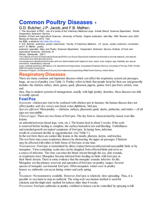
Common poultry diseases 1
1. Common Poultry Diseases 1
G.D. Butcher, J.P. Jacob,and F.B. Mather2
1. This document is PS47, one of a series of the Veterinary Medicine-Large Animal Clinical Sciences Department, Florida
Cooperative Extension Service,
Institute of Food and Agricultural Sciences, University of Florida. Original publication date May 1999. Revised June 2003.
Review ed February 2012. Visit
the EDIS w ebsite at http://edis.ifas.ufl.edu.
2. G.D. Butcher, extension poultry veterinarian, Faculty of Veterinary Medicine, J.P. Jacob, poultry extension coordinator,
and F. B. Mather, poultry
extension specialist, Dairy and Poultry Sciences Department, Cooperative Extension Service, Institute of Food and
Agricultural Sciences, University of
Florida, Gainesville, 32611.
The Institute of Food andAgricultural Sciences(IFAS) is an Equal Opportunity Institutionauthorized to provide research, educational
informationand other servicesonly to
individualsand institutionsthat function withnon-discriminationwith respect to race, creed, color, religion, age, disability, sex, sexual
orientation, marital status, national
origin, political opinionsor affiliations. U.S. Departmentof Agriculture, Cooperative ExtensionService,University of Florida,IFAS, Florida
A&M University Cooperative
Extension Program, andBoardsof County CommissionersCooperating. Millie Ferrer-Chancy, Interim Dean
RespiratoryDiseases
There are many common and important diseases which can affect the respiratory system (air passages,
lungs, air sacs) of poultry (see Table 1). Poultry refers to birds that people keep for their use and generally
includes the chicken, turkey, duck, goose, quail, pheasant, pigeon, guinea fowl, pea fowl, ostrich, emu
and
rhea. Due to modern systems of management, usually with high poultry densities, these diseases are able
to readily spread.
Fowl Pox
Synonyms :chicken pox (not to be confused with chicken pox in humans; the human disease does not
affect poultry and vice versa),sore head, avian diphtheria, bird pox
Species affected :Most poultry -- chickens, turkeys, pheasants,quail, ducks, psittacine, and ratites -- of all
ages are susceptible.
Clinical signs :There are two forms of fowl pox. The dry form is characterized by raised, wart-like
lesions
on unfeathered areas (head,legs, vent, etc.). The lesions heal in about 2 weeks. If the scab
is removed before healing is complete, the surface beneath is raw and bleeding. Unthriftiness
and retarded growth are typical symptoms of fowl pox. In laying hens, infection
results in a transient decline in egg production (see Table 1 ).
In the wet form there are canker-like lesions in the mouth, pharynx, larynx, and trachea.
The wet form may cause respiratory distress by obstructing the upper air passages. Chickens
may be affected with either or both forms of fowl pox at one time.
Transmission :Fowl pox is transmitted by direct contact between infected and susceptible birds or by
mosquitos. Virus-containing scabs also can be sloughed from affected birds and serve as
a source of infection. The virus can enter the blood stream through the eye, skin wounds,
or respiratory tract. Mosquitos become infected from feeding on birds with fowl pox in
their blood stream. There is some evidence that the mosquito remains infective for life.
Mosquitos are the primary reservoir and spreaders of fowl pox on poultry ranges. Several
species of mosquito can transmit fowl pox. Often mosquitos winter-over in poultry
houses so, outbreaks can occur during winter and early spring.
2
Treatment : No treatment is available. However,fowl pox is relatively slow-spreading. Thus, it is
possible to vaccinate to stop an outbreak. The wing-web vaccination method is used for
chickens and the thigh-stick method for turkeys older than 8 weeks.
Prevention: Fowlpox outbreaks in poultry confined to houses can be controlled by spraying to kill
2. mosquitos. However,if fowl pox is endemic in the area,vaccination is recommended. Do
not vaccinate unless the disease becomes a problem on a farm or in the area. Refer to the
publication PS-36 (Vaccination of Small Poultry Flocks) for more information on fowl pox
vaccinations.
Newcastle Disease
Synonyms: pneumoencephalitis
The highly contagious and lethal form of Newcastle disease is known as viscerotropic (attacks
the internal organs) velogenic Newcastle disease,VVND,exotic Newcastle disease,
or Asiatic Newcastle disease. VVND is not present in the United States poultry industry at
this time.
Species affected: Newcastle disease affects allbirds of all ages. Humans and other mammals are also
susceptible to Newcastle. In such species, it causes a mild conjunctivitis.
Clinical signs: There are three forms of Newcastle disease -- mildly pathogenic (lentogenic), moderately
pathogenic (mesogenic) and highly pathogenic (velogenic). Newcastle disease is characterized
by a sudden onset of clinical signs which include hoarse chirps (in chicks), watery
discharge from nostrils, labored breathing (gasping), facial swelling, paralysis, trembling,
and twisting of the neck (sign of centralnervous system involvement). Mortality ranges
from 10 to 80 percent depending on the pathogenicity. In adult laying birds, symptoms
can include decreased feed and water consumption and a dramatic drop in egg production
(see Table 1).
Transmission: The Newcastle virus can be transmitted short distances by the airborne route or
introduced on contaminated shoes, caretakers,feed deliverers, visitors, tires, dirty equipment,
feed sacks,crates,and wild birds. Newcastle virus can be passed in the egg, but
Newcastle-infected embryos die before hatching. In live birds, the virus is shed in body
fluids, secretions, excreta,and breath.
Treatment: There is no specific treatment for Newcastle disease. Antibiotics can be given for 3-5 days
to prevent secondary bacterial infections (particularly E. coli ). For chicks, increasing the
brooding temperature 5°F may help reduce losses.
Prevention: Prevention programs should include vaccination (see publication PS-36, Vaccination of
Small Poultry Flocks), good sanitation, and implementation of a comprehensive biosecurity
program.
Infectious Bronchitis
Synonyms: IB,bronchitis, cold
Species affected: Infectious bronchitis is a disease of chickens only. A similar disease occurs in bobwhite
quail (quail bronchitis), but it is caused by a different virus.
Clinical signs: The severity of infectious bronchitis infection is influenced by the age and immune status
of the flock, by environmental conditions, and by the presence of other diseases. Feed and
water consumption declines. Affected chickens will be chirping, with a watery discharge
from the eyes and nostrils, and labored breathing with some gasping in young chickens.
Breathing noises are more noticeable at night while the birds rest. Egg production drops
dramatically. Production will recover in 5 or 6 weeks,but at a lower rate. The infectious
bronchitis virus infects many tissues of the body, including the reproductive tract (see
3
Table 1). Eggshells become rough and the egg white becomes watery. (See publication
PS-24, Egg Quality, for other causes of poor egg quality.)
Transmission: Infectious bronchitis is a very contagious poultry disease. It is spread by air, feed bags,
infected dead birds, infected houses, and rodents. The virus can be egg-transmitted,
however, affected embryos usually will not hatch.
Treatment: There is no specific treatment for infectious bronchitis. Antibiotics for 3-5 days may aid in
combating secondary bacterial infections. Raise the room temperature 5°F for broodingage
3. chickens until symptoms subside. Baby chicks can be encouraged to eat by using a
warm, moist mash.
Prevention: Establish and enforce a biosecurity program. Vaccinations are available.
Quail Bronchitis
Synonyms: none
Species affected: Bobwhite quail are affected. Japanese corturnix quail are resistant. The disease is
prevalent
in the southern states where bobwhite quail are common. Quail bronchitis occurs
seasonally as new hatches and broods come along each year.
Clinical signs: Respiratory distress occurs with trachealrales (rattles), sneezing, and coughing. Feed and
water consumption declines dramatically. There can also be conjunctivitis (inflammation
of the eye). Loose watery feces are seen in older and sub-acutely affected birds. Nasal
discharges are not seen,differentiating quail bronchitis from similar diseases in other
poultry (see Table 1).
Transmission: Once infected,quail bronchitis remains on the farm for the duration of the breeding
season,infecting each successive brood.
Treatment: There is no specific treatment against quail bronchitis. Quail bronchitis infections are
often complicated by concurrent mycoplasma infections. Antibiotics can be used to
combat secondary infections. Add tylosin (500g/ton) to the feed for 10 days, withhold
the medication for 5 days, and then repeat medication for 5 days. Alternate medication
regimens are tylosin (Tylan) or erythromycin (Gallimycin) in the drinking water for the
same period of time.
Prevention: There is no commercial vaccine on the market. It is necessary to break the cycle by
depopulating and thoroughly cleaning and disinfecting pens and equipment, followed by
a 30-90 day quarantine of the facilities.
Avian Influenza
Synonyms: AI,flu, influenza, fowl plague
Species affected: Avian influenza can occur in most, if not all, species of birds.
Clinical signs: Avian influenza is categorized as mild or highly pathogenic. The mild form produces
listlessness, loss of appetite, respiratory distress, diarrhea, transient drops in egg production,
and low mortality. The highly pathogenic form produces facial swelling, blue comb
and wattles, and dehydration with respiratory distress. Dark red/white spots develop in
the legs and combs of chickens. There can be blood-tinged discharge from the nostrils.
Mortality can range from low to near 100 percent. Sudden exertion adds to the total
mortality. Egg production and hatchability decreases. There can be an increase in
production of soft-shelled and shell-less eggs (see Table 1).
Transmission: The avian influenza virus can remain viable for long periods of time at moderate
temperatures
and can live indefinitely in frozen material. As a result, the disease can be spread
through improper disposal of infected carcasses and manure. Avian influenza can be
spread by contaminated shoes, clothing, crates,and other equipment. Insects and rodents
may mechanically carry the virus from infected to susceptible poultry.
Treatment: There is no effective treatment for avian influenza. With the mild form of the disease,
good husbandry, proper nutrition, and broad spectrum antibiotics may reduce losses from
secondary infections. Recovered flocks continue to shed the virus. Vaccines may only be
used with special permit.
Prevention: A vaccination program used in conjunction with a strict quarantine has been used to
control mild forms of the disease. With the more lethal forms, strict quarantine and rapid
destruction of all infected flocks remains the only effective method of stopping an avian
influenza outbreak. If you suspect you may have Avian Influenza in your flock, even the
4. mild form, you must report it to the state veterinarian’s office. A proper diagnosis of avian
influenza is essential. Aggressive action is recommended even for milder infections as this
virus has the ability to readily mutate to a more pathogenic form.
For more information on avian influenza, refer to publication PS-38 (Avian Influenza in
Poultry Species).
Infectious Coryza
Synonyms: roup,cold, coryza
Species affected: chickens, pheasants,and guinea fowl. Common in game chicken flocks.
Clinical signs: Swelling around the face,foul smelling, thick, sticky discharge from the nostrils and eyes,
labored breathing, and rales (rattles -- an abnormal breathing sound) are common clinical
signs. The eyelids are irritated and may stick together. The birds may have diarrhea and
growing birds may become stunted (see Table 1).
Mortality from coryza is usually low, but infections can decrease egg production and
increase the incidence and/or severity of other diseases. Mortality can be as high as 50
percent, but is usually no more than 20 percent. The clinical disease can last from a few
days to 2-3 months, depending on the virulence of the pathogen and the existence of other
infections such as mycoplasmosis.
Transmission: Coryza is primarily transmitted by direct bird-to-bird contact. This can be from infected
birds brought into the flock as well as from birds which recover from the disease which
remain carriers of the organism and may shed intermittently throughout their lives.. Birds
risk exposure at poultry shows, bird swaps,and live-bird sales. Inapparent infected adult
birds added into a flock are a common source for outbreaks. Within a flock, inhalation of
airborne respiratory droplets, and contamination of feed and/or water are common modes
of spread.
Treatment: Water soluble antibiotics or antibacterials can be used. Sulfadimethoxine (Albon®, Di-
Methox™) is the preferred treatment. If it is not available, or not effective,sulfamethazine
(Sulfa-Max®, SulfaSure™), erythromycin (gallimycin®), or tetracycline (Aureomycin®) can
be used as alternative treatments. Sulfa drugs are not FDA approved for pullets older than
14 weeks of age or for commercial layer hens. While antibiotics can be effective in reducing
clinical disease, they do not eliminate carrier birds.
Prevention: Good management and sanitation are the best ways to avoid infectious coryza. Most
outbreaks occur as a result of mixing flocks. All replacement birds on “coryza-endemic”
farms should be vaccinated. The vaccine (Coryza-Vac) is administered subcutaneously
(under the skin) on the back of the neck. Each chicken should be vaccinated four times,
starting at 5 weeks of age with at least 4 weeks between injections. Vaccinate again at 10
months of age and twice yearly thereafter.
5
Infectious Laryngotracheitis
Synonyms: LT,ILT, trach,laryngo
Species affected: Chickens and pheasants are affected by LT. Chickens 14 weeks and older are more
susceptible
than young chickens. Most LT outbreaks occur in mature hens. In recent years,LT
has also caused significant respiratory problems in broilers greater than 3 weeks of age,
especially during the cooler seasons of the year. This is believed to be due to unwanted
spread of LT vaccines between poultry flocks.
Clinical signs: The clinical sign usually first noticed is watery eyes. Affected birds remain quiet because
breathing is difficult. Coughing, sneezing, and shaking of the head to dislodge exudate
plugs in the windpipe follow. Birds extend their head and neck to facilitate breathing
(commonly referred to as “pump handle respiration”). Inhalation produces a wheezing
and gurgling sound. Blood-tinged exudates and serum clots are expelled from the trachea
5. of affected birds. Many birds die from asphyxiation due to a blockage of the trachea when
the trachealplug is freed (see Table 1).
Transmission: LT is spread by the respiratory route. LT is also spread from flock to flock by
contaminated
clothing, shoes, tires, etc. Birds that recover should be considered carriers for life.
LT may be harbored in speciality poultry such as exhibition birds and game fowl.
Treatment: Incinerate dead birds, administer antibiotics to control secondary infection, and vaccinate
the flock. Mass vaccination by spray or drinking water method is not recommended for
large commercial or caged flocks. Individual bird administration by the eye-drop route
is suggested. Follow manufacturers instructions. In small poultry flocks, use a swab to
remove plug from gasping birds, and vaccinate by eye-drop method.
Prevention: Vaccinate replacement birds for outbreak farms. Vaccination for LT is not as successful
as for other disease, but is an excellent preventive measure for use in outbreaks and in
epidemic areas. Refer to the publication PS-36 (Vaccination of Small Poultry Flocks) for
more information on LT vaccinations.
TurkeyRhinotracheitis
Synonyms: TRT,rhino tracheitis
Species affected: Turkeys of all ages are susceptible, but the disease is most severe in young poults.
Chickens
are susceptible to the virus. Experimentally, guinea fowl and pheasants are susceptible,
but waterfowl and pigeons are resistant.
Clinical signs: Respiratory signs in poults include snicking, rales, sneezing, nasal exudates (often frothy),
foamy conjunctivitis, and sinusitis. Drops in egg production can be as much as 70 percent
(see Table 1).
Transmission: Spread is primarily by contact with contaminated environments, feed and water,recovered
birds, equipment, and personnel.
Treatment: No drugs are available to combat the virus. Antibiotic therapy is recommended to control
secondary bacterial infections.
Prevention: No vaccines are currently available. Prevention is dependent on a comprehensive biosecurity
program.
Chlamydiosis
Synonyms: ornithosis, psittacosis, parrot fever.
The disease was called psittacosis or parrot fever when diagnosed in psittacine (curvebeaked)
birds, and called ornithosis when diagnosed in all other birds or in humans.
Currently, the term chlamydiosis is used to describe infections in any animal.
6
Species affected: Affected species include turkeys, pigeons, ducks, psittacine (curve-beaked) birds,
captive
and aviary birds, many other bird species, and other animals. Chickens are not commonly
affected. Humans are susceptible, especially older and immunosuppressed individuals
who are at a higher risk. Chlamydiosis in humans is an occupational disease of turkey
growers, haulers, and processing workers in the live-bird areas and of workers in pet-bird
aviaries although the incidence is rare. For more information, refer to publication PS-23
(Avian Diseases Transmissible to Humans).
Clinical signs: Clinical signs in most birds include nasal-ocular discharge, conjunctivitis, sinusitis,
diarrhea, weakness,loss of body weight, and a reduction in feed consumption. In turkeys
there is also respiratory distress and loose yellow to greenish-yellow colored droppings.
Chylamydiosis runs rather slowly through turkey flocks, with a maximum incidence of
around 50 percent (see Table 1).
Transmission: The primary means of transmission is through inhalation of fecaldust and respiratory
6. tract secretions. It can also be transmitted on contaminated clothing and equipment.
Recovered birds remain carriers and will continue to intermittently shed the infective
agent for long periods after clinical signs have subsided. Environmental stress may
provoke a reoccurrence of the disease.
Treatment: Chlorotetracycline can be given in the feed (200-400 g/ton) for 3 weeks. Other antibiotics
are usually ineffective. Recovered birds are safe for processing. Permanent lesions on the
heart and liver are not infectious. FDA withdrawal periods for medications used must be
strictly observed to avoid residual chemicals in the tissues.
Prevention: There is no vaccine. Have a good biosecurity program, excluding wild birds as much as
possible.
Swollen Head Syndrome
Synonyms: Facialcellulitis, thick head, Dikkop, SHS
Species affected: Chickens and turkeys are the known natural hosts. Experimentally, guinea fowl and
pheasants are susceptible but pigeons, ducks, and geese are resistant to the infection.
SHS does not presently occur in the United States,but is present in most countries of the
world.
Clinical signs: In chicks and poults, there is initial sneezing, followed by reddening and swelling of the
tear ducts and eye tissue. Facial swelling will extend over the head and down the jaw
and wattles. Adult chickens have mild respiratory disease followed by a few birds having
swollen heads. Other signs include disorientation, twisting of the neck, and a significant
drop in egg production (see Table 1).
Transmission: The infection spreads by direct contact with infected birds or indirectly by exposure to
infectious material.
Treatment: There is no proven medication for swollen head syndrome. The disease is caused by a
virus classified as a pneumovirus. A disease closely mimicking SHS is caused by a mixed
infection of respiratory viruses and specific bacteria. Antibiotic therapy may be helpful
against the bacterial component.
Prevention: A commercialvaccine is available. Swollen head syndrome is considered an exotic disease
and a live vaccine is not approved for use in the United States.
Synonyms: MG,chronic respiratory disease (CRD),infectious sinusitis, mycoplasmosis
Species affected: chickens,turkeys, pigeons, ducks, peafowl and passerine birds.
Clinical signs: Clinical symptoms vary slightly between species. Infected adult chickens may show no
outward signs if infection is uncomplicated. However,sticky, serous exudate from nostrils,
foamy exudate in eyes,and swollen sinuses can occur, especially in broilers. The air sacs
7
may become infected. Infected birds can develop respiratory rales and sneeze. Affected
birds are often stunted and unthrifty (see Table 1).
There are two forms of this disease in the turkey. With the “upper form” the birds
have watery eyes and nostrils, the infraorbitals (just below the eye) become swollen,
and the exudate becomes caseous and firm. The birds have respiratory rales and show
unthriftiness.
With the “lower form”, infected turkeys develop airsacculitis. As with chickens, birds
can show no outward signs if the infection is uncomplicated. Thus, the condition may
go unnoticed until the birds are slaughtered and the typical legions are seen. Birds with
airsacculitis are condemned.
MG in chicken embryos can cause dwarfing, airsacculitis, and death.
Transmission: MGcan be spread to offspring through the egg. Most commercial breeding flocks,
however,
are MG-free. Introduction of infected replacement birds can introduce the disease to
MG-negative flocks. MG can also be spread by using MG-contaminated equipment.
Treatment : Outbreaks of MG can be controlled with the use of antibiotics. Erythromycin, tylosin,
7. spectinomycin, and lincomycin all exhibit anti-mycoplasma activity and have given good
results. Administration of most of these antibiotics can be by feed,water or injection.
These are effective in reducing clinical disease. However,birds remain carriers for life.
Prevention: Eradication is the best control of mycoplasma disease. The National Poultry Improvement
Plan monitors all participating chicken and turkey breeder flocks.
Mycoplasmasynoviae
Synonyms: MS,infectious synovitis, synovitis, silent air sac
Species affected: chickens and turkeys.
Clinical signs: Birds infected with the synovitis form show lameness, followed by lethargy, reluctance to
move, swollen joints, stilted gait, loss of weight, and formation of breast blisters. Birds
infected with the respiratory form exhibit respiratory distress. Greenish diarrhea is common
in dying birds (see Table 1). Clinically, the disease in indistinguishable from MG.
Transmission: MS is transmitted from infected breeder to progeny via the egg. Within a flock, MS is
spread by direct contact with infected birds as well as through airborne particles over
short distances.
Treatment: Recovery is slow for both respiratory and synovitis forms. Several antibiotics are variably
effective. The most effective are tylosin, erthromycin, spectinomycin, lincomycin,
and chlorotectracycline. These antibiotics can be given by injection while some can be
administered in the feed or drinking water. These treatments are most effective when the
antibiotics are injected.
Prevention: Eradication is the best and only sure control. Do not use breeder replacements from
flocks that have had MS. The National Poultry Improvement Plan monitors for MS.
Mycoplasmameleagridis
Synonyms: MM,N strain, H strain
Species affected: MM affects turkeys of all ages, although poults are affected more severely than mature
turkeys. Recently, MM has been shown to infect pigeon, quail and peafowl.
Clinical signs: A drop-off in production and hatchability can be expected in breeder flocks. There can be
very high mortality in young poults. Unthriftiness, respiratory distress, stunting, crooked
neck with deformity of cervical vertebrae,and leg deformation are common in young
birds (see Table 1).
Transmission: Egg transmission is low in the early breeding period, but rises as the the age of the flock
increases. Infections can be introduced into a flock by contaminated equipment, shoes,
and clothing of workers and visitors.
Treatment: Severalantibiotics have been effective including tylosin, erythromycin, spectinomycin,
and linco-spectinomycin.
Prevention: The best preventive measure is to keep MM-free breeders. The MM-free status of breeders
can be confirmed by periodic blood tests through the National Poultry Improvement Plan.
Aspergillosis
Synonyms: brooder pneumonia, mycotic pneumonia, fungal pneumonia, Aspergillus . When the
source of the disease is the hatchery,the disease is called brooder pneumonia. In older
birds, the disease is called aspergillosis.
Species affected: Allbirds (domestic poultry, pigeons, canary and zoo bird species), animals, humans, and
plants are susceptible.
Clinical signs: Aspergillosis occurs as an acute disease of young birds and a chronic disease in mature
birds. Young birds have trouble breathing and gasp for air. Characteristically, there are
no rales or respiratory sounds associated with aspergillosis. Feed consumption decreases.
Occasionally there is paralysis or convulsions caused by the fungal toxin. Mortality in
young birds averages 5-20 percent,but may be as high as 50 percent. Mature birds also
have respiratory distress, reduced feed consumption, and may have a bluish and dark
8. color of the skin (cyanosis). Nervous disorders, such as twisted necks, may occur in a few
birds (see Table 1). Mortality in mature birds is usually less than 5 percent.
Transmission: Aspergillosis is caused by a fungus. The fungus grows well at room temperature and
higher. All litter and nest materials (peat moss, peanut hulls, sawdust, peat, bark, straw)
have been known to have been contaminated with aspergillus. Feed and water should be
suspect when attempting to identify the source of contamination.
Treatment : There is no cure for infected birds. The spread can be controlled by improving ventilation,
eliminating the source of the infection, and adding a fungistat (mycostatin, mold curb,
sodium or calcium propionate, or gentian violet) to the feed and/or copper sulfate or
acidified copper in the drinking water for 3 days. The litter can be sprayed lightly with an
oil-base germicide to control dust and air movement of fungal spores.
Prevention: It is important to thoroughly clean and disinfect the brooding area between broods. Use
only clean litter, preferably soft wood shavings. Do not use sawdust, litter high in bark
content, or shavings that have been wet.
Viral Diseases (nonrespiratory)
Marek’s Disease
Synonyms: acute leukosis, neural leukosis, range paralysis, gray eye (when eye affected)
Species affected: Chickens between 12 to 25 weeks of age are most commonly clinically affected.
Occasionally
pheasants,quail, game fowl and turkeys can be infected.
Clinical signs: Marek’s disease is a type of avian cancer. Tumors in nerves cause lameness and paralysis.
Tumors can occur in the eyes and cause irregularly shaped pupils and blindness. Tumors
of the liver, kidney, spleen, gonads, pancreas,proventriculus, lungs, muscles, and skin
can cause incoordination, unthriftiness, paleness, weak labored breathing, and enlarged
feather follicles. In terminal stages, the birds are emaciated with pale, scaly combs and
greenish diarrhea (see Table 2).
Marek’s disease is very similar to Lymphoid Leukosis, but Marek’s usually occurs in
chickens 12 to 25 weeks of age and Lymphoid Leukosis usually starts at 16 weeks of age.
Transmission: The Marek’s virus is transmitted by air within the poultry house. It is in the feather
dander, chicken house dust, feces and saliva. Infected birds carry the virus in their blood
for life and are a source of infection for susceptible birds.
Treatment: none
Prevention: Chicks can be vaccinated at the hatchery. While the vaccination prevents tumor formation,
it does not prevent infection by the virus.
LymphoidLeukosis
Synonyms: visceralleukosis, leukosis, big liver, LL
Species affected: Although primarily a disease of chickens, lymphoid leukosis can infect turkeys, guinea
fowl, pheasants, and doves, but not on a large scale.
Clinical signs: The virus involved has a long incubation period (4 months or longer). As a result, clinical
signs are not noticeable until the birds are 16 weeks or older. Affected birds become
progressively weaker and emaciated. There is regression of the comb. The abdomen
becomes enlarged. Greenish diarrhea develops in terminal stages (see Table 2).
Transmission: The virus is transmitted through the egg to offspring. Within a flock, it is spread by birdto-
bird contact and by contact with contaminated environments. The virus is not spread
by air. Infected chicken are carriers for life.
Treatment: none
Prevention: The virus is present in the yolk and egg white of eggs from infected hens. Most national
and international layer breeders have eradicated lymphoid leukosis from their flocks.
Most commercial chicks are lymphoid-leukosis negative because they are hatched from
9. LL-free breeders. The disease is still common in broiler breeder flocks.
Infectious BursalDisease
Synonyms: Gumboro, IBD,infectious bursitis, infectious avian nephrosis
Species affected: chickens
Clinical signs: In affected chickens greater than 3 weeks of age, there is usually a rapid onset of the
disease with a sudden drop in feed and water consumption, watery droppings leading to
soiling of feathers around the vent, and vent pecking. Feathers appear ruffled. Chicks are
listless and sit in a hunched position. Chickens infected when less than 3 weeks of age do
not develop clinical disease, but become severely and permanently immunosuppressed
(see Table 2).
10
Transmission: The virus is spread by bird-to-bird contact, as well as by contact with contaminated people
and equipment. The virus is shed in the bird droppings and can be spread by air on dust
particles. Dead birds are a source of the virus and should be incinerated.
Treatment: There is no specific treatment. Antibiotics, sulfonamides, and nitrofurans have little or no
effect. Vitamin-electrolyte therapy is helpful. High levels of tetracyclines are contraindicated
because they tie up calcium, thereby producing rickets. Surviving chicks remain
unthrifty and more susceptible to secondary infections because of immunosuppression.
Prevention: A vaccine is commercially available.
Equine Encephalitis
Synonyms: EE,EEE, WEE
Note: This disease should not be confused with St. Louis Encephalits (SLE). Chickens are used
as sentinels (test animals) in SLE suspect areas,such as southern Florida. While SLE is
also carried by mosquitos, that is where the similarities between the two encephalitis
diseases end. Chickens do not get SLE. Refer to Factsheet VM71 (St. Louis Encephalitis -
The Role of Chickens) for more information on SLE.
Species affected: Equine encephalitis is a contagious disease of birds (especially pheasants),mammals
(especially horses), and people. Birds are the major source of the virus.
Clinical signs: Two forms affect birds: eastern equine encephalitis (EEE) and western equine encephalitis
(WEE). The clinical signs are identical and include reduced feed consumption, staggering,
and paralysis. Surviving birds may be blind, have muscle paralysis, and have difficulty
holding their head up. Damage to the bird’s nervous system varies with species. In
pheasants,there is pronounced leg paralysis, twisting of the neck, and tremors. Mortality
is high. Chukar partridges and turkeys show drowsiness, paralysis, weakness,and death
(see Table 2).
Transmission: Infected mosquitoes are the primary source of the virus. The Culiseta melanuria mosquito
is the primary transmitter of the virus to poultry. Other mosquito species transmit
the disease too, but feed mostly on other animals. Cannibalism of sick or dead birds by
penmates is a major source of transmission within pens.
Treatment: none
Prevention: Remove the source of infection by establishing mosquito control: keep weeds mowed in
a 50-foot strip around bird pens. This removes cover and resting areas for mosquitos.
Eliminate mosquito breeding areas. Fog areas with malathion.
It is possible to immunize birds, especially pheasants, with the vaccine prepared for
horses. The recommended dose is one-tenth of a horse dose per bird.
Avian Encephalomyelitis
Synonyms: epidemic tremor, AE
Species affected: The disease is most prevalent in chickens less than 6 weeks of age. Pheasants,corturnix
quail, and turkeys are natural hosts as well, but less susceptible than chickens. Ducklings,
young pigeons, and guinea fowl can be experimentally infected.
10. Clinical signs: Signs commonly appear during the first week of life and between the second and third
weeks. Affected chicks may first show a dull expression of the eyes,followed by progressive
incoordination, sitting on hocks, tremors of the head and neck, and finally paralysis
or prostration. Affected chicks are inactive. Some may refuse to walk or will walk on their
hocks. In advanced cases,many chicks will lie with both feet out to one side (prostrate)
and die. All stages (dullness, tremors, prostration) can usually be seen in an affected
flock. Feed and water consumption decreases and the birds lose weight. In adult birds,
11
a transitory drop (5-20 percent) in egg production may be the only clinical sign present.
However,in breeding flocks, a corresponding decrease in hatchability is also noted as
the virus is egg- transmitted until hens develop immunity. Chickens which survive the
clinical disease may develop cataracts later in life (see Table 2).
Transmission: The virus can be transmitted through the egg from infected hen to chick, accounting for
disease during the first week of life. The disease can also be spread through a flock by
direct contact of susceptible hatchlings with infected birds, accounting for the disease
at 2-3 weeks of age. Indirect spread can occur through fecalcontamination of feed and
water. Recovered birds are immune and do not spread the virus.
Treatment: There is no treatment for outbreaks. Infected birds should be removed, killed and incinerated.
Recovered chicks are unthrifty.
Prevention: A vaccine is available.
Egg Drop Syndrome
Synonyms: egg drop, egg drop syndrome 76, EDS-76
Species affected: The naturalhosts for EDS virus are ducks and geese,but EDS has become a major cause
of reduced egg production in chickens in many parts of the world. No illness has been
observed in ducks or geese. Chickens of all ages and breeds are susceptible. The disease is
most severe in broiler-breeders and brown-egg layer strains.
Clinical signs: There are no reliable signs other than the effects on egg production and egg quality.
Healthy-appearing hens start laying thin-shelled and shell-less eggs. Once established,
the condition results in a failure to achieve egg production targets. Transient diarrhea and
dullness occur prior to egg shell changes. Fertility and hatchability are not affected (see
Table 2).
Transmission: It is believed that the syndrome was first introduced into chickens from contaminated
vaccine. Vertical transmission occurs from infected breeders to chicks. Newly hatched
chicks excrete the virus in the feces.
Treatment: There is no successfultreatment. Induced molting will restore egg production.
Prevention: Prevention involves a good biosecurity program.
Infectious Tenosynovitis
Synonyms: viral arthritis, tenosynovitis, teno, reovirus enteritis, reovirus septicemia, malabsorption
syndrome, helicopter disease
Species affected: turkeys and chickens
Clinical signs: Severalserotypes of the reovirus have been identified. Some localize in the joints
(tenosynovitis)
while others target respiratory or intestinal tissues (septicemic form) (see Table 2).
The principal sign of tenosynovitis is lameness with swelling of the tendon sheaths of the
shank and area extending above the hock (see Table 2). Affected birds are lame, sit on
their hocks, and are reluctant to move. Rupture of the tendon can occur in older roaster
birds, resulting in permanent lameness of the affected leg. If more than two joints are
affected,the entire carcass will be condemned.
Infection can also play a part in broiler stunting, the result of malabsorption syndrome. In
chicks, malabsorption due to viral enteritis is called “helicopter disease” because feathering
11. is affected. Wing feathers protrude at various angles. A reovirus is believed to play
only a secondary role in this syndrome.
In commercial layer flocks, increased mortality may be the first sign of the septicemia
form (see Table 2). Egg production will decrease by about two to three times the
NonrespiratoryBacterial Diseases
Fowl Cholera
Synonyms: avian pasteurellosis, cholera, avian hemorrhagic septicemia.
Species affected: Domestic fowlof all species (primarily turkeys and chickens), game birds (especially
pheasants and ducks), cage birds, wild birds, and birds in zoological collections and
aviaries are susceptible.
Clinical signs: Fowlcholera usually strikes birds older than 6 weeks of age. In acute outbreaks, dead
birds may be the first sign. Fever, reduced feed consumption, mucoid discharge from
the mouth, ruffled feathers,diarrhea, and labored breathing may be seen. As the disease
progresses birds lose weight, become lame from joint infections, and develop rattling
noises from exudate in air passages. As fowlcholera becomes chronic, chickens develop
abscessed wattles and swollen joints and foot pads. Caseous exudate may form in the
sinuses around the eyes. Turkeys may have twisted necks (see Table 3).
Transmission: Multiple means of transmission have been demonstrated. Flock additions, free-flying
birds, infected premises, predators, and rodents are all possibilities.
Treatment: A flock can be medicated with a sulfa drug (sulfonamides, especially sulfadimethoxine,
sulfaquinonxalene, sulfamethazine, and sulfaquinoxalene) or vaccinated, or both, to stop
mortality associated with an outbreak. It must be noted, however, that sulfa drugs are not
FDA approved for use in pullets older than 14 weeks or for commercial laying hens. Sulfa
drugs leave residues in meat and eggs. Antibiotics can be used, but require higher levels
and long term medication to stop the outbreak.
Prevention: On fowlcholera endemic farms, vaccination is advisable. Do not vaccinate for fowl
cholera unless you have a problem on the farm. Rodent control is essential to prevent
future outbreaks.
Omphalitis
Synonyms: navelill, mushy chick disease
Species affected: chickens
Clinical signs: Affected chicks may have external navel infection, large unabsorbed yolk sacs,peritonitis
with fetid odor, exudates adhering to the navel, edema of the skin of ventral body area,
septicemia and dehydration (see Table 3).
Transmission: Infection occurs at the time of hatching or shortly thereafter,before navels are healed.
Chicks from dirty hatching eggs or eggs with poor quality shells, or newly hatched
chicks placed in dirty holding boxes, are most susceptible. Chicks removed prior to
complete healing of the navel due to improper temperature and/or humidity are also more
susceptible. Eggs that explode in the hatching tray contaminate other eggs in the tray and
increase the incidence.
Treatment: There is no specific treatment for omphalitis. Most affected birds die in the first few days
of life. Unaffected birds need no medication.
Prevention: Controlis by prevention through effective hatchery sanitation, hatchery procedures,
breeder flock surveillance, and proper preincubation handling of eggs. Mushy chicks
should be culled from the hatch and destroyed. If chick mortality exceeds 3 percent,the
breeder flocks and egg handling and hatching procedures should be reviewed.
14
Pullorum
12. Synonyms: bacillary white diarrhea, BWD
Species affected: Chickens and turkeys are most susceptible, although other species of birds can become
infected. Pullorum has never been a problem in commercially grown game birds such as
pheasant, chukar partridge and quail. Infection in mammals is rare.
Clinical signs: Death of infected chicks or poults begins at 5-7 days of age and peaks in another 4-5 days.
Clinical signs including huddling, droopiness, diarrhea, weakness,pasted vent, gasping,
and chalk-white feces,sometimes stained with green bile. Affected birds are unthrifty and
stunted because they do not eat (see Table 3 ). Survivors become asymptomatic carriers
with localized infection in the ovary.
Transmission: Pullorum is spread primarily through the egg, from hen to chick. It can spread further by
contaminated incubators, hatchers,chick boxes, houses, equipment, poultry by-product
feedstuffs and carrier birds.
Treatment: Treatment is for flock salvage only. Several sulfonamides, antibiotics, and antibacterials
are effective in reducing mortality, but none eradicates the disease from the flock. Pullorum
eradication is required by law. Eradication requires destroying the entire flock.
Prevention: Pullorum outbreaks are handled, on an eradication basis, by state/federalregulatory agencies.
As part of the National Poultry Improvement Program, breeder replacement flocks
are tested before onset of production to assure pullorum-free status. This mandatory
law includes chickens, turkeys, show birds, waterfowl, game birds, and guinea fowl. In
Florida, a negative pullorum test or certification that the bird originated from a pullorumfree
flock is required for admission for exhibit at shows and fairs. Such requirements have
been beneficial in locating pullorum-infected flocks of hobby chickens.
Necrotic Enteritis
Synonyms: enterotoxemia, rot gut
Species affected: Rapidly growing young birds, especially chickens and turkeys 2-12 weeks of age, are
most
susceptible. Necrotic enteritis is a disease associated with domestication and is unlikely
to threaten wild bird populations. Necrotic enteritis is primarily a disease of broilers,
roasters and turkeys. Ulcerative enteritis, on the other hand, commonly affects pullets
and quail.
Clinical signs: Initially there is a reduction in feed consumption as well as dark, often blood-stained,
feces. Infected chickens will have diarrhea. Chronically affected birds become emaciated.
The bird, intestines, and feces emit a fetid odor (see Table 3).
Transmission: Necrotic enteritis does not spread directly from bird to bird. Bacteria are ingested along
with infected soil, feces,or other infected materials. The bacteria then grow in the intestinal
tract. Infection commonly occurs in crowded flocks, immuno-suppressed flocks, and
flocks maintained in poor sanitary conditions.
Treatment: The clostridia bacteria involved in necrotic enteritis is sensitive to the antibiotics bacitracin,
neomycin, and tetracycline. However,antibiotics such as penicillin, streptomycin,
and novobiocin are also effective. Bacitracin is the most commonly used drug for control
of necrotic enteritis. As with all drugs, legality and withdrawal time requirements must be
observed.
Prevention: Prevention should be directed toward sanitation, husbandry, and management.
15
Ulcerative Enteritis
Synonyms: quail disease
Species affected: Captive quail are extremely susceptible and must be maintained on wire-bottom pens or
on preventive medications. Chickens, turkeys, partridges, grouse, and other species are
occasionally clinically affected.
Clinical signs: In quail, the disease is acute with high mortality. In chickens, signs are less dramatic.
13. Acute signs are extreme depression and reduction in feed consumption. Affected birds sit
humped with eyes closed. Other signs included emaciation, watery droppings streaked
with urates,and dull ruffled feathers (see Table 3). Accumulated mortality will reach 50
percent if the flock is not treated.
Transmission: Birds become infected by direct contact with carrier birds, infected droppings or
contaminated
pens, feed and water. Bacteria are passed in the droppings of sick and carrier
birds. Infection can be spread mechanically on shoes, feed bags, equipment, and from
contamination by rodents and pets.
Treatment: Bacitracin and neomycin can be used singly or in combination. Other antibiotics and
drugs such as tetracyclines, penicillin, Lincomycin, and Virginomycin are also effective.
Consult a veterinarian for dose, route, and duration of treatment.
Prevention: Ulcerative enteritis is difficult to prevent in quail. When quail have access to their own
droppings, this disease commonly occurs. To eradicate,depopulate stock, thoroughly
clean and disinfect, and start over with young, clean stock.
Botulism
Synonyms: limberneck, bulbar paralysis, western duck sickness, alkali disease
Species affected: Allfowl of any age, humans, and other animals are highly susceptible. The turkey
vulture
is the only animal host known to be resistant to the disease.
Clinical signs: Botulism is a poisoning causing by eating spoiled food containing a neurotoxin produced
by the bacterium Clostridiumbotulinum . Paralysis,the most common clinical sign,
occurs within a few hours after poisoned food is eaten. Pheasants with botulism remain
alert, but paralyzed. Legs and wings become paralyzed, then the neck becomes limp.
Neck feathers become loose in the follicle and can be pulled easily (see Table 3).
If the amount eaten is lethal, prostration and death follow in 12 to 24 hours. Death is a
result of paralysis of respiratory muscles. Fowl affected by sublethal doses become dull
and sleepy.
Transmission: Botulism is common in wild ducks and is a frequent killer of waterfowlbecause the
organisms multiply in dead fish and decaying vegetation along shorelines.
Decaying bird carcasses on poultry ranges,wet litter or other organic matter, and fly
maggots from decaying substances may harbor botulism. There is no spread from bird to
bird.
Treatment: Remove spoiled feed or decaying matter. Flush the flock with Epsom salts (1 lb/1000
hens) in water or in wet mash. It has been reported that potassium permanganate
(1:3000) in the drinking water is helpful. Affected birds can be treated with botulism
antitoxin injections.
Prevention: Incinerate or bury dead birds promptly. Do not feed spoiled canned vegetables. Control
flies. Replace suspected feed.
16
Staphylococcus
Synonyms: staph infection, staph septicemia, staph arthritis, bumblefoot .
Species affected:Allfowl, especially turkeys, chickens, game birds, and waterfowl,are susceptible.
Clinical signs: Staphylococcal infections appear in three forms -- septicemia (acute),arthritic (chronic),
and bumblefoot. The septicemia form appears similar to fowl cholera in that the birds are
listless, without appetite, feverish, and show pain during movement. Black rot may show
up in eggs (the organism is passed in the egg). Infected birds pass fetid watery diarrhea.
Many will have swollen joints (arthritis) and production drops (see Table 3).
The arthritic form follows the acute form. Birds show symptoms of lameness and breast
blisters, as well as painful movement (see Table 3). Birds are reluctant to walk, preferring
14. to sit rather than stand.
Bumblefoot is a localized chronic staph infection of the foot, thought to be caused by
puncture injuries. The bird becomes lame from swollen foot pads (see Table 3).
Transmission: Staphylococcus aureus is soil-borne and outbreaks in flocks often occur after storms when
birds on range drink from stagnant rain pools.
Treatment: Novobiocin (350 g/ton) can be given in the feed for 5-7 days. Erythromycin and penicillin
can be administered in the water for 3-5 days or in the feed (200 g/ton) for 5 days.
Other antibiotics and drugs are only occasionally effective.
Prevention: Remove objects that cause injury. Isolate chronically affected birds. Provide nutritionally
balanced feed.