Human Immune System Overview in 40 Characters
•
1 like•107 views

An integrated and brief overview of the function of Human Immune System.

Recommended
More Related Content
What's hot
What's hot (20)
Similar to Human Immune System Overview in 40 Characters
Similar to Human Immune System Overview in 40 Characters (20)
Recently uploaded
Recently uploaded (20)
Human Immune System Overview in 40 Characters
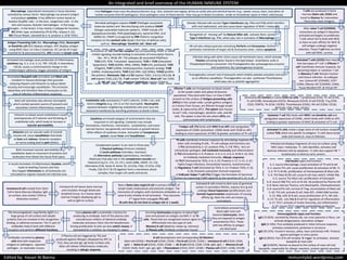
- 1. First Pathogen must cross the physical barriers (e.g.: skin, stomach and vagina; all has an acidic pH) and chemical barrier (e.g.: sweat, mucus, tears, and saliva; all contain enzymes that kill pathogens). Once pathogens cross all those barrier, they may go to blood circulation, reside at intracellular space or infect cells/tissues. Microbial pathogens express PAMP (Pathogen associated molecular patter) and Necrotic/dying cells produce DAMP (Danger associated molecular pattern). PAMP include LPS (pipopolysaccharide), PGN (peptidoglycan), bacterial DNA, viral dsRNA etc. PAMP is recognized by PRR (Pattern recognition receptor) on the sentinel cells (body’s first line of defense cells), such as: Macrophage Dendritic cell , Mast cell Macrophage (specialized monocyte) in tissue becomes activated by various factor. Macrophage can present antigen and produce cytokine. It has different names based on location (Kupffer cells - in the liver, Langerhans cells - in the skin and mucosa, Alveolar macrophages - in the lungs, Microglia – in the brain). It has mainly 2 subtype for function: M1 (Killer type, activated by LPS & IFNγ, release IL-12), M2 (Tissue Repair, elevated by IL-4, produce IL-10 & TGFβ ) Already infected cells secrete Type-I interferons (eg- IFNα and IFNβ which interfere with viral replication), and also cause down regulation of MHC-I molecules. Antigen Presenting Cells like- Macrophage (Mφ), Neutrophil or Dendritic cell (DC) displays antigen. APC displays antigen using MHC Class I or Class II molecule. DC can be of 2 type (myeloid DC & plasmacytoid DC). APC travels to lymph node. Activated macrophage cause production of inflammatory cytokines (eg. IL-1, IL-6, IL-12, TNF, CXCL8). A chemokine, CXCLB, causes a conformational change in the ICAM/integrin adhesion, trigger activation of Integrin. Granulated Basophil cells (circulate) and Mast cells (resident in tissues) discharge their granules releasing Heparin and Histamin to reduce blood viscosity and encourage vasodilation. This increases blood flow and therefore flow of leucocytes to the area of infection (Inflammatory response). T helper cell (Th) binds with MHC II on APC and upregulates expression of CD40L (activation). CD40L binds with CD40 on APC, leading to more expression of MHC & greater activation of Th cell. Cytokines are broad category of small proteins that are important in cell signaling. Cytokines may include chemokines, interferons, interleukins, lymphokines, and tumor necrosis factors; but generally not hormones or growth factors. Other effects of cytokines involve: Activation of Complement system, cause Fever and Vasodilation. Endothelial cells overexpress P and E-selectin, ICAM-1 etc; and bind to integrins (e.g. LFA-1) on the neutrophils. Neutrophils squeeze between neighboring endothelial cells and cross the basement membrane by secreting proteases to break it down. NK cell also release granules containing Perforin and Granzymes. Perforin perforates membrane of target cell & Granzymes enter, induce apoptosis Prostaglandins convert into Prostacyclin which inhibits platelet activation and acts as an effective vasodilator. Prostaglandins can also synthesize Thromboxane which play role as platelet aggregation. Recognition of ‘missing self’ by Natural Killer cell, activate them, secrete Type II interferon (eg- IFNγ, which play role in activation of Macrophage ) Infection along with other stress cause cell membrane injury, which activate Platelet activating factor found in the lipid bilayer. Arachidonic acids in Phospholipid bilayer converted into Prostaglandins (by cyclooxygenase enzyme) and Leukotriene (by Lipooxygenase enzyme) Mast cell activation also attracts Eosinophils which combat parasitic worms (if present) and neutralize/ control inflammatory response Complement system: It can start in three way- i) Classical pathway (immune complex) ii) Lectin pathway (microbes, mannose) iii) Alternative pathway (spontaneous, microbes) Molecules that play role in the complement cascade are: Classical (C1q,C1r, C1s, C2, C4 ), Lectin (MBL, MASP, C2, C4), Alternative (C3b, factor B, factor D), C3 (C3a, C3b), C5 (C5a, C5b) Finally, C5b C6 C7 & C9 together form a membrane attack complex, then target cell swells and bursts Histamin acts on vascular walls of smooth muscle cell, cause vasodilation that leads to heat and redness. Histamin receptors on nerve ending lead to pain (Dolor). Histamin on blood vessel endothelium cause overexpression of P-selectin and Shrinking of endothelial cells, which leads to increase in vascular permeability. Both increased vascular permeability & vasodilation leads to movement of proteins and leukocytes from blood into tissue fluid space. Cytotoxic T cell (Tc) binds with MHC I on Dendritic cell and upregulates expression of CD40L, which binds with CD40 on APC, leading to greater expression of MHCI & activation of Tc cell. Infected cell displays fragments of virus on surface using MHC class I molecules. Tc cells identifies, activates and destroys infected cells by apoptosis. Tc cells also support NK T lymphocytes to destroy early cancers. It results formation of inflammatory Exudate, which leads to Swelling (Tumor) and Pain. Also happen Chemotaxis ie, all leukocytes are stimulated to migrate towards the infected area. Activated T cells (CD25) then mature into two types of T cell: i) Effector T cell: Coordinate immune response, by becoming helper or cytotoxic T cell ii) Memory T cell: Remain inactive until future infection. Its subtypes are- Central MT (CD45RO, CCR7 CD44), Effector MT (CD45RO, CD44), Tissue Resident MT, & Virtual MT T cells are produced in bone marrow (Stem cell, CD34) and travel to thymus for maturation. There they meet Antigens. After naïve T cell (CD45RA) encounters an antigen it becomes activated and begins to proliferate. T cells which fail to bind with MHC or have an over affinity for MHC & self-antigen undergo negative selection. Those T cell that can bind with self-MHC, survive. Activated Tc cells create a large clone of cell-surface receptors (called TCR) which are specific to antigens. Tc cells leave lymph node and travels to area of infection. Th cells coordinate immune response by stimulating response of other cells including B cells. Th cell subtype and functions are: i) Th1 (Activated by IL-12; produce IFNγ, IL-2 & TNFα; Acts on intracellular pathogen, Cell mediated immunity and inflammation) ii) Th2 (Activated by IL-4; Produce IL-4, IL-5, IL-6, IL-10 & IL-13; Acts on Antibody mediated immunity, Allergic response) iii) Th17 (Activated by TGFβ, IL-6, IL-23; Produce IL-17, IL-21, IL-22; Fights fungal infections, Extracellular bacteria, Autoimmunity) iv) Regulatory T cell (Activated by TGFβ, IL-12; Produce TGFβ, IL-10, IL-35; Prevents overactive immune response) v) Follicular helper T cell (Tfh) (Trigger the formation of Germinal Centers by expressing CD40 Ligand & by the secretion of IL-21, IL-4) Immature B cell leaves bone marrow and circulates through blood and lymphoid tissue. As it leaves bone marrow it begins displaying IgD as well as IgM on surface. Surviving Centrocytes progress to Apical light zone and present an antigen via MHC II to Th cells. Those that are recognized receive signal to differentiate into two type of cells: Memory B cell (detector, keeps sp. memory) & Plasma cells (Antibody production house) Activated B cells travel to dark zone of germinal centers in secondary follicles, express less Ig and undergo Clonal Expansion (proliferation) and Somatic hypermutation (expression of varying affinity Ig). Now the cells are known as centroblasts. Antibodies secreted by plasma cells bind with respective antigens or pathogens; opsonize pathogen , or neutralize viruses. Centroblasts proceed to Basal Light zone and become Centrocytes. Here they are exposed to antigen presented by FDCs. Those with low affinity to antigen are destroyed. Every plasma cell is essentially a factory for producing an antibody. Each of the plasma cells manufactures millions of identical antibody molecules and pours them into the bloodstream. During proliferation B cells can also switch classes, if stimulated by a cytokine, by changing Fc region. The immunoglobulin superfamily (IgSF) is a large group of cell surface and soluble proteins that are involved in the recognition, binding, or adhesion processes. The Fc part of antibodies help to bind with different receptors and perform different functions. Immature B cell created from Stem Cell in Bone Marrow (displays IgM on surface, also contain CD34 as distinctive marker) Now a Naïve (aka virgin) B cell in primary follicles of lymph nodes endocytoses and presents antigen. The interaction of antigens with membrane antibodies on naive B cells initiates B cell activation. They becomes 2nd signal from conjugate Th1 cell. (B cells that do not bind to antigen die in 1 week) Edited by: Hasan Al Banna immunitybd.wordpress.com An integrated and brief overview of the HUMAN IMMUNE SYSTEM Stimulation of TLR (Toll like receptor) by corresponding PAMP or DAMP initiate signaling cascade leading to activation of transcription factors, like- AP-1, NFκB, IRF. TLR subtypes (& associated PAMP)are: TLR1 (LPS, PGN, Triacylated lipoprotein), TLR2 + TLR6 (Diacylated lipoprotein), TLR3 (dsRNA, tRNA, siRNA), TLR4 (LPS, paclitaxel), TLR5 (Flagelin), TLR7 (ssRNA, Imidazoquinolon, Guanosin analog), TLR8 (ssRNA, Imidazoquinolin), TLR9 (CpG DNA, CpG ODNs), TLR10 (profilin- like protein). Monocyte, Mφ and DC express TLR(1, 2,4,5,6,7,8,9,10), B cell express TLR(1,2,6,7,9), T cell contain TLR(3,9), Mast cell has TLR(4, 8), NK cell has TLR3, Intestinal Epithelium express TLR(4,5) Effector T cells are transported via blood vessels to the lymph nodes and spleen & become specialized. They bind with Class I or Class II MHC present on the surface of Antigen-presenting cells (APCs) in the Lymph node. Lymph gathers antigens as it drains from tissues, are filtered through lymph nodes, & captured by APCs. The spleen also houses B and T cells, macrophages, dendritic cells, and NK cells. The spleen is also the site where APCs can communicate with lymphocytes. Distinctive CD markers in some CD45+ve cells: T lymphocyte (CD3), Th cell (CD4), Tc cell (CD8), Granulocyte (CD15), Monocyte (CD14), B cell (CD19), Treg (CD4, CD25, FOXP3), Tfh (CD4, CXCR5), Thrombocyte (CD61), NK cell (CD56, CD16), NKT(CD4, CD8, CD56, CD161) Immunoglobulin types and function: IgG (75-85%): secreted by Plasma cell, can cross placenta in fetus, can activate phagocytosis, neutralizes antigen. IgM (5-10%): First antibody produced in initial immune response, activates complement, pentamar in structure. IgA (10-15%): Found in mucous, saliva, tears and breast milk. Protects against pathogen in entry points. IgD (0.001%): Part of B cell receptor & trigger its activation. Activate basophils & mast cells IgE (0.002%): Remain as bound to the surface of mast cell and basophils, responsible for allergic reactions, protect against parasites Interleukin types and function: IL-1: source Macrophage; cause activation of Th and B cell IL-2: source Th1 cell; cause proliferation & activation of NK, Tc IL-3: Th Tc & NK; proliferation of Hematopoietic & Mast cells IL-4: Th2 Mast & NK cell; cause B cell class switch, inhibit Mφ IL-5: source Th2 Mast cell; proliferation of Eosinophil IL-6: source Mφ Th2; acts on B cell, Ab produced by Plasma cell IL-8: Bone marrow Thymus; acts Neutrophils, Chemoattractant IL-9: sourceTh2 cell; survival of Treg, accumulation of Mast cell IL-10: Th2 cell; activate B cell, help APC, inhibit Mφ function IL-12: Mφ, mDC & B cell; cause proliferation of Tc, NK cells IL-13: Th cell; acts Mφ & B cell for regulation of inflammation IL-17: Th17; activate of innate immunity, pro-inflammation IL-22: source Th17; regulate autoantibody production B cell development and corresponding CD Markers: Stem cell (CD34) > Pro B cell (CD34, CD19) > Pre B cell (CD19, CD20) > Immature B cell (CD19, CD20, IgM) > Naïve B cell (CD19, CD20, CD38) > GC B cell (CD19, CD20, CD38, IgM, IgD) > Memory B cell (CD19, CD20, CD27, IgA, IgG, IgE) > Plasmablast (CD19, CD27, CD38) > Plasma cell (CD27, CD38, CD138) If Plasma cell are triggered by Th2 and produced against Allergen (displayed by APC to Th2), they secrete IgE. IgE binds to Mast cells. Mast cell release inflammatory molecules, resulting in allergic response.