
Recommended
Recommended
More Related Content
What's hot
What's hot (20)
Similar to PRINCIPLE OF ARTHROSCOPY n.pptx
Similar to PRINCIPLE OF ARTHROSCOPY n.pptx (20)
Recently uploaded
Recently uploaded (20)
PRINCIPLE OF ARTHROSCOPY n.pptx
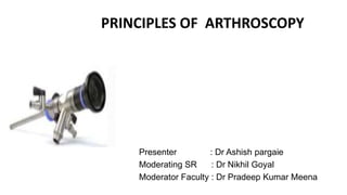
- 1. PRINCIPLES OF ARTHROSCOPY Presenter : Dr Ashish pargaie Moderating SR : Dr Nikhil Goyal Moderator Faculty : Dr Pradeep Kumar Meena
- 2. Learning Objectives: Instruments and equipment Anesthesia Documentation Advantages & disadvantages Indication & contraindication Basic arthroscopic techniques Complication
- 4. MEANING OF ARTHROSCOPY • Greek: “Arthro”: Joint “Skopein” : To look literally means; to look within the joint Offers; high degree of accuracy low morbidity for making diagnosis and offering treatment
- 5. Main monitor Camera unit device Video recording & image device Power box for radiofrequency instruments Irrigatation pump Light source device
- 7. ARTHROSCOPE - Rigid optical instrument Optical characteristics : determined by diameter ,angle of inclination and field of view diameter : 1.7-7 mm 4 mm (most commonly used , especially for knee joint 1.9 & 2.7 mm usefull for tighter joints like wrist & ankle
- 8. • Angle of inclination- is angle between axis of arthroscope and line perpendicular to surface of lens , varies from 0-120◦ . 25- 30◦ most commonly used 70- 90 ◦ seeing around corners
- 9. • Field of view- refers to viewing angle encompassed by lens and varies according to type of arthroscope • 1.9 mm scope has a 65 * field of view • 2.7 mm scope has 90 * field of view • Wider viewing angles make orientation by observer much easier. • Two designs – viewing Operating developed by O’Conner allows direct viewing , with a channel for the placement of operative instruments in line with the arthroscope.
- 10. TELEVISION CAMERAS • McGinty and Johnson the first to introduce a television camera to the arthroscopy system. • Advantages More comfortable operating position for the surgeon Avoidance of contamination of the operative field by the surgeon’s face Involvement of the rest of the surgical team in the procedure
- 11. Improvement offered by latest three chip technology Decresae size of camera Increase resolution of image Cableless arthroscopic systems in which video signal is transmitted from an arthroscope with its own light source
- 12. Figure : Rotation of arthroscope with 30-degree angle of inclination, which causes scanning effect that increases field of view by about three times. Dotted circle shows field of view and is compared at lower left with small circle that shows field of view of 0-degree arthroscope.
- 13. Figure : ROTATION OF ARTHROSCOPE WITH 70-DEGREE ANGLE OF INCLINATION. THIS SCANS A LARGE CIRCLE BUT CREATES A BLIND AREA DIRECTLY AHEAD OF IT IN WHICH NOTHING CAN BE SEEN.
- 14. ACCESSORY INSTRUMENTS • Basic instrument kit : – Arthroscopes (30- and 70-degree) – Probe – Scissors – Basket forceps – Grasping forceps – Arthroscopic knives – Motorized meniscus cutter and shaver – Electrosurgical – Laser/ Radiofrequency instruments – Miscellaneous epuipment
- 15. Arthroscopic instruments for shoulder procedures.
- 16. PROBE : • The extension of the arthroscopist’s finger. • Used – • To feel the consistensy of a structure • To determine the depth • To identify and palpate loose structures • To maneuver loose bodies into more accessible grasping position • To probe fossae & recess • To maneuver intraarticular structure • To elevate meniscus
- 17. • Most are right – angle • 2 mm fixed tap size . This is used to measure length of structure inside joint cavity . • Use the elbow of the probes for palpation • Magnification occurs with the arthroscope : closer it is the higher the magnification • So it can be placed close or far depending on the observer’s desire.
- 18. FIGURE - Arthroscopic probe used in exploring intraarticular structures during arthroscopic triangulation techniques.
- 19. (A) Arthroscopic view of a right knee medial compartment with the 30 arthroscope in the lateral viewing portal using a 5-mm probe and physician-applied valgus. Notice the normal relationship of the femur (F), meniscus (M), and tibia (T), despite the relatively large physiologic gapping of the joint space. (B) Arthroscopic view of a right knee lateral compartment with the 30 arthroscope in the lateral viewing portal using a 5-mm probe and a physician-applied varus stress. Notice the abnormal separation of the meniscus from the tibia, " drive-through " sign, and inflamed synovium of the posterior capsule consistent with posterolateral corner insufficiency.
- 20. SCISSORS : • 3-4 mm in diameter • JAWS : straight / hooked • Hooked scissors preferred as jaws hook tissue & pull it between cutting edges of scissors rather than pushing materials as in straight scissors. • CURVES : right / left • ANGLES : right/left ,usuallly with a rotating of jaw mechanism, actually cut at an angle to shaft of the scissors. • Useful in detaching difficult to reach meniscal fragments .
- 21. FIGURE - Commonly used arthroscopic instruments scissors
- 22. BASKET FORCEPS : • One of the most commonly used arthroscopic intruments. • Open base that permits the tissue to drop free within the joint & don’t require instrument to be removed from the joint & cleaned with each bite The debris is subsequently removed from the joint by suction . • 3-5 mm sizes with straight or curved shaft • Usually used for trimming the peripheral rim of the meniscus
- 23. • Basket forceps specialized for meniscus are wide , low – profile basket with hooked configuration. Shaft : straight / curved Jaws : straight / hooked Basket in assortment of 30 ,45,90 degree Also as 15 degree up & down – biting
- 24. GRASPING FORCEPS • Retrieve material from the joint generally loose bodies from the joints . • Grasping tissue to cutting used to retrieve material from the joint, or to hold other tissue under tension to facilitate cutting • Rachet closure system for better hold • Jaws : single / double action with regular serrated interdigitating teeth /1-2 sharp teeth • Usually double side serrrated forcep used for securing loose bodies – doesn’t slip from it.
- 25. ELECTROSURGICAL LASERS • Electrocautery : for cutting & hemostasis . • Works in a non – electrolyte medium like distilled water , co2 or glycine. • Newer coated tip function in both NS / RL • Laser: role under investigation . • Co2 laser, YAG laser , excimer laser.
- 26. RADIOSURGICAL SYSTEM • Radio frequency system used for tissue ablation, electrocautery & capsular shrinkage. • Monopolar: grounding pad & draw energy through the body • Bipolar :b/w electrodes at the site of treatment • Used for cuttting and haemostasis for arthroscopic synovectomies and subacromial decompression . • Complications include- articular cartilage damage, Osteonecrosis Tissue damage
- 27. KNIFE BLADES • Should be inserted through cannula sheaths and cutting portion be exposed only when it enters the arthroscopic field • Available varities are – hooked or retrograde blades , regular down – cutting blades – straight and curved • Magnetic properties of blades : helpful in retrieving them when broken
- 28. MOTORISED SHAVING SYSTEMS Consisting of • Outer hollow sheath • Inner hollow rotating cannula with corresponding windows & diameter of cutting tip usually 3-5.5 mm • Principle: the window of inner sheath function as a two edges cylindrical blade ,that spins within the outer hollow tube. • Suction through the cylinder bring the fragment of soft tissue in window and as the blade rotates , the fragments are amputated , sucked to the outside , and collected in the suction trap.
- 30. IMPLANTS • Suture anchors • Meniscal repair devices • Devices for tendon and ligament fixation • Articular cartilage repair
- 31. SUTURE ANCHORS • Used to attached ligaments and tendons to bone without bony tunnel passage sutures • Desirable characteristics Must fixed the suture to the bone permit an easy surgical technique Not cause long – term problems
- 33. MENISCAL REPAIR DEVICES • Allow an all – inside meniscal repair without the need for arthroscopic knot- tying • 3 categories • Arrows • Darts • Meniscal screws
- 34. IRRIGATION SYSTEMS Irrigation and distension Essential to all arthroscopic procedure Joint distension is maintained better with RL than NS. Inflow is via arthroscopic sheath : 6.2 mm diameter with the cannula in separate portal with 68 mm of pressure of water Usually two 3-5 lit plastic bags of RL, interconnevted with a Y – connector are suspended for use with the arthroscopy pump. Continuous irrigation : keep clear viewing maintain hydrostatic pressure and distension
- 36. IRRIGATION SYSTEMS & ARTHROSCOPY SETUP
- 37. DISTENSION PRESSURE • Optimal pressure required to distension the joint . • Ingress = egress to maintain hydrostatic pressure & distention within joint. • For each foot of elevation of solution bag above joints = 22 mm of hg pressure • Varied according to joints as follows: Knee 60-80 mm of hg Shoulder 30 mm of hg below systolic pressure Elbow 40- 60 mm of hg Ankle 40-60 mm of hg
- 38. TORNIQUET Contraindication history of thrombophlebitis significant peripheral vascular disease Advantages increased visibility Disadvantages blanching of the synovium difficulty to diagnosis synovial disorders • Ischemic damages if prolonged toruniquet time ( 90 – 120 min)
- 39. LEG HOLDERS The biggest advantages of leg holders is that they permit application of stress primarily to open the posteriomedial compartment for viewing or manipulation of the meniscus and posterior horn meniscus surgery The post does not confine knee and offers unlimited number of positions for the knee to be placed . Disadvantages obstruct the operations in lateral compartment
- 40. LEG HOLDERS
- 41. METHOD OF STERILIZATION • Ethylene oxide ( best method) • Low temprature sterilization process • CIDEX is used for cold disinfection of equipments between sucessive procedures during whole day • Knives, forceps etc : by steam autoclaving • Fibreoptic materials , camera , motorised instruments : by soaking in cidex sol. For 10 min . Or STERIS for 30 min.
- 42. ANESTHESIA ARTHROSCOPY can be performed under • Local anesthesia • Regional anesthesia • General anesthesia
- 43. REGIONAL ANESTHESIA • Usually used in lower extremities – epidural or spinal anesthesia femoral and sciatic block feature of peripheral blocks- • immediate ambulation • require experince anesthesiologst • longer time to prepare • generally use a 1:1 mixture of 1 % lignocaine and 0.25 % bupivacaine • Upper extremities brachial block
- 44. GENERAL ANESTHESIA Used – • Not cooperative patient • Allergy to local anesthesia • Less experienced surgeon • Increased pain (acutely injured knee)
- 45. INDICATION AND CONTRAINDICATIONS • No absolute indication • Diagnostic arthroscopy preoperative evaluation and confirmation of the clinial diagnosis documentation of specific lesions • Contraindication: Risk of joint sepsis , remote infection Ankylosis around the joint Capsular disruption
- 46. POST – OP PAIN • Oral NSAIDS or IM,IV administration Reduced swelling Increase ROM in early postoperative period • 30 ml of 0.25 % bupivacaine +/- morphine 3 mg intrarticular or subacromial flow Excellent post operative pain relief Cathers should be removed in 48 hours
- 47. DOCUMENTATION • Drawings and documentation are very essential • 35- mm reflex camera photos • Digital video recordings
- 48. INDICATION OF ARTHROSCOPY DIAGNOSTIC For pre operative evaluation & Confirmation of clinical diagnosis For documentation in medicolegal cases THERAPEUTIC Smoothening of torn cartilage Damaged ligaments reconstruction Loose bodies removal Joint effusion Biopsy procedures Fracture fixation Sports related injuries
- 49. ADVANTAGES OF ARTHROSCOPY • Reduced postoperative morbidity • Smaller incision • Less intense inflammatory response • Improved thrughness of diagnosis • Absense of secondary effects neuromas, scars
- 50. • Reduced hospital cost • Reduced complication rate • Imroved follow-up evaluation : second – look • Possibility of performing surgical procedures that are difficult to perform through open arthrotomy
- 51. DISADVANTAGES OF ARTHROSCOPY • Skill and temperament to perform arthroscopic surgery • Need to maneuver within the tight confines of the intraarticular space • Time – consuming procedures in cases of inexperienced surgeons and follows a steep learning curve • Expensive equipment
- 52. HOW IS ARTHROSCOPY PERFORMED ? Under anesthesia make small incision in the skin around joint. Eg . Anteromedial and Anterolateral entry points in the knee jnt. A sterile fluid is pumped into joint and then the arthroscope is inserted. Examine joint by images from arthroscope If necessary , other instruments inserted for procedure i.e. repair any damage or remove material that causes symptoms. Afterwards, the fluid is drained out , cuts are closed &dressed
- 53. BASIC ARTHROSCOPIC TECHNIQUES • Patience and persistence • Techniques are mostly self – taught • Artificial models or amputated specimens for initial practice • Perform arthroscopic procedures in the company of an experienced arthroscopist . • It has a steep learning curve • Keep in mind that open arthrotomy is to be preferred over poorly performed arthroscopic procedures
- 54. TRIANGULATION TECHNIQUE • Involves use of one or more instuments inserted through separate portals and brought into the optical field of the arthroscope • Tip of the instuments and arthroscope forming apex of a triangle • When the instrument is located , the scope and instrument are advanced together towards the intended area , reducing the field of vision and increasing the magnification
- 55. • If disoriented and difficulty in triangulation the instrument may be brought into the joint to contact the sheath and sliding to the tip • Stereoscopic sense and two – handed ability are developed gradually
- 57. MOST COMMON CONDITIONS FOUND DURING ARTHROSCOPY Acute or chronic injury shoulder : rotator cuff tendon tears impingement syndrome recurrent dislocation knee : meniscal (cartilage) tears chondromalacia (wearing or injury of cartilage cushion) ACL & PCL tear with instability
- 58. Wrist : carpal tunnel syndrome Loose bodies of bone & or cartilage : example : knee shoulder elbow ankle wrist • Some problems associated with arthritis also can be treated
- 59. knee arthroscopy: ACL repair
- 62. COMMONLY DONE ARTHROSCOPIC SURGERIES • Rotator cuff injury • Repair or resection of torn cartilage (meniscus) from knee or shoulder • Reconstruction of anterior cruciate ligament in knee • Removal of inflamed (synovium) in knee ,shoulder,elbow, wrist & ankle • Release of carpal tunnel • Repair of torn ligaments • Removal of loose bone or cartilage in knee , shoulder, elbow, ankle & wrist
- 63. COMPLICATIONS • Damage to intraarticular structure : most common • Damage to menisci and fat pad • Damage to cruciate ligaments • Damage to extraarticular structure • Hemathrosis • Thrombophelebitis • Infection • Torniquet paresis • Synovial herniation and fistulas • Instrument breakage
- 64. FOLLOW – UP AFTER AETHROSCOPIC SURGERIES RECOVERY TIME DEPENDS UPON MANY FACTORS : • Severity of disease • Type of surgery • Supports for 3 to 7 days , weight bearing on the operated leg as tolerated. • Analgesics • Rest, ice packs, and limb elevation also recommended
- 65. • Physiotherapy not required in all patients, should be individualised • Sitting job can be resumed one week after surgery • 3-4 weeks to recover fully for routine daily activities • 3 months before one can confortably return to sports .
- 66. Elsevier, February 2022 LEVEL OF EVIDENCE Case series: IV
- 67. PURPOSE • Determine the rate of intraoperative and early postoperative (90- day) complications of multiligamentous knee reconstruction surgeries • Both medical and surgical and associated variables from the 15- year experience of a single academic institution
- 68. METHOD • Patients treated at a single academic institution between 2005 and 2019 who underwent multiligament knee surgery were identified • Inclusion criteria : – 2+ ligament reconstructions performed concurrently – > 90 days postoperative follow-up.
- 69. • Exclusion criteria : – Included revision ligamentous knee surgery. – Patient demographics, mechanism of injury – Associated injuries of patients with intraoperative and postoperative complications – Time from injury to multiligamentous knee reconstruction – Surgical data: • including tourniquet time • procedure time, • type of procedures performed were retrospectively recorded.
- 70. • Results : – 301 knees in 296 patients met the eligibility criteria. – There were 11 intraoperative complications in 9 knees (rate of 3%) and 136 postoperative complications in 90 knees (rate of 30%) – Shorter time from injury to date of surgery was associated with arthrofibrosis (P = .001) – superficial wound infections (P = .015). – Concurrent head injuries were associated with less complications (P = .029). – Procedural time >300 minutes was associated with intraoperative blood transfusions (P > .05), – deep infections (P = .003) – arthrofibrosis (P = .012).
- 71. Inside-out meniscal reair was associated with •Superficial and deep infections (P = .006 and .0004). •Tibial-based posterolateral corner (PLC) reconstruction was associated with symptomatic hardware (P = .037) • Arthrofibrosis (P = .019) in comparison with fibular-based PLC reconstruction. •Posterior cruciate ligament (PCL) reconstruction was associated with deep infections (P = .015), •arthrofibrosis (P = .003), • postoperative blood transfusions (P = .018).
- 72. Complications associated with • Longer procedure times • Inside-out meniscal repair • Tibia-based PCL reconstruction • Shorter time to surgery
- 73. THANK YOU
Editor's Notes
- The axons are single nerve fibers, much like the “wiring” of the nerve. The endoneurium exists between individual axons. Many axons form groups called fascicles. The fascicles are surrounded by perineurium. The whole nerve is surrounded by epineurium.
- The epineurium is the external covering of the nerve. It is a loose connective tissue encasing the nerve and also dividing fascicles groups. The amount of epineurium is variable from nerve to nerve as well as along the course of an individual nerve. Epineurial fibroblasts are involved in the inflammatory response and proliferate in response to injury. This can cause 2-3 mm thickening of a nerve in response to chronic inflammation
- The perineurium is an extension of the blood brain barrier, controlling the environment within a fascicle. The perineurium surrounds the nerve fascicle. Removal of the perineurium results in loss of nerve function. The perineurium resists longitudinal traction and provides the elastic properties of peripheral nerve