IgG4 Related Disease: Case Presentation And Literature Review
•
2 likes•225 views

IgG4 related disease is a rare disease in which cases delayed diagnosis is common. The recent development of diagnostic criteria is helpful for early diagnosis.

Recommended
More Related Content
What's hot
What's hot (20)
Similar to IgG4 Related Disease: Case Presentation And Literature Review
Similar to IgG4 Related Disease: Case Presentation And Literature Review (20)
Recently uploaded
Recently uploaded (20)
IgG4 Related Disease: Case Presentation And Literature Review
- 1. IgG4 Related Disease: Case Presentation And Literature Review Lanzillotta et al. 2020
- 2. Why we need to learn and discuss about IgG4 related disease?
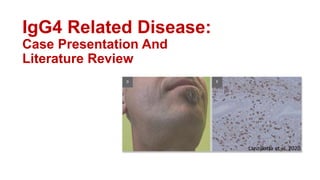
- 3. Case summery of Mr. A. Hossain Photo-taken with permission
- 4. Case summery Mr. A Hossain, 30-year-old, has been suffering from recurrent episode of obstructive jaundice for 2 years. Jaundice was associated with itching but no history of fever or abdominal pain. No history of ascites. After evaluation, distal biliary stricture was found on 2019. He underwent ERCP with plastic biliary stent placement on January 2020 with subsequent resolving of jaundice. On December 2020, biliary stent was removed. Unfortunately, after 3 months of removing of the stent, his jaundice reappears. This time IgG4 level is done which comes positive (Patient: 330mg/dl, normal value ≤120mg/dl). His IgE level is 1245. CT scan shows pancreato-duodenal groove pancreatitis(localized) causing biliary obstruction resulting proximal dilatation. MRCP shows distal biliary stricture.
- 5. December, 2019 December, 2019 March, 2021 January, 2020 1st episode of jaundice ERCP with biliary stenting Biliary stent-removed Jaundice- reappeared IgG4-330mg/dl, IgE- 1245 Localized pancreatitis on CT Distal biliary stricture June, 2021 Timeline history
- 6. IgG4 Related Disease: Literature Review BMJ 2020;369:m1067 | doi: 10.1136/bmj.m1067
- 7. IgG4- related disease- Phenotypes Lanzillotta et al. 2020 31% 24% 24% 22%
- 8. IgG4- related disease- Diagnosis Lanzillotta et al. 2020
- 9. • Serum IgG4: elevate in neoplastic, infectious, and autoimmune diseases • Histological examination- mainstay investigation
- 10. Pathologies associated to serum IgG4 elevation Cancer Pancreatic adenocarcin, Bile duct cancer/cholangiocarcinoma, Intraductal papillary mucinous neoplasm Autoimmune diseases Systemic lupus erythematosus , Antiphospholipid syndrome, Autoimmune hepatitis, Rheumatoid arthritis, Systemic sclerosis, Sjögren's syndrome, Polymyositis/dermatomyositis ANCA-related vasculitis Churg-Strauss syndrome*, Behcet's disease, Microscopic polyangiitis Infections Parasitic infections, Bacterial infections, Viral infections Others Multicentric Castleman's disease*, Eosinophilic disorders (fasciitis, pneumonia, and hypereosinophilic syndrome) , Chronic hepatitis, Liver (doi: 10.1155/2012/602809)
- 11. All of the above type Lanzillotta et al. 2020
- 12. IgG4- related disease- treatment response Lanzillotta et al. 2020
- 13. IgG4- related disease- Outcome Lanzillotta et al. 2020
- 14. • Rituximab is the most widely used biological agent in IgG4-RD and allowing early tapering of glucocorticoid therapy.
- 15. Traditional potential biomarkers of IgG4 related disease (IgG4-RD) Lanzillotta et al. 2020
- 16. Biomarkers for disease activity Lanzillotta et al. 2020
- 17. Lanzillotta et al. 2020 Novel potential biomarkers of IgG4 related disease (IgG4-RD)
- 18. A: abdominal computed tomography scan showing a “sausage-like” pancreas (*) with a surrounding rim of hypodense tissue (arrowheads), classic radiological features of autoimmune pancreatitis. Lanzillotta et al. 2020
- 19. Hegade VS et al. Frontline Gastroenterology 2019;10:275–283.
- 20. Hegade VS et al. Frontline Gastroenterology 2019;10:275–283.
- 21. D-F: classic histopathological and immunohistochemical features of IgG4-RD in a pancreatic biopsy: areas of storiform fibrosis (*; (D);, sequential sections shows an IgG4/IgG ratio >40%(E) Hematoxylin and eosin(D) Immunohistochemistry for IgG4 and IgG (E) E Lanzillotta et al. 2020
- 22. The 2019 American College of Rheumatology/European League Against Rheumatism Classification Criteria for IgG4-Related Disease (DOI 10.1002/art.41120)
- 23. The 2019 American College of Rheumatology/European League Against Rheumatism Classification Criteria for IgG4-Related Disease (DOI 10.1002/art.41120)
- 24. The 2019 American College of Rheumatology/European League Against Rheumatism Classification Criteria for IgG4-Related Disease (DOI 10.1002/art.41120)
- 25. Step 1. Entry criteria Yes† or No Characteristic* clinical or radiologic involvement of a typical organ (e.g., pancreas, salivary glands, bile ducts, orbits, kidney, lung, aorta, retroperitoneum, pachymeninges, or thyroid gland [Riedel’s thyroiditis]) OR pathologic evidence of an inflammatory process accompanied by a lympho-plasmacytic infiltrate of uncertain etiology in one of these same organs The 2019 American College of Rheumatology/European League Against Rheumatism Classification Criteria for IgG4-Related Disease (DOI 10.1002/art.41120)
- 26. The 2019 American College of Rheumatology/European League Against Rheumatism Classification Criteria for IgG4-Related Disease (DOI 10.1002/art.41120)
- 27. QUESTIONS ABOUT MR. HOSSAIN’S CONDITION •How many points our patient get? Is there anything else that can we do for further evaluation? 1. IgG4 2.5 times high- 6points 2. Localized(?) pancreatic involvement+ Biliary tree involve- 19 points Total: 25 points (more thant 20 points meets the criteria)
- 28. Thank You
- 29. The 2019 American College of Rheumatology/European League Against Rheumatism Classification Criteria for IgG4-Related Disease (DOI 10.1002/art.41120)
- 30. Timeline: 1st episode of jaundice-2019 MRCP(2019)- Biliary stricture January, 2020- ERCP with stent placement December, 2020- stent is removed Reappear jaundice on March-2021
- 31. • Compared with white patients, Asian patients are at higher risk of developing IgG4-RD in the head and neck region • Patients with head and neck involvement also seem to more often have atopic manifestations • Patients with Mikulicz’s/systemic disease have more organs involved, higher values of IgG4-RD responder index (RI), and serum IgE concentrations. • Patients with retroperitoneal and head and neck involvement seem more prone to have a fibrotic outcome than do those with other IgG4-RD phenotypes, and are thus more challenging to treat
- 32. • Induction of remission Glucocorticoids Glucocorticoids represent the first line agent for inducing remission in all patients with active IgG4- RD.21 Induction of remission should aim to resolve symptoms and biochemical and radiological abnormalities, and improvement should typically be observed within days to several weeks • The starting dose of corticosteroids typically consists of 30- 40 mg/day (0.6-1 mg/kg) of prednisone or steroid equivalent.105 110 One retrospective study and one randomized controlled trial reported no differences in terms of remission rate between patients with IgG4- RD treated with high dose (0.8-1 mg/kg) and medium dose (0.5-0.6 mg/kg) corticosteroids • Immunosuppressive drugs When predictors of relapse—such as multi-organ involvement, elevation of serum IgG4 and IgE at baseline, and peripheral blood eosinophilia—are present, disease modifying anti-rheumatic drugs (DMARDs) can be added to first line steroid therapy to improve the likelihood of obtaining disease remission. Azathioprine, mycophenolate mofetil, methotrexate, leflunomide, tacrolimus, ciclospor
- 33. Available therapeutic strategies for inducing and maintaining remission of IgG4 related disease (IgG4-RD) Lanzillotta et al. 2020
- 34. Available therapeutic strategies for inducing and maintaining remission of IgG4 related disease (IgG4-RD) Lanzillotta et al. 2020
- 35. Available therapeutic strategies for inducing and maintaining remission of IgG4 related disease (IgG4- RD) Lanzillotta et al. 2020
- 36. Established, emerging, and novel potential biological therapies for IgG4 related disease Lanzillotta et al. 2020
- 37. Novel diagnostic biomarkers • Increased ratios of serum IgG4 to total IgG (>10%) • IgG4:IgG RNA ratio on peripheral blood seemed to accurately distinguish IgG4 related cholangitis from hepatobiliary malignancies and inflammatory processes • Multicolor flow cytometry, next generation sequencing, and gene expression analyses led to the identification of disease specific B cell and T cell
- 38. • FDG-PET: fluorodeoxyglucose- positron emission topography