More Related Content
Similar to Art. jessica (20)
More from anaelishockey (20)
Art. jessica
- 1. Genetically encoded norbornene directs
site-specific cellular protein labelling via a
rapid bioorthogonal reaction
Kathrin Lang1†
, Lloyd Davis1†
, Jessica Torres-Kolbus2
, Chungjung Chou2
, Alexander Deiters2
* and
Jason W. Chin1
*
The site-specific incorporation of bioorthogonal groups via genetic code expansion provides a powerful general strategy
for site-specifically labelling proteins with any probe. However, the slow reactivity of the bioorthogonal functional groups
that can be encoded genetically limits the utility of this strategy. We demonstrate the genetic encoding of a norbornene
amino acid using the pyrrolysyl tRNA synthetase/tRNACUA pair in Escherichia coli and mammalian cells. We developed a
series of tetrazine-based probes that exhibit ‘turn-on’ fluorescence on their rapid reaction with norbornenes. We
demonstrate that the labelling of an encoded norbornene is specific with respect to the entire soluble E. coli proteome and
thousands of times faster than established encodable bioorthogonal reactions. We show explicitly the advantages of this
approach over state-of-the-art bioorthogonal reactions for protein labelling in vitro and on mammalian cells, and
demonstrate the rapid bioorthogonal site-specific labelling of a protein on the mammalian cell surface.
T
here is a pressing need for general methods to site-specifically
label proteins, in diverse contexts, with user-defined probes.
Current protein-labelling methods involve the use of fluor-
escent protein fusions1–4
, self-labelling proteins (for example,
SNAPtag, HALOtag and CLIPtag)5–8, ligases (such as biotin
ligase, lipolic acid ligase, sortase and phosphopantetheinyl transfer-
ase)9–15
and self-labelling tags (for example, tetracysteine and tetra-
serine)16,17
. Although some of these approaches allow rapid labelling
and have had substantial impact on biological studies, they require
the use of protein fusions and/or the introduction of additional
sequences into the protein of interest. This may disturb the structure
and function of the protein and can make it challenging to place
probes at any position in a protein. Moreover, the range of probes
that can be incorporated by some of these techniques is limited3,4,18.
Ideal methods for protein labelling would (i) allow probes to be
placed easily at any position in any protein expressed in prokaryotic
and eukaryotic cells and organisms, (ii) be rapid and quantitative,
(iii) be specific for a user-defined site in a protein, (iv) require no
toxic reagents and generate no toxic by-products, (v) show ‘turn-
on’ fluorescence with minimal off-site or background labelling
and (vi) allow for labelling with diverse probes. In principle, the
genetically encoded, site-specific incorporation of unnatural
amino acids that bear bioorthogonal functional groups would
allow the labelling of specific proteins at defined sites with
essentially any probe.
Bioorthogonal groups, including azides, alkynes, ketones, ani-
lines, alkenes, tetrazoles and 1,2-aminothiols, have been genetically
encoded using amber suppressor aminoacyl tRNA synthetase/
tRNACUA pairs19–29. For established reactions that have been demon-
strated on proteins, the rate constants for the corresponding model
reactions30
are in the range 1022
M21
s21
to 1024
M21
s21
(although for emerging approaches higher rates have been
reported)29,31,32. Clearly, the rates of established reactions are
sufficient to allow useful labelling of metabolically incorporated
azido- and keto-bearing glycan analogues presented at high density
on the cell surface, and the labelling of amino acid analogues incor-
porated nonspecifically throughout the proteome33–35
. However, the
sluggishness of established bioorthogonal reactions often makes it
challenging to label proteins quantitatively at defined sites in vitro
and may account for the fact that there are currently no examples
of labelling proteins expressed on the mammalian cell surface
using genetically encoded unnatural amino acids.
Recent advances in bioorthogonal chemistry demonstrate that
strained alkenes, including norbornenes and trans-cyclooctenes,
react rapidly and specifically with tetrazines in inverse electron-
demand Diels–Alder cycloaddition reactions to form stable
adducts. The rate constants of these reactions are orders of magni-
tude faster than those for other established bioorthogonal reac-
tions36–38
. Genetically encoding a component of these reactions is
therefore an attractive strategy for realizing rapid and site-specific
protein labelling (Fig. 1a).
Results and discussion
Synthesis and genetic encoding of a norbornene-containing
amino acid. The pyrrolysyl-tRNA synthetase/tRNACUA pair
(PylRS/tRNACUA) from Methanosarcina species (M. barkeri (Mb)
and M. mazei (Mm)), which naturally incorporates pyrrolysine
(1, Fig. 1b), is orthogonal to endogenous tRNAs and aminoacyl-
tRNA synthetases in Escherichia coli and eukaryotic cells39–42
.
Using this PylRS/tRNACUA pair, and its synthetically evolved
derivatives, we and others directed the efficient incorporation of
unnatural amino acids, including post-translationally modified
amino acids, chemical handles and photocaged amino acids, at
specific sites in desired proteins in E. coli, yeast and mammalian
cells27,28,39,40,43–46. Moreover, recently we demonstrated the
incorporation of unnatural amino acids, using this pair, in a
whole animal42
. We envisioned that this synthetase/tRNA pair
might be used to site-specifically and quantitatively incorporate a
1
Medical Research Council Laboratory of Molecular Biology, Hills Road, Cambridge CB2 0QH, UK, 2
Department of Chemistry, North Carolina State University,
Raleigh, North Carolina 27695-8204, USA, †
These authors contributed equally to this work. *e-mail: alex_deiters@ncsu.edu; chin@mrc-lmb.cam.ac.uk
ARTICLES
PUBLISHED ONLINE: 5 FEBRUARY 2012 | DOI: 10.1038/NCHEM.1250
NATURE CHEMISTRY | VOL 4 | APRIL 2012 | www.nature.com/naturechemistry298
© 2012 Macmillan Publishers Limited. All rights reserved.
- 2. norbornene-containing amino acid into proteins produced in
diverse organisms, and that the norbornene-containing protein
could be labelled rapidly and selectively with tetrazine-based probes.
We designed the norbornene-containing amino acid N1-5-
norbornene-2-yloxycarbonyl-L-lysine (2, Fig. 1b) and synthesized it
in three steps and 77% overall yield (Supplementary Information
and Supplementary Scheme S1). To investigate whether 2 is a sub-
strate for the MbPylRS/tRNACUA pair we transformed E. coli with
pBKPylS (which encodes MbPylRS) and psfGFP150TAGPylT-His6
(which encodes MbtRNACUA and a C-terminally hexahistidine
(His6)-tagged superfolder green fluorescent protein (sfGFP) gene
with an amber codon at position 150). In the presence of 2
(1 mM), full-length sfGFP was isolated in good yield (Fig. 2,
4 mg l21
of culture), comparable to the yields obtained for other
well-incorporated unnatural amino acids28,32,45
. GFP expression
was clearly amino acid-dependent. Similarly, myoglobin (Myo)
bearing an amber codon at position 4 and T4 lysozyme (T4L)
bearing an amber codon at position 83 produced good yields of
protein in the presence, but not absence, of 2 (Fig. 2 and
Supplementary Fig. S1). The incorporation of 2 was further con-
firmed by electrospray ionization mass spectrometry (ESI–MS) of
purified proteins (Fig. 2 and Supplementary Fig. S1).
Synthesis of biocompatible tetrazines. To create unsymmetrical
tetrazines that contain a unique reactive group for functionalization
with biophysical probes (Fig. 1c, Supplementary Scheme S2
and Supplementary Information) we reacted equimolar quantities
of 5-amino-2-cyanopyridine and 2-cyanopyridine (or 2-
cyanopyrimidine) with an excess of aqueous hydrazine to obtain
s-dihydrotetrazines S5a and S6a (ref. 36). Treatment of these
dihydrotetrazines with a mixed anhydride formed in situ from
isobutylchloroformate and N-tert-butyloxycarbonylglycine afforded
compounds S5b and S6b, respectively, which were oxidized readily
to their corresponding tetrazines 5 and 6 with sodium nitrate in
acetic acid (direct oxidation of dihydrotetrazines S5a and S6a
produced tetrazines that bear amino groups with markedly reduced
reactivity, as expected based on increased p-conjugation of the
amino group lone pair in these tetrazines). Acidic deprotection of
the tert-butyloxycarbonyl groups afforded tetrazines S5c and S6c.
The primary amino group in these tetrazine derivatives provides a
handle for further functionalization with biophysical probes.
We envisioned that analogues of 5 and 6 that bear a carboxy
group in place of the amine would be more electron deficient and
potentially more reactive in inverse electron-demand cycloaddition
reactions with norbornenes. To create tetrazines 7 and 8, we reacted
N-tert-butyloxycarbonylethylenediamine with 6-cyanopyridine-3-
carboxylic acid under standard amide-coupling conditions. The
resulting nitrile S7a was reacted with acetonitrile or 2-cyanopyrimi-
dine in aqueous hydrazine to give dihydrotetrazines S7b and S8b,
respectively, which after oxidation with sodium nitrate afforded tet-
razines 7 and 8, respectively. Deprotection of 8 under acidic con-
ditions gave tetrazine S8c. The primary amino group in this
tetrazine derivative provides a handle for further functionalization
with biophysical probes. All the tetrazines synthesized were stable
in MeOH/H2O and dimethylsulfoxide (DMSO)/H2O at room
temperature for several days, as judged by liquid chromato-
graphy–mass spectrometry (data not shown).
Kinetic analysis of the rapid tetrazine Diels–Alder cycloaddition.
Tetrazines 5–8 react readily with 5-norbornene-2-ol to form the
corresponding dihydropyridazines S15 and its isomeric forms S16
in protic solvents in .96% conversion (Supplementary Fig. S2 and
Supplementary Information). The rate constants for these
reactions were determined under pseudo first-order conditions by
following the exponential decay over time in the ultraviolet
absorbance of the tetrazine at 320 or 300 nm (Supplementary
Fig. S3). The reactions were faster in more polar solvent systems,
that is in solvent mixtures of higher water content, as expected36,47
.
Tetrazine 8 displays the highest activity towards 5-norbornene-2-ol
with second-order rate constants of approximately 9 M21
s21
in
H2O/MeOH (95:5) at 21 8C, whereas 5 reacts with a rate constant
of approximately 1 M21
s21
under the same conditions
(Supplementary Table S1 and Supplementary Information). This
confirms that the tetrazine–norbornene reaction is orders of
magnitude faster than established bioorthogonal reactions30
.
Tetrazine-based fluorophores: ‘turn-on’ fluorogenic probes. To
create fluorescent probes based on 5, 6 and 8, the primary amino
groups of S5c, S6c and S8c were conjugated to succinimidylesters
O
O
N
N N
N
R'
O
N
NH
R'
O
O
H
N
OCOOH
H2N
O
H
N
OCOOH
H2N
H
N
OCOOH
H2N
N
O
H
N
OCOOH
H2N
N3
N
N
N N
N
X N
HN
O
+
N
H
NO
N
N N
N
N
H
NO
N
N N
N
N N
NHR
NHR NHR
1
2
3
4
r.t.,
aqueous
a
cb
7 R = Boc
9 X = CH, R = TAMRA-X
10 X = N, R = TAMRA-X
11 X = CH, R = BODIPY TMR-X
12 R = TAMRA-X
13 R = BODIPY-FL
14 R = fluorescein
8 R = Boc5 X = CH, R = Boc
6 X = N, R = Boc
Figure 1 | Scheme to label proteins via an inverse electron-demand Diels–
Alder cycloaddition, and structural formulae of relevant compounds.
a, Genetically encoded norbornenes react rapidly with tetrazines, bearing
probes (red star), in aqueous solution at ambient temperature and pressure
to site-specifically label proteins. b, Amino acid structures of pyrrolysine (1),
N1-5-norbornene-2-yloxycarbonyl-L-lysine (2), N1-tert-butyloxycarbonyl-L-
lysine (3) and N1-2-azidoethyloxycarbonyl-L-lysine (4). c, Structures (5–14)
of tetrazines and tetrazine–fluorophores used in this study. TAMRA-X,
BODIPY TMR-X and BODIPY-FL are common names for fluorophores: their
structural formulae are shown in Supplementary Fig. S4. Red boxes denote
parent tetrazines. r.t. ¼ room temperature.
PylRS
a-His6
sfGFP
27,975.5
16,000 32,000
i ii100
0
Mass (Da)
Intensity(%)
3
2
+ + +
+
+
Myo
+ + +
––––
–– – –
+
+
12,000 20,000
100
0
18,532.5
Intensity(%)
Mass (Da)
a b
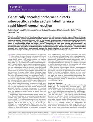
Figure 2 | Efficient, genetically-encoded incorporation of 2 using the
PylRS/tRNACUA pair in E. coli. a, Amino acid dependent expression of sfGFP
that bears an amber codon at position 150 and myoglobin that bears an
amber codon at position 4. b, Mass spectrometry characterization of amino
acid incorporation. i, sfGFP-2-His6, found: 27,975.5+1.5 Da, calculated:
27,977.5 Da. ii, Myo-2-His6, found: 18,532.5+1.5 Da, calculated: 18,532.2 Da.
NATURE CHEMISTRY DOI: 10.1038/NCHEM.1250 ARTICLES
NATURE CHEMISTRY | VOL 4 | APRIL 2012 | www.nature.com/naturechemistry 299
© 2012 Macmillan Publishers Limited. All rights reserved.
- 3. or isothiocyanates of fluorescein, tetramethylrhodamine (TAMRA) and
boron dipyrromethene (BODIPY) dyes (Supplementary Information,
Supplementary Figs S4 and S5, Supplementary Table S2).
The fluorescence of the visible light emitting tetrazine conjugate 9
and BODIPY tetrazine conjugate 13 was reduced substantially with
respect to the fluorescence of the succinimidyl or isothiocyanate
derivatives of the parental fluorophores. This is in agreement with
recent work that shows fluorophores can be quenched by energy trans-
fer to a proximal tetrazine chromophore that absorbs between 510 and
530 nm (ref. 48). However, even though 5, 6 and 8 have very similar
absorption spectra, the reduction in fluorescence of the dye conjugates
was dependent on the specific combination of tetrazine and fluoro-
phore. For example, 9 (5-TAMRA-X) showed a much greater
reduction in fluorescence with respect to the parent TAMRA-X than
did 10 (6-TAMRA-X) and 12 (8-TAMRA-X). Fluorescein (emission
maximum at 518 nm) was quenched minimally by conjugation to 8.
The fluorescence of 9, 11 and 13 was turned on upon cycloaddition
with 5-norbornene-2-ol, leading to a 5–10 fold gain in fluorescence
intensity (Fig. 3a, Supplementary Fig. S5).
Rapid labelling of norbornene-containing proteins with
tetrazine-based probes. To demonstrate that the tetrazine–dye
probes react efficiently and specifically with recombinant proteins
that bear site-specifically incorporated 2, purified sfGFP-2, Myo-2
and T4L-2 were incubated overnight with fluorophore 9 (10 equiv.)
at room temperature. Sodium dodecyl sulfate polyacrylamide gel
electrophoresis (SDS–PAGE)-based fluorescence imaging and ESI–
MS analysis (Fig. 3a and Supplementary Fig. S7) confirmed the
quantitative labelling of the proteins that contained 2, whereas no
nonspecific labelling was detected with the control proteins that
contained N1-tert-butyloxycarbonyl-L-lysine (3) in place of 2 at the
same site. In additional experiments we showed, using mass
spectrometry, the specific and quantitative labelling of proteins that
contained 2 with tetrazine derivatives 5, 6 and 8, as well as with
tetrazine fluorophores 12, 13 and 14 (Supplementary Figs S6
and S7). Previous labelling experiments of proteins containing
unnatural amino acids with specific fluorophores required washing
steps to remove free dye non-covalently associated with the protein.
We found that we could image the specific labelling of proteins
that contained 2 without washing the sample or the gel; this
improvement may, at least in part, result from the ‘turn-on’
fluorescence of the tetrazine fluorophores.
To further probe the specificity of the reaction between the
genetically encoded norbornene and the tetrazine-based fluoro-
phores, we performed the labelling reaction on the proteome of
E. coli expressing either sfGFP-2-His6 or Myo-2-His6 (Fig. 3c and
Supplementary Fig. S8). We controlled the level of recombinant
protein expression so that it was equal to or less than that of
many endogenous proteins by modulating the concentration of 2
added to cells. This ensures that any specific labelling of the target
protein versus native proteins was not an artefact of the abundance
of the target protein. Cells were harvested 3.5 hours after the
induction of protein expression, washed with PBS and incubated
with fluorophore probes (12 or 13) at room temperature. After
washing the cell pellets, the cells were lysed and the reaction mix-
tures were analysed by SDS–PAGE to assess the protein levels.
Fluorescence scanning of SDS–PAGE gels revealed that the tetra-
zine–norbornene cycloaddition is highly specific for 2 with
respect to other soluble E. coli proteins. The low background fluor-
escence suggests that any nonspecific labelling of cellular proteins is
minimal. Cells that incorporate amino acid 3, or no amino acid, in
place of 2 at the amber codon show a comparable background
500 600 700
λ (nm)
Fluorescence(a.u.)
9
9 + Nor
450 550 650
λ (nm)
sfGFP
24,000 32,000
27,975.5
28,783.0
0
100
Before conjugation
After conjugation
Intensity(%)
a
Fluorescence(a.u.)
9
13
13 + Nor
b i iii ii
+
Time (minutes)
1 2 3 5 10 60300 0.5 120
c
Coomassie
+
3
2
Conc. (mM)
+
– –
– –
––
++
+3
2
+
– – – – – – – –
––––––––––––––––
– – – –
+++
1 1 2 5
+
+++
1 1 2 5
+
+++
1 1 2 5
+
+++
1 1 2 5
d
Mass (Da)
Coomassie
Fluorescence
α-His 12 13
1 2 3 54 6 7 8 109 11 12 13 1514 16 17 18 2019
Fluorescence
Figure 3 | Characterization of tetrazine–norbornene reactions. a, ‘Turn-on’ fluorescence of tetrazine fluorophores 9 (i) and 13 (ii) on reaction with
5-norbornene-2-ol (Nor). b, Specific and quantitative labelling of sfGFP that bears 2, demonstrated by SDS–PAGE (Coomassie staining and in-gel fluorescence)
(i) and mass spectrometry (ii) before bioconjugation (red spectrum, found 27,975.5+1.5 Da, expected 27,977.5 Da) and after bioconjugation (blue spectrum,
found 28,783.0+1.5 Da, expected 28,784.4 Da). c, Specificity of labelling 2 in sfGFP versus the E. coli proteome. Lanes 1–5: Coomassie-stained gel showing
proteins from E. coli producing sfGFP in the presence of the indicated concentration of unnatural amino acids 2 or 3. Lanes 6–10: The expressed protein was
detected in lysates using an anti-His6 antibody. Lanes 11–20: fluorescence images of protein labelled with the indicated fluorophore 12 or 13. d, Labelling of
myoglobin that bears 2 at position 4 with fluorophore 12: fluorescence imaging (top) and Coomassie-stained loading control (bottom).
ARTICLES NATURE CHEMISTRY DOI: 10.1038/NCHEM.1250
NATURE CHEMISTRY | VOL 4 | APRIL 2012 | www.nature.com/naturechemistry300
© 2012 Macmillan Publishers Limited. All rights reserved.
- 4. labelling profile (compare lanes 11 and 12 to lanes 13–15 and lanes
16 and 17 to lanes 18–20 in Fig. 3c). This suggests that background
fluorescence arising from the reaction of tetrazine probes with 2
incorporated at the C-terminus of endogenous proteins in response
to amber codons is a minimal contributor to the observed back-
ground in these cellular labelling experiments.
To show that the high rate constants measured on small mol-
ecules translate into rapid protein labelling, we labelled myoglobin
bearing 2 at position 4 with 12 (10 equiv.). In-gel fluorescence
imaging of the labelling reaction as a function of time (Fig. 3d)
demonstrates that the reaction is complete in approximately
30 minutes. Rapid labelling of proteins that incorporate 2 was also
observed with probes 9 and 12 (Supplementary Fig. S9).
In contrast, the labelling of an alkyne-containing amino acid at
the same site in myoglobin required 50 equiv. of azide fluorophore
and 18 hours to reach completion in a copper-catalysed [3 þ 2] click
reaction28. This demonstrates that the labelling method we report
here has a clear advantage for the labelling of recombinant proteins.
Site-specific protein labelling on the mammalian cell surface.
Although it has been possible to label abundant molecules at mul-
tiple chemical handles on cell surfaces via metabolic incorporation
of bioorthogonal functional groups33–35
, there are no reports of lab-
elling single, genetically defined sites on proteins on the mammalian
cell surface using any of the unnatural amino acids that currently
can be genetically encoded.
We demonstrated that 2 can be encoded genetically with
high efficiency into proteins in mammalian cells using the
MmPylRS/tRNACUA pair by western blot, fluorescence imaging
and mass spectrometry46
(Fig. 4a,b and Supplementary Fig. S10).
To show the site-specific labelling of a mammalian protein, we
introduced an amber codon into an epidermal growth factor recep-
tor (EGFR)-GFP fusion gene at position 128 in the extracellular
portion of the receptor in a vector that contained MmPylRS, creating
pMmPylRS-EGFR(128TAG)-GFP-HA. We transfected HEK293 cells
with pMmPylRS-EGFR(128TAG)-GFP-HA and p4CMVE-U6-PylT
that encodes four copies of the MmPyltRNACUA. In the presence
eGFP TAMRA Merged + DIC
2 (1 mM)
3 (1 mM)
a
mCherry eGFP
mCherry eGFP
+2
–2
eGFP TAMRA Merged + DIC
mCherry eGFPTAG HA
anti-FLAG
anti-HA
3 (1mM)
2 (1mM)
MmPylT
MmPylRS/
mCherry-TAG-eGFP
+
– – – –
– –
–
–
– ––
+ + + + +
+ + + +
+ +
+
b
c
Figure 4 | Site-specific incorporation of 2 into proteins in mammalian cells and the specific labelling of EGFR-GFP on the cell surface with 9. a, Cells that
contain the PylRS/tRNACUA pair and the mCherry(TAG)eGFP-HA reporter produced GFP only in the presence of 2. b, Western blots confirm that the
expression of full length mCherry(TAG)eGFP-HA is dependent on the presence of 2. c, Specific and rapid labelling of a cell surface protein in live mammalian
cells. EGFR-GFP that bears 2 or 3 at position 128 is visible as green fluorescence at the membrane of transfected cells (left panels). Treatment of cells with 9
(200 nM) leads to selective labelling of EGFR that contains 2 (middle panels). Right panels show merged green and red fluorescence images, DIC ¼ differential
interference contrast. Cells were imaged four hours after the addition of 9.
NATURE CHEMISTRY DOI: 10.1038/NCHEM.1250 ARTICLES
NATURE CHEMISTRY | VOL 4 | APRIL 2012 | www.nature.com/naturechemistry 301
© 2012 Macmillan Publishers Limited. All rights reserved.
- 5. of 2 or 3, cells produced full-length EGFR-GFP that can be visual-
ized at the cell membrane by fluorescence microscopy. To demon-
strate the specific labelling of EGFR-GFP that contained 2 with
tetrazine fluorophores we treated cells with 9 (200 nM), washed
the cells and imaged the red fluorescence arising from TAMRA lab-
elling as well as the green fluorescence arising from expression of
full-length EGFR-GFP, in which the C-terminal GFP is intracellular.
Clear labelling of cells that bear EGFR-2-GFP was observed within
two hours and TAMRA fluorescence clearly co-localized with
cell-surface EGFR-GFP fluorescence. No labelling was observed
for cells in the same sample that did not express EGFR-GFP, and
cells bearing EGFR-3-GFP were not labelled with 9. These
observations confirm that 2 at position 128 of EGFR was labelled
specifically with the tetrazine–TAMRA conjugate 9 (Fig. 4c and
Supplementary Figs S11–S14).
Next we aimed to compare the site-specific tetrazine labelling of
2 on the surface of mammalian cells with the labelling of a site-
specifically incorporated azide, using a cyclooctyne, via a reaction
previously employed to successfully label azides installed into cell-
surface glycans and throughout the proteome33,34
. We first demon-
strated that an azide-containing amino acid, N1-(2-azidoethyloxy-
carbonyl-L-lysine) (4) (Fig. 1b), can be incorporated efficiently
into proteins in mammalian cells using the PylRS/tRNACUA pair
(Supplementary Fig. S15). We then incorporated 4 into EGFR-
GFP at position 128 with an efficiency comparable to that for the
incorporation of 2, as judged by GFP fluorescence. However,
when we attempted to label 4 with a cyclooctyne-based fluorophore
(S17, TAMRA-DIBO-alkyne, commercially available from
Invitrogen, Supplementary Fig. S4) under conditions identical to
those used to label 2 with tetrazine fluorophores, we did not
observe specific labelling (Supplementary Fig. S16). Similarly,
when we attempted to label 4 under conditions provided by the sup-
plier we did not observe specific labelling of cell-surface EGFR
(Supplementary Fig. S17). These results suggest that the norbor-
nene–tetrazine reaction provides a clear advantage over established
bioconjugation reactions with unnatural amino acids for protein
labelling on the mammalian cell surface.
Conclusions and outlook
In conclusion, we report the efficient synthesis and site-specific,
genetically encoded incorporation of the norbornene-containing
amino acid 2 into proteins in E. coli and mammalian cells. We
describe the development of a series of tetrazine-based probes that
exhibit ‘turn-on’ fluorescence on their rapid reaction with norbor-
nenes. We demonstrate that proteins that bear 2 can be specifically
labelled in vitro, in complex mixtures and on the surface of mamma-
lian cells, and explicitly demonstrate the advantages of this approach
for site-specific protein labelling. We are currently exploring exten-
sions of the approaches described here for imaging site-specifically
labelled proteins in cells and whole organisms to provide new
biological insights. In addition, we are pursuing the discovery and
genetic encoding of new, rapid bioorthogonal chemistries
in proteins.
Methods
Protocols for the chemical syntheses of norbornene lysine 2 and various tetrazine
probes, as well as protocols on mass spectrometry, determination of kinetic rate
constants and cloning for mammalian cells are given in the Supplementary
Information.
Protein expression and purification. To express sfGFP with an incorporated
unnatural amino acid, we transformed E. coli DH10B cells with pBKPylS (which
encodes MbPylRS) and psfGFP150TAGPylT-His6 (which encodes MbtRNACUA and
a C-terminally hexahistidine tagged sfGFP gene with an amber codon at position
150). Cells were recovered in 1 ml of suboptimal broth media (supplemented with
0.2% glucose) for one hour at 37 8C before incubation (16 hours, 37 8C, 230
revolutions per minute (r.p.m.)) in 100 ml of Luria broth (LB) that contained
kanamycin (50 mg ml21
) and tetracycline (25 mg ml21
). Of this overnight culture,
20 ml was used to inoculate one litre of LB supplemented with kanamycin
(25 mg ml21
) and tetracycline (12 mg ml21
), and then incubated at 37 8C. At D600 ¼
0.4 to 0.5, a solution of 2 in H2O was added to give a final concentration of 2 mM.
After 30 minutes, protein expression was induced by the addition of arabinose to a
final concentration of 0.2%. After three hours of induction, cells were harvested by
centrifugation and frozen at 280 8C until required. Cells were thawed on ice and
suspended in 30 ml of lysis buffer (Tris-HCl (10 mM), imidazole (20 mM), NaCl
(200 mM), pH 8, phenylmethanesulfonylfluoride (1 mM), lysozyme (1 mg ml21
),
deoxyribonuclease A (100 mg ml21
), Roche protease inhibitor).
Proteins were extracted by sonication at 4 8C. The extract was clarified by
centrifugation (20 minutes, 21,000g, 4 8C), Ni2þ
-NTA beads (Qiagen) (600 ml) were
added to it and the mixture was incubated with agitation for one hour at 4 8C. Beads
were collected by centrifugation (ten minutes, 1,000g). The beads were resuspended
three times in 30 ml of wash buffer (Tris-HCl (10 mM), imidazole (20 mM), NaCl
(200 mM), pH 8) and spun down at 1,000g. Subsequently, the beads were
resuspended in 10 ml of wash buffer and transferred to a column. The protein was
eluted with 3 ml of wash buffer supplemented with 200 mM imidazole and further
purified by size-exclusion chromatography employing a HiLoad 16/60 Superdex 75
Prep Grade column (GE Life Sciences) at a flow rate of 1 ml min21
(buffer: Tris-HCl
(20 mM), NaCl (100 mM), pH 7.4). Fractions that contained the protein were
pooled and concentrated with an Amicon Ultra-15 3 kDa MWCO centrifugal filter
device (Millipore). Purified proteins were analysed by 4–12% SDS–PAGE and their
mass confirmed by mass spectrometry (see Supplementary Information). Sperm-
whale myoglobin and T4 lysozyme with incorporated 2 were prepared in the same
way, except that cells were transformed with pMyo4TAGPylT-His6 (which encodes
MbtRNACUA and a C-terminally hexahistidine-tagged sperm-whale myoglobin gene
with an amber codon at position 4) and pBKPylS or pT4L83TAGPylT-His6 (which
encodes MbtRNACUA and a C-terminally hexahistidine-tagged T4 lysozyme gene
with an amber codon at position 83) and pBKPylS. Yields of purified proteins were
up to 4 mg l21
.
Protein labelling via tetrazine–norbornene cycloaddition. For in vitro labelling of
purified proteins with tetrazine–dye conjugates, purified recombinant proteins with
site-specifically incorporated 2, sfGFP-2, Myo-2, T4L-2 (all 10 mM in Tris-HCl
(20 mM), NaCl (100 mM), pH 7.4) were incubated with 10 equiv. of the tetrazine–
dye conjugate 9 (2 mM in DMSO). The solution was incubated at room temperature
and aliquots were taken after 12 hours and analysed by SDS–PAGE and (after
desalting with a C4-ZIPTIP) by ESI-MS. The SDS–PAGE gels were either stained
with Coomassie or scanned with a Typhoon imager to visualize in-gel fluorescence.
For in vitro labelling of purified proteins with tetrazine–dye conjugates as a
function of time, 2 nmol of purified Myo-2 (10 mM in Tris-HCl (20 mM), NaCl
(100 mM), pH 7.4) was incubated with 20 nmol of tetrazine–dye conjugate 12 (10 ml
of a 2 mM solution in DMSO). At different time points (0, 30 s, 1 min, 2 min, 3 min,
5 min, 10 min, 30 min, 1 h, 2 h) 8 ml aliquots were taken from the solution,
quenched with a 200-fold excess of 5-norbornene-2-ol and plunged into liquid
nitrogen. Samples were mixed with NuPAGE lithium dodecyl sulfate (LDS) sample
buffer supplemented with 5% b-mercaptoethanol, heated for ten minutes to 90 8C
and analysed by 4–12% SDS–PAGE. The amounts of labelled proteins were
quantified by scanning the fluorescent bands with a Typhoon Trio phosphoimager
(GE Life Sciences). Bands were quantified with the ImageQuant TL software
(GE Life Sciences) using rubber-band background subtraction. In-gel fluorescence
showed that labelling was complete within 30 minutes using 10 equiv. tetrazine–
fluorophore 12 (Fig. 3d). In a similar experiment, sfGFP-2 was incubated with
tetrazine–fluorophore 12 or 9 and samples analysed at different time points
(Supplementary Fig. S9).
To label the whole E. coli proteome with tetrazine–dye conjugates, E. coli DH10B
cells that contained either psfGFP150TAGPylT-His6 and pBKPylS or
pMyo4TAGPylT-His6 and pBKPylS were inoculated into LB that contained
kanamycin (50 mg ml21
) and tetracycline (25 mg ml21
). The cells were incubated
with shaking overnight at 37 8C at 250 r.p.m. Of this overnight culture, 2 ml were
used to inoculate 100 ml of LB supplemented with kanamycin (25 mg ml21
) and
tetracycline (12 mg ml21
) and incubated at 37 8C. At OD600 ¼ 0.5, 3 ml culture
aliquots were removed and supplemented with different concentrations (1 mM,
2 mM and 5 mM) of 2 or 1 mM of 3. After 30 minutes of incubation with shaking at
37 8C, protein expression was induced by the addition of 30 ml of 20% arabinose.
After 3.5 hours of expression, cells were collected by centrifugation (16,000g, five
minutes) of 1 ml of cell suspension. The cells were resuspended in PBS buffer, spun
down again and the supernatant discarded. This process was repeated twice more.
Finally, the washed cell pellet was suspended in 100 ml PBS and incubated with 3 ml
of tetrazine–dye conjugate 12 or 13 (2 mM in DMSO) at room temperature
overnight. The cells were collected again by centrifugation and washed twice with
1 ml PBS by suspending and centrifugation. Finally, the cells were resuspended in
100 ml of NuPAGE LDS sample buffer supplemented with 5% b-mercaptoethanol,
heated at 90 8C for ten minutes and centrifuged at 16,000g for ten minutes. The
crude cell lysate was analysed by 4–12% SDS–PAGE to assess protein levels. Gels
were either Coomassie stained or scanned with a Typhoon imager to make
fluorescent bands visible. Western blots were performed with antibodies against the
hexahistidine tag (Cell Signaling Technology, His tag 27E8 mouse mAb #2366).
Incorporation of 2 in mammalian cells. HEK293 cells were seeded onto a corning
96-well plate and grown to approximately 90% confluence in 10% fetal bovine serum
ARTICLES NATURE CHEMISTRY DOI: 10.1038/NCHEM.1250
NATURE CHEMISTRY | VOL 4 | APRIL 2012 | www.nature.com/naturechemistry302
© 2012 Macmillan Publishers Limited. All rights reserved.
- 6. Dulbecco’s modified eagle medium (FBSDMEM) with penicillin/streptomycin.
Cells were transfected with two plasmids, pMmPylRS-mCherry-TAG-EGFP-HA and
p4CMVE-U6-PylT, which contain four copies of the wild-type pyrrolysyl tRNA (see
Supplementary Information). Transfection was carried out using the Lipofectamine
2000 Transfection Reagent from Invitrogen, according to the manufacturer’s
protocol. The growth media in which the cells were transfected was 10% FBS
DMEM, and it contained either 2 (1 mM), 3 (1 mM) or no additional amino acid, as
indicated. Cells were imaged on a Zeiss 710 laser-scanning microscope to assay eGFP
and mCherry expression after 16–24 hours. Cells were then lysed using 1X Reporter
Lysis Buffer (Promega) supplemented with CompleteMini protease inhibitor
cocktail (Roche). After lysis the cell debris was pelletted and the supernatant that
contained soluble proteins removed and added to 4X NuPage LDS sample buffer
(Invitrogen). Samples were loaded and run out by SDS–PAGE. Western blotting was
carried out to detect full-length reporter protein using rabbit anti-HA (Sigma)
antibody, detected with an anti-rabbit HRP conjugate (Cell Signaling). As a
transfection control, Western blotting was also carried out to detect the synthetase
using a mouse anti-FLAG antibody (AbFrontier) detected by an HRP-conjugated
anti-mouse secondary antibody (Cell Signaling). MS/MS analysis was carried out to
show the incorporation of 2 (see Supplementary Information).
Labelling in mammalian cells. Cells were seeded and grown on 35 mm microdishes
(Ibidi) coated with poly-L-lysine (Sigma). At 90% confluence, cells were
transfected using Lipofectamine 2000 (Invitrogen) with two plasmids, p4CMVE-U6-
PylT and pMmPylRS-EGFR(128TAG)-GFP-HA. The transfection was carried out in
DMEM with 0.1% FBS and containing 1 mM of 2, 3 or 4, as indicated. After
transfection, cells were grown for 16 hours and then incubated in amino acid-free
DMEM with 0.1% FBS for 2–5 hours. Then, the hEGFR–eGFP fusion was labelled
with 200 nm of tetrazine–dye conjugate 9 (tet1-TAMRA-X) for 2–16 hours, as
indicated, washed for ten minutes in DMEM with 0.1% FBS and imaged on a Zeiss
LSM 780 or Zeiss LSM 710 laser scanning microscope with a Plan Apochromat 63×
oil-immersion objective and using a 1× or 2× scan zoom, averaging 16. An Argon
laser (488 nm) was used to excite eGFP, which was detected between 493 nm and
554 nm. TAMRA was excited using a diode–pumped solid state 561 nm laser and
detected at 566–685 nm. Cells transfected in the presence of amino acid 4 were
grown for 16 to 24 hours after transfection. According to the suppliers
protocols, cells were washed in Dulbecco’s phosphate buffered saline (DPBS)
with 1% FBS, incubated with DiBO-TAMRA dye (Invitrogen) in DPBS with 1%
FBS for 16 hours, washed four times in DPBS 1% FBS and imaged in
DPBS 1% FBS.
Received 4 October 2011; accepted 13 December 2011;
published online 5 February 2012
References
1. Chalfie, M., Tu, Y., Euskirchen, G., Ward, W. W. & Prasher, D. C. Green
fluorescent protein as a marker for gene expression. Science 263,
802–805 (1994).
2. Heim, R., Prasher, D. C. & Tsien, R. Y. Wavelength mutations and
posttranslational autoxidation of green fluorescent protein. Proc. Natl Acad. Sci.
USA 91, 12501–12504 (1994).
3. Giepmans, B. N., Adams, S. R., Ellisman, M. H. & Tsien, R. Y. The fluorescent
toolbox for assessing protein location and function. Science 312,
217–224 (2006).
4. Shaner, N. C., Steinbach, P. A. & Tsien, R. Y. A guide to choosing fluorescent
proteins. Nature Methods 2, 905–909 (2005).
5. Los, G. V. et al. HaloTag: a novel protein labeling technology for cell imaging and
protein analysis. ACS Chem. Biol. 3, 373–382 (2008).
6. Keppler, A. et al. A general method for the covalent labeling of fusion proteins
with small molecules in vivo. Nature Biotechnol. 21, 86–89 (2003).
7. Kosaka, N. et al. In vivo stable tumor-specific painting in various colors using
dehalogenase-based protein-tag fluorescent ligands. Bioconjug. Chem. 20,
1367–1374 (2009).
8. Gautier, A. et al. An engineered protein tag for multiprotein labeling in living
cells. Chem. Biol. 15, 128–136 (2008).
9. George, N., Pick, H., Vogel, H., Johnsson, N. & Johnsson, K. Specific labeling of
cell surface proteins with chemically diverse compounds. J. Am. Chem. Soc. 126,
8896–8897 (2004).
10. Zhou, Z., Koglin, A., Wang, Y., McMahon, A. P. & Walsh, C. T. An eight residue
fragment of an acyl carrier protein suffices for post-translational introduction of
fluorescent pantetheinyl arms in protein modification in vitro and in vivo. J. Am.
Chem. Soc. 130, 9925–9930 (2008).
11. Yin, J. et al. Genetically encoded short peptide tag for versatile protein labeling
by Sfp phosphopantetheinyl transferase. Proc. Natl Acad. Sci. USA 102,
15815–158120 (2005).
12. Fernandez-Suarez, M. et al. Redirecting lipoic acid ligase for cell surface
protein labeling with small-molecule probes. Nature Biotechnol. 25,
1483–1487 (2007).
13. Uttamapinant, C. et al. A fluorophore ligase for site-specific protein labeling
inside living cells. Proc. Natl Acad. Sci. USA 107, 10914–10919 (2010).
14. Popp, M. W., Antos, J. M., Grotenbreg, G. M., Spooner, E. & Ploegh, H. L.
Sortagging: a versatile method for protein labeling. Nature Chem. Biol. 3,
707–78 (2007).
15. Antos, J. M. et al. Site-specific N- and C-terminal labeling of a single polypeptide
using sortases of different specificity. J. Am. Chem. Soc. 131,
10800–10801 (2009).
16. Griffin, B. A., Adams, S. R. & Tsien, R. Y. Specific covalent labeling of
recombinant protein molecules inside live cells. Science 281, 269–272 (1998).
17. Halo, T. L., Appelbaum, J., Hobert, E. M., Balkin, D. M. & Schepartz, A. Selective
recognition of protein tetraserine motifs with a cell-permeable, pro-fluorescent
bis-boronic acid. J. Am. Chem. Soc. 131, 438–439 (2009).
18. Hinner, M. J. & Johnsson, K. How to obtain labeled proteins and what to do with
them. Curr. Opin. Biotechnol. 21, 766–776 (2010).
19. Chin, J. W. et al. Addition of p-azido-L-phenylalanine to the genetic code of
Escherichia coli. J. Am. Chem. Soc. 124, 9026–9027 (2002).
20. Zhang, Z., Wang, L., Brock, A. & Schultz, P. G. The selective incorporation of
alkenes into proteins in Escherichia coli. Angew. Chem. Int. Ed. 41,
2840–2842 (2002).
21. Chin, J. W. et al. An expanded eukaryotic genetic code. Science 301, 964–967 (2003).
22. Deiters, A. et al. Adding amino acids with novel reactivity to the genetic code of
Saccharomyces cerevisiae. J. Am. Chem. Soc. 125, 11782–11783 (2003).
23. Deiters, A., Cropp, T. A., Summerer, D., Mukherji, M. & Schultz, P. G. Site-
specific PEGylation of proteins containing unnatural amino acids. Bioorg. Med.
Chem. Lett. 14, 5743–5745 (2004).
24. Mehl, R. A. et al. Generation of a bacterium with a 21 amino acid genetic code.
J. Am. Chem. Soc. 125, 935–939 (2003).
25. Wang, L., Zhang, Z., Brock, A. & Schultz, P. G. Addition of the keto functional
group to the genetic code of Escherichia coli. Proc. Natl Acad. Sci. USA 100,
56–61 (2003).
26. Carrico, Z. M., Romanini, D. W., Mehl, R. A. & Francis, M. B. Oxidative coupling
of peptides to a virus capsid containing unnatural amino acids. Chem. Commun.
1205–1207 (2008).
27. Fekner, T., Li, X., Lee, M. M. & Chan, M. K. A pyrrolysine analogue for protein
click chemistry. Angew. Chem. Int. Ed. 48, 1633–1635 (2009).
28. Nguyen, D. P. et al. Genetic encoding and labeling of aliphatic azides and alkynes
in recombinant proteins via a pyrrolysyl-tRNA synthetase/tRNA(CUA) pair
and click chemistry. J. Am. Chem. Soc. 131, 8720–8721 (2009).
29. Wang, Y., Song, W., Hu, W. J. & Lin, Q. Fast alkene functionalization in vivo by
photoclick chemistry: HOMO lifting of nitrile imine dipoles. Angew. Chem. Int.
Ed. 48, 5330–5333 (2009).
30. Agard, N. J., Baskin, J. M., Prescher, J. A., Lo, A. & Bertozzi, C. R. A comparative
study of bioorthogonal reactions with azides. ACS Chem. Biol. 1, 644–648 (2006).
31. Wang, J. et al. A biosynthetic route to photoclick chemistry on proteins. J. Am.
Chem. Soc. 132, 14812–14818 (2010).
32. Nguyen, D. P., Elliott, T., Holt, M., Muir, T. W. & Chin, J. W. Genetically
encoded 1,2-aminothiols facilitate rapid and site-specific protein labeling via a
bio-orthogonal cyanobenzothiazole condensation. J. Am. Chem. Soc. 133,
11418–11421 (2011).
33. Laughlin, S. T. & Bertozzi, C. R. Imaging the glycome. Proc. Natl Acad. Sci. USA
106, 12–17 (2009).
34. Prescher, J. A. & Bertozzi, C. R. Chemical technologies for probing glycans. Cell
126, 851–854 (2006).
35. Johnson, J. A., Lu, Y. Y., Van Deventer, J. A. & Tirrell, D. A. Residue-specific
incorporation of non-canonical amino acids into proteins: recent developments
and applications. Curr. Opin. Biotechnol. 14, 774–780 (2010).
36. Blackman, M. L., Royzen, M. & Fox, J. M. Tetrazine ligation: fast bioconjugation
based on inverse-electron-demand Diels–Alder reactivity. J. Am. Chem. Soc. 130,
13518–13519 (2008).
37. Devaraj, N. K., Weissleder, R. & Hilderbrand, S. A. Tetrazine-based cycloadditions:
application to pretargeted live cell imaging. Bioconjug. Chem. 19, 2297–2299 (2008).
38. Devaraj, N. K. & Weissleder, R. Biomedical applications of tetrazine
cycloadditions. Acc. Chem. Res. 44, 816–827 (2011).
39. Mukai, T. et al. Adding L-lysine derivatives to the genetic code of mammalian
cells with engineered pyrrolysyl-tRNA synthetases. Biochem. Biophys. Res.
Commun. 371, 818–822 (2008).
40. Neumann, H., Peak-Chew, S. Y. & Chin, J. W. Genetically encoding N(epsilon)-
acetyllysine in recombinant proteins. Nature Chem. Biol. 4, 232–234 (2008).
41. Hancock, S. M., Uprety, R., Deiters, A. & Chin, J. W. Expanding the genetic code
of yeast for incorporation of diverse unnatural amino acids via a pyrrolysyl-
tRNA synthetase/tRNA pair. J. Am. Chem. Soc. 132, 14819–14824 (2010).
42. Greiss, S. & Chin, J. W. Expanding the genetic code of an animal. J. Am. Chem.
Soc. 133, 14196–14199 (2011).
43. Polycarpo, C. R. et al. Pyrrolysine analogues as substrates for pyrrolysyl-tRNA
synthetase. FEBS Lett. 580, 6695–6700 (2006).
44. Li, X., Fekner, T., Ottesen, J. J. & Chan, M. K. A pyrrolysine analogue for site-
specific protein ubiquitination. Angew. Chem. Int. Ed. 48, 9184–9187 (2009).
45. Nguyen, D. P., Garcia Alai, M. M., Kapadnis, P. B., Neumann, H. & Chin, J. W.
Genetically encoding N(epsilon)-methyl-L-lysine in recombinant histones.
J. Am. Chem. Soc. 131, 14194–14195 (2009).
NATURE CHEMISTRY DOI: 10.1038/NCHEM.1250 ARTICLES
NATURE CHEMISTRY | VOL 4 | APRIL 2012 | www.nature.com/naturechemistry 303
© 2012 Macmillan Publishers Limited. All rights reserved.
- 7. 46. Gautier, A. et al. Genetically encoded photocontrol of protein localization in
mammalian cells. J. Am. Chem. Soc. 132, 4086–4088 (2010).
47. Wijinen, J. W., Zavarise, S., Engberts, J. B. F. N, Cahrton, Ml. J. Substituent
effects on an inverse electron demand hetero Diels–Alder reaction in aqueous
solution and organic solvents: cycloaddition of substituted styrenes to
di(2-pyridyl)-1,2,4,5-tetrazine. J. Org. Chem. 61, 2001–2005 (1996).
48. Devaraj, N. K., Hilderbrand, S., Upadhyay, R., Mazitschek, R. & Weissleder, R.
Bioorthogonal turn-on probes for imaging small molecules inside living cells.
Angew. Chem. Int. Ed. 49, 2869–2872 (2010).
Acknowledgements
We thank the Medical Research Council (U105181009, UD99999908), the European
Research Council and the National Institutes for Health (GM079114) for funding.
J.T.K. was supported by the National Science Foundation Graduate Research Fellowship
under Grant No. NSF 0750733. We thank R. Mehl, who pointed out developments in
inverse electron-demand Diels–Alder reactions while on sabbatical in the Chin lab.
Author contributions
K.L, L.D. J.T.K., C.C., A.D. & J.W.C. designed the research and analysed the data. K.L, L.D.,
J.T.K. and C.C. performed the experiments. K.L. and J.W.C co-wrote the paper with input
from the co-workers.
Additional information
The authors declare no competing financial interests. Supplementary information and
chemical compound information accompany this paper at www.nature.com/
naturechemistry. Reprints and permission information is available online at http://www.
nature.com/reprints. Correspondence and requests for materials should be addressed to
A.D. and J.W.C.
ARTICLES NATURE CHEMISTRY DOI: 10.1038/NCHEM.1250
NATURE CHEMISTRY | VOL 4 | APRIL 2012 | www.nature.com/naturechemistry304
© 2012 Macmillan Publishers Limited. All rights reserved.