Imperfecta syndromes for orthodontist by almuzian
•Download as DOCX, PDF•
3 likes•760 views

Osteogenesis imperfecta is a genetic disorder characterized by brittle bones that fracture easily from minor trauma or stress. It is caused by mutations in the COL1A1 and COL1A2 genes which encode type 1 collagen. Common features include bone fractures, spinal curvature, loose joints, bluish sclera, early hearing loss, and translucent teeth. Dentinogenesis imperfecta is a related condition that causes discolored, opalescent teeth that are prone to wear, breakage, and loss. Amelogenesis imperfecta is another related condition that results in abnormal enamel formation and causes teeth to have abnormal color, increased sensitivity, and rapid wear.

Recommended
Recommended
More Related Content
What's hot
What's hot (20)
Viewers also liked
Viewers also liked (20)
Similar to Imperfecta syndromes for orthodontist by almuzian
Similar to Imperfecta syndromes for orthodontist by almuzian (20)
More from University of Sydney and Edinbugh
More from University of Sydney and Edinbugh (20)
Recently uploaded
Recently uploaded (19)
Imperfecta syndromes for orthodontist by almuzian
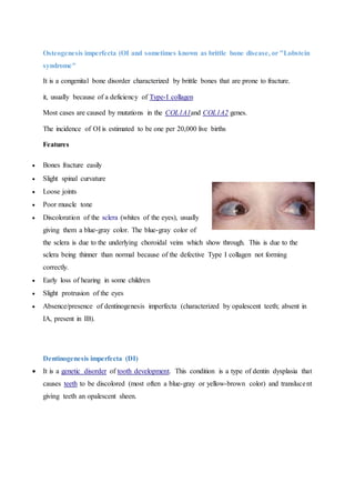
- 1. Osteogenesis imperfecta (OI and sometimes known as brittle bone disease, or "Lobstein syndrome" It is a congenital bone disorder characterized by brittle bones that are prone to fracture. it, usually because of a deficiency of Type-I collagen Most cases are caused by mutations in the COL1A1and COL1A2 genes. The incidence of OI is estimated to be one per 20,000 live births Features Bones fracture easily Slight spinal curvature Loose joints Poor muscle tone Discoloration of the sclera (whites of the eyes), usually giving them a blue-gray color. The blue-gray color of the sclera is due to the underlying choroidal veins which show through. This is due to the sclera being thinner than normal because of the defective Type I collagen not forming correctly. Early loss of hearing in some children Slight protrusion of the eyes Absence/presence of dentinogenesis imperfecta (characterized by opalescent teeth; absent in IA, present in IB). Dentinogenesis imperfecta (DI) It is a genetic disorder of tooth development. This condition is a type of dentin dysplasia that causes teeth to be discolored (most often a blue-gray or yellow-brown color) and translucent giving teeth an opalescent sheen.
- 2. Mohammed Almuzian, University of Glasgow, 2013 Page 1 Teeth are also weaker than normal, making them prone to rapid wear, breakage, and loss. These problems can affect both primary (deciduous) teeth and permanent teeth. This condition is inherited in an autosomal dominant pattern, which means one copy of the altered gene in each cell is sufficient to cause the disorder. Dentinogenesis imperfecta affects an estimated 1 in 6,000 to 8,000 people. Clinical features The teeth may be gray to yellowish brown. They exhibit translucent or opalescent hue. Enamel is usually lost early due to loss of scalloping at the dentoenamel junction (DEJ). However, the teeth are not more susceptible to dental caries than normal ones. However, certain patients with dentinogenesis imperfecta will suffer from multiple periapical abscesses apparently resulting from pulpal strangulation secondary to pulpal obliteration or from pulp exposure due to extensive coronal wear. Radiographic features Type I and II show total obliteration of the pulp chamber. Amelogenesis imperfecta (AI) Arkutu et al 2012 and Gadia2012 0.5% AI is caused by mutations or altered expression in five genes: AMEL (amelogenin),ENAM (enamelin), Systems used for classifying AI: 1. Mode of inheritance: autosomal dominant, autosomal recessive, X-linked, isolated case 2. Phenotype: hypoplastic, hypocalcified, hypomaturation, hypomaturation-hypoplastic with taurodontism.
- 3. Mohammed Almuzian, University of Glasgow, 2013 Page 2 Characterisitcsof hypoplasticAI Enamel of reduced thickness due to a defect in the formation of normal matrix Pitting and grooves Hard and translucent enamel Radiographically, the enamel contrasts normally from dentine. Characteristicsof hypocalcified AI Defect in enamel calcification Enamel of normal thickness Weak in structure Appears opaque or chalky Teeth become stained and rapidly wear down Radiographically, enamel is less radio-opaque than dentine. Characterisitcsof hypomaturation AI Enamel of normal thickness but mottled in appearance Slightly softer than normal and vulnerable to tooth wear, but not as severe as the hypocalcified type Radiographically, similar radiodensity as dentine. Characteristicsof hypomaturation-hypoplasiawith taurodontism Mixed hypomaturation and hypoplasia appearance Taurodontism: body and pulp chamber enlarged, and the floor of pulp chamber and furcation is moved apically down the root. Diagnosis Factors to consider during diagnosis include family history, pedigree plotting (a diagram of a family health history tree), clinical observations and radiographic assessment. Further laboratory-based genetic diagnosis can be done, but this is more useful as a research tool. Complications It presents with a rare abnormal formation of the enamel or external layer of the crown of teeth.
- 4. Mohammed Almuzian, University of Glasgow, 2013 Page 3 People afflicted with amelogenesis imperfecta have teeth with abnormal color: yellow, brown or grey; this disorder can afflict any number of teeth of both dentitions. The teeth have a higher risk for dental cavities and are hypersensitive to temperature changes as well as rapid attrition, excessive calculus deposition, and gingival hyperplasia. The developmental age of the patient should be used when assessing AI patients for tooth eruption. Occasionally, space maintainers may be indicated to prevent tipping of adjacent teeth into the space. Impaction Hypodontia Root malformations have been considered a risk factor for orthodontic apical root resorption. Taurodontism Maxillay hypoplasia and AOB X bite Orthodontic Problems Bond strengths are lower than ideal, leading to multiple bond failures in treatment and the need to step back to 'pick up' these teeth, thereby increasing treatment duration. Debonding of the appliance can cause factures to the fragile enamel and must therefore be performed with caution. The lack of uniformity of enamel coverage means that the second and third order bends (which are part of a pre–adjusted appliance prescription) are not uniformly expressed and more detailing bends at the end stage of treatment are needed to counteract this when final restorations using veneers and crowns are not indicated. Light force to reduce resorption Taurodontism: This feature of AI can increase the susceptibility to root resorption during orthodontic treatment. The use of removable appliances in the correction of malocclusion Solutions Glass ionomer cement-based adhesives are thought to improve appliance retention as they are less reliant on microtag formation, and also help in the reduction of further enamel demineralisation.
- 5. Mohammed Almuzian, University of Glasgow, 2013 Page 4 Plastic brackets can be used instead of metal brackets because they can be removed with a hand piece at debond without damaging the enamel surface. Traditional banded appliances are old-fashioned, but may also be used to overcome some of these problems. If the clinical crown height is minimal and banding is not possible, the use of preformed stainless steel crowns with welded tubes or brackets is recommended. Functional with HG to control AOB