SlideShare a Scribd company logo
1 of 28
Download to read offline
Annual Review of Biomedical Engineering
Biology and Models of the
Blood–Brain Barrier
Cynthia Hajal,1,∗
Baptiste Le Roi,2,∗
Roger D. Kamm,1,3
and Ben M. Maoz2,4,5
1
Department of Mechanical Engineering, Massachusetts Institute of Technology, Cambridge,
Massachusetts 02139, USA
2
Department of Biomedical Engineering, Tel Aviv University, Tel Aviv 6997801, Israel
3
Department of Biological Engineering, Massachusetts Institute of Technology, Cambridge,
Massachusetts 02139, USA
4
Sagol School of Neuroscience, Tel Aviv University, Tel Aviv 6997801, Israel;
email: bmaoz@tauex.tau.ac.il
5
Center for Nanoscience and Nanotechnology, Tel Aviv University, Tel Aviv 6997801, Israel
Annu. Rev. Biomed. Eng. 2021. 23:359–84
The Annual Review of Biomedical Engineering is
online at bioeng.annualreviews.org
https://doi.org/10.1146/annurev-bioeng-082120-
042814
Copyright © 2021 by Annual Reviews. This work is
licensed under a Creative Commons Attribution 4.0
International License, which permits unrestricted
use, distribution, and reproduction in any medium,
provided the original author and source are credited.
See credit lines of images or other third-party
material in this article for license information
∗
These authors contributed equally to this article
Keywords
blood–brain barrier, organ-on-a-chip, tissue engineering, self-assembly,
organoids, neurological diseases
Abstract
The blood–brain barrier (BBB) is one of the most selective endothelial bar-
riers. An understanding of its cellular, morphological, and biological prop-
erties in health and disease is necessary to develop therapeutics that can be
transported from blood to brain. In vivo models have provided some insight
into these features and transport mechanisms adopted at the brain, yet they
have failed as a robust platform for the translation of results into clinical out-
comes. In this article, we provide a general overview of major BBB features
and describe various models that have been designed to replicate this bar-
rier and neurological pathologies linked with the BBB. We propose several
key parameters and design characteristics that can be employed to engineer
physiologically relevant models of the blood–brain interface and highlight
the need for a consensus in the measurement of fundamental properties of
this barrier.
359
Annu.
Rev.
Biomed.
Eng.
2021.23:359-384.
Downloaded
from
www.annualreviews.org
Access
provided
by
37.237.170.14
on
04/06/23.
See
copyright
for
approved
use.
Contents
1. INTRODUCTION . . . . . . . . . . . . . . . . . . . . . . . . . . . . . . . . . . . . . . . . . . . . . . . . . . . . . . . . . . . 360
2. THE BLOOD–BRAIN BARRIER. . . . . . . . . . . . . . . . . . . . . . . . . . . . . . . . . . . . . . . . . . . . . . 360
2.1. Development and Biology . . . . . . . . . . . . . . . . . . . . . . . . . . . . . . . . . . . . . . . . . . . . . . . . . 360
2.2. Characteristics of the Blood–Brain Barrier . . . . . . . . . . . . . . . . . . . . . . . . . . . . . . . . . . 362
3. MODELS OF THE BLOOD–BRAIN BARRIER . . . . . . . . . . . . . . . . . . . . . . . . . . . . . . . 367
3.1. In Vivo Models . . . . . . . . . . . . . . . . . . . . . . . . . . . . . . . . . . . . . . . . . . . . . . . . . . . . . . . . . . . 367
3.2. Cell Source . . . . . . . . . . . . . . . . . . . . . . . . . . . . . . . . . . . . . . . . . . . . . . . . . . . . . . . . . . . . . . . 368
3.3. In Vitro Models . . . . . . . . . . . . . . . . . . . . . . . . . . . . . . . . . . . . . . . . . . . . . . . . . . . . . . . . . . . 370
4. BLOOD–BRAIN BARRIER–RELATED DISEASES: CURRENT MODELS
AND IMPROVEMENTS . . . . . . . . . . . . . . . . . . . . . . . . . . . . . . . . . . . . . . . . . . . . . . . . . . . . . . 374
4.1. Alzheimer’s Disease Models. . . . . . . . . . . . . . . . . . . . . . . . . . . . . . . . . . . . . . . . . . . . . . . . 374
4.2. Stroke Models . . . . . . . . . . . . . . . . . . . . . . . . . . . . . . . . . . . . . . . . . . . . . . . . . . . . . . . . . . . . 375
4.3. Brain Cancer Models . . . . . . . . . . . . . . . . . . . . . . . . . . . . . . . . . . . . . . . . . . . . . . . . . . . . . . 375
4.4. Infectious Disease Models . . . . . . . . . . . . . . . . . . . . . . . . . . . . . . . . . . . . . . . . . . . . . . . . . 376
4.5. Other Diseases: In Vitro and Computational Models . . . . . . . . . . . . . . . . . . . . . . . . 376
5. CHALLENGES AND FUTURE DIRECTIONS . . . . . . . . . . . . . . . . . . . . . . . . . . . . . . 377
1. INTRODUCTION
The blood–brain barrier (BBB), the interface between blood and parenchyma at the brain, fea-
tures a unique cellular architecture of endothelial cells lining the blood vessels, pericytes associ-
ated with the basement membrane, and astrocytes extending their endfeet to the abluminal side
of the vessels. Owing to the organization of the BBB, transport across it is considerably reduced
relative to other tissues. To facilitate selective transport, the BBB draws upon transcytosis, the ac-
tive receptor-mediated transport of specific molecules through the endothelial cells. While this
barrier protects neural tissue from toxins in the blood, it conversely limits the passage of thera-
peutics. The complexity of the BBB has impeded the development of models that can recapitulate
its properties in health and disease. In this review, we first provide an overview of the cellular,
morphological, and functional features of the BBB, and then discuss the new tools that can pro-
vide quantitative measurements of its function in health and disease. Next, we describe several
BBB models with increasing levels of complexity, with a focus on the assessment of their barrier
properties. Finally, we discuss models of neurological diseases in terms of current approaches and
potential improvements, and we explore critical questions that remain unaddressed in the field.
2. THE BLOOD–BRAIN BARRIER
2.1. Development and Biology
Brain development and function require an effective BBB, a tight and selective interface between
the blood circulation and brain parenchyma. Moreover, studies show that several brain diseases
are rooted in dysfunction of the BBB. In the following subsections, we describe the development,
biology, and functionality of the BBB, laying the foundation for addressing its role in the patho-
genesis of brain diseases.
2.1.1. A brief history of the blood–brain barrier. In 1921, Lina Stern and Raymond Gautier
(1) formally named the biological structure observed by Paul Ehrlich, Edwin Goldmann, and Max
360 Hajal et al.
Annu.
Rev.
Biomed.
Eng.
2021.23:359-384.
Downloaded
from
www.annualreviews.org
Access
provided
by
37.237.170.14
on
04/06/23.
See
copyright
for
approved
use.
BE23CH14_Maoz ARjats.cls November 9, 2021 14:0
Lewandowsky 20 years earlier “la barrière hémato-encéphalique.” Using trypan blue and potas-
sium ferrocyanide, Goldmann, following Ehrlich’s previous works (2), had found that the blood
compartment is hermetically separated from brain tissue by a barrier capable of preventing the
passage of nutrients or drugs. Later, Hugo Spatz (3) observed the presence of nonfenestrated en-
dothelial cells responsible for the properties of the blood–tissue interface. The current paradigm
of the BBB was finally settled in 1967 by Reese & Karnovsky (4), following their observation, by
electron microscopy, of the accumulation of horseradish peroxidase exclusively in the lumens of
brain capillaries. They concluded that endothelial tight junctions (TJs) were responsible for this
phenomenon and conferred on the BBB its unique barrier properties (5).
2.1.2. Embryogenesis of the blood–brain barrier. The nervous and brain vascular systems
are coupled not only in their functionality but also in their developmental process. Development
of the BBB occurs through two major events. First, the newly formed brain vasculature invades the
future central nervous system (CNS) structures (6). Second, barrier properties are acquired via the
canonical Wingless–Int1 (Wnt)/β-catenin signaling pathway, which is responsible for the upregu-
lation of TJs in brain endothelial cells and thus the induction of barrier properties in the invading
vasculature (7). Then, barriergenesis enables endothelial cells to acquire a full BBB phenotype
by increasing their expression of glucose transporter 1 (GLUT-1), claudins, and P-glycoprotein
following the sustained secretion of different factors by cells of the CNS, such as platelet-derived
growth factor B (PDGF-B) from pericytes (8) and retinoic acid from astrocytes (9).
2.1.3. Vasculogenesis of the blood–brain barrier. Endothelial cells of the perineural vascu-
lar plexus are the main component of the BBB and typically develop TJs during early embry-
onic development. Endothelial cell differentiation and BBB maintenance genes are expressed at
different time points during the embryonic developmental process. They include the β-catenin
gene Ctnnb1, which promotes the differentiation and maintenance of the BBB via the canonical
Wnt signaling pathway (10). The development of the CNS vasculature begins via vasculogenesis,
whereby mesoderm-derived angioblasts invade the head region and coalesce to form the perineu-
ral vascular plexus. This process is followed by the angiogenic formation of vessel sprouts through
the secretion of vascular endothelial growth factor (VEGF) by neurons of the subventricular neu-
roectoderm. This angiogenic phase, characterized by interactions between vascular sprouts and
neural precursor cells, is followed by the acquisition of a BBB phenotype, as evidenced by an
increase in the expression of TJ proteins on endothelial cell membranes (7).
2.1.4. Perivascular cells. Astrocytes and pericytes are the two main perivascular cell types of
the BBB and play key roles in its formation and properties. Both originate from the neural crest
following three signaling pathways, the Delta/Notch and neuregulin/ErbB pathways (11) for as-
trocytes and the PDGF-B/PDGF receptor β pathway for pericytes (12). Pericytes can also derive
from tissue myeloid progenitor cells or from mature macrophages in the early vascular develop-
ment of the CNS (13). This double embryological origin infers the two major roles of pericytes in
the BBB: barrier maturation and protection. The two cell types differ in spatial organization at the
BBB. Pericytes fully wrap around the brain capillaries formed by endothelial cells, and astrocytes
physically connect their endfeet to the abluminal side of the endothelium covering a small area of
the vessel.
Pericytes and astrocytes have complementary functions in the BBB. Pericytes enable the ex-
pression and organization of several TJ proteins and key transporter receptors (TRs) (14) and
participate in the secretion of the basement membrane surrounding BBB capillaries. Conversely,
astrocytes help refine the BBB phenotype during development via their regulation of efflux TRs
www.annualreviews.org • Blood–Brain Barrier Models 361
Annu.
Rev.
Biomed.
Eng.
2021.23:359-384.
Downloaded
from
www.annualreviews.org
Access
provided
by
37.237.170.14
on
04/06/23.
See
copyright
for
approved
use.
VASOMOTOR CONTROL AT THE BLOOD–BRAIN BARRIER: PERICYTES,
ASTROCYTES, OR SOMETHING ELSE?
Although the major elements of the BBB have been extensively studied both anatomically and histologically, their
roles in the regulation of cerebral blood flow by vasomotor tuning remain ambiguous. On the one hand, Hall et al.
(17) propose that pericyte contractions, resulting from smooth muscle actin contractile fibers, are activated prior
to those of arteriole smooth muscle cells because neurons, which are closer to the capillaries than they are to arte-
rioles, come in contact first with pericytes. On the other hand, Hill et al. (18) suggest that pericytes do not express
smooth muscle actin, which they deem essential to generate cellular contractions. Similarly, the function of astro-
cytes remains debated in the context of vasomotor control in the brain. While Mishra et al. (19) demonstrated that
astrocytes are a mediator of Ca+
2 -dependent nitric oxide generation by interneurons and do not directly generate
capillary contractions, others propose that caveolae expressed on astrocytes are mediators of neurovascular coupling
(20) or indicate the presence of interpericyte tunneling nanotubes that mediate communication among pericytes
(21). As evidenced by continuous disagreements in the field, the observed phenomena are still not fully understood
at the cellular and molecular levels.
(15). In the mature BBB, astrocytes act as a filter between the contents of BBB capillaries and
glial/neuronal cells in the brain parenchyma. They also maintain homeostasis between blood and
brain tissue, in part via their expression of aquaporin 4 (AQP-4) (16). The full importance of per-
icytes and astrocytes regarding their impact on vasomotor tone at the BBB remains to be fully
elucidated (see the sidebar titled Vasomotor Control at the Blood–Brain Barrier: Pericytes, Astro-
cytes, or Something Else?)
2.1.5. The neurovascular unit. The BBB is part of a wider structure, the neurovascular unit
(NVU), which contains the BBB capillaries as well as polarized neurons and supporting cells found
in the brain stroma, such as microglia, resident macrophages of the CNS, and other glial cells,
along with their basement membrane. In addition to their individual functions, which involve
relaying information for neurons and immune defense for microglia, these two cell types play a
nonnegligible role in homeostasis, as microglial cells control proliferation and differentiation of
neurons in the parenchyma and maintain BBB integrity via claudin-5 expression through their
physical interactions with endothelial cells (22). Conversely, during brain injury or immunolog-
ical stimulation, microglia exhibit a counterproductive response through their secretion of reac-
tive oxygen species (ROS) to disrupt the BBB and weaken the overall integrity of the CNS (23).
Neurons also play a similar dual role at the BBB. On the one hand, they regulate blood flow pro-
portionally to their activity (24) via astrocyte engagement. On the other hand, neurons can be
detrimental to the integrity of the BBB during prolonged ischemia, as they can activate astrocytes
and disrupt the barrier (25).
2.2. Characteristics of the Blood–Brain Barrier
Several morphological features of the BBB inform its functional properties, ultimately governing
the controlled transport of nutrients, molecules, and therapeutics from blood to brain (Figure 1).
2.2.1. Hemodynamics. Fluid flow dynamics at the BBB play an important role in determining
the functional properties of this barrier. The intravascular pressure gradient between pre-BBB
capillary arterioles and postcapillary venules is the primary regulator of BBB luminal flow.Dilation
362 Hajal et al.
Annu.
Rev.
Biomed.
Eng.
2021.23:359-384.
Downloaded
from
www.annualreviews.org
Access
provided
by
37.237.170.14
on
04/06/23.
See
copyright
for
approved
use.
In vivo rat BBB
Vascular basement
membrane
Junctional
proteins
Red blood
cell
Astrocytic
endfoot
Pericyte
Endothelial
cell
Parenchymal basement
membrane
In vivo human BBB In vitro human BBB
Lumen
Lumen
500 µm 100 µm
20 µm
Pericyte
Pericyte
Tight
junction
Tight
junction
Endothelial
blood vessels
Basal lamina
Basal lamina
Neuron
Neuron
Blood
vessels
Blood
vessels
Lumen
Lumen
a b
1
2
3
1 µm
1 µm
µm
1 µm
Astrocyte
endfoot
Astrocyte
endfoot
CD31
F-actin
GFAP
Nuclei
10 µm
Figure 1
Comparison between in vivo and in vitro BBB capillaries. (a) Schematic representation of the BBB. (b) Characteristic features of the
BBB. (Top left) SEM section of rat BBB capillaries. (Bottom left) Corresponding confocal image of immunostained vessels. (Center)
Microvascular corrosion cast of a fresh human brain where pial arteries 1
 connect to cortical arterioles in the cerebral cortex 2
, which
give rise to the BBB capillary network 3
. (Top right) SEM section of in vitro human BBB capillaries. (Bottom right) Corresponding
confocal image of immunostained vessels. Abbreviations: BBB, blood–brain barrier; GFAP, glial fibrillary acidic protein; SEM, scanning
electron microscope. Panel a adapted from Reference 26, copyright 2019 Elsevier. Panel b adapted from (left) Reference 27, copyright
2008 Elsevier; (center) Reference 28, copyright 1998 Wiley-Liss; and (right) Reference 29, copyright 2018 Elsevier.
of resistance arterioles increases this pressure gradient, thus increasing BBB capillary flow (30). In
addition,owing to the abundance of caveolae,neurovascular coupling in arteriolar endothelial cells
begins with increased neural activity and ends with smooth muscular cell relaxation surrounding
the arterioles. This process ultimately causes arteriolar vasodilation and increased capillary blood
flow (20). Red blood cell velocity in the BBB of rodents is remarkably high (0.5–2.0 mm/s), owing
primarily to the small diameters of BBB capillaries (around 4 μm in rodents) and brain arteriolar
vasodilation (31). These rapid responses and changes in blood flow in BBB capillaries are espe-
cially critical for the regulation of gas transport across the barrier and for overall brain function
(20).
Increased blood flow velocities in BBB capillaries correspond to large wall shear stresses
(20–40 dyn/cm2
) (32), which maintain the BBB phenotype of brain capillary endothelial cells
through junctional protein upregulation and transport regulation. In cerebral arteries, however,
this pulsatility is typically damped once fluid reaches the BBB, which then translates to reduced in-
terstitial flow in the parenchyma (33, 34). In addition, the absence of endothelial cell fenestrations
and highly controlled transport at the brain suggest that BBB capillaries must employ an alternate
system to regulate fluid flow in the interstitial space. Indeed, the discovery of the meningeal lym-
phatic system surrounding BBB capillaries, through which astrocytes regulate cerebrospinal and
interstitial fluid flows via their AQP-4 channels, has elucidated some of the mechanisms employed
by the BBB to regulate flow in the brain parenchyma (see section 1 of the Supplemental Material)
(35).
2.2.2. Functionality. The upregulation of several TJ,adherens junction (AJ),and TR proteins at
the BBB governs its reliance on selective modes of transport and informs its restricted endothelial
www.annualreviews.org • Blood–Brain Barrier Models 363
Annu.
Rev.
Biomed.
Eng.
2021.23:359-384.
Downloaded
from
www.annualreviews.org
Access
provided
by
37.237.170.14
on
04/06/23.
See
copyright
for
approved
use.
TRANSCELLULAR MODES OF TRANSPORT
Transcellular transport is governed primarily by the size and chemical composition of the solute:
1. Passive diffusion. Ions, gases, and other lipophilic molecules passively cross the BBB by diffusing through
the lipid bilayer and cytosol of endothelial cells (46). Highly soluble lipids with low molecular weight are
better able to diffuse through the barrier (37, 47).
2. Active ABC transporter efflux. Lipophilic substances that cannot passively diffuse through the BBB, such
as cholesterol and long-chain fatty acids,rely instead on energy-dependent ABC transport through specific
efflux pumps (P-glycoprotein, breast cancer resistance protein, multidrug resistance–associated protein).
3. Active solute carrier transport or carrier-mediated transport. Specific solute carriers expressed on BBB en-
dothelial cells (e.g., GLUT-1, monocarboxylic acid transporters, l-type amino acid transporter 1) can ac-
tively bind and transport solutes (e.g., glucose, monocarboxylates, amino acids) into the brain parenchyma
(37, 46).
4. Active transcytosis with receptor- or adsorptive-mediated transport (RMT or AMT). Although the ma-
jority of large molecules are significantly impeded in their transport across the BBB, some enter the brain
via active vesicular transcytosis (46). In RMT, surface receptors bind to macromolecules, form caveolae,
and exocytose the compounds on the opposite pole of the cell (46). AMT occurs in the case of nonspecific
interactions between solutes and cell surfaces, such as electrostatic interactions (38).
permeability to solutes and other compounds. This section is summarized in the sidebar titled
Transcellular Modes of Transport.
2.2.2.1. Junctions and modes of transport. The maintenance of homeostasis at the BBB via
solute transport is unique in that it is highly regulated via junctional surface proteins (36). These
proteins can be categorized as TJs connecting the plasma membranes of cells and regulating para-
cellular transport, AJs joining the actin filaments of neighboring cells and stabilizing TJs, or TRs
governing transcellular shuttling across the barrier (Supplemental Table 1) (37).Transport across
the BBB adopts two major routes: paracellular and transcellular. Owing to smaller gaps between
endothelial cells, paracellular transport at the BBB is impeded in comparison to other endothelial
barriers (38). A few small molecules, such as osmotic or biologically active agents, transiently relax
junction regulation in order to passively cross the BBB (38, 39). In general, molecules that adopt
the paracellular route are smaller than ∼70 kDa, as evidenced by low permeability measurements
for 70 kDa dextran at the BBB (40, 41). The production of ROS and certain proteins, such as
cytochalasin, also increases barrier permeability in intact microvessels, suggesting that several fac-
tors can influence transport. Limited paracellular transport at the BBB has resulted in the reliance
on regulated modes of transcellular transport to maintain homeostasis (42).
During inflammation, diapedesis of mononuclear cells, such as leukocytes and monocytes, is
highly upregulated across the BBB through the opening of TJs following interleukin-8 secretion
(43). Tumor cells from various primary sites have also been observed to extravasate from the BBB
via the paracellular route following secretion of serine proteases that disrupt TJs and AJs (44).
Importantly, although transcellular exchange across the BBB is believed to be crucial, the limited
resolution offered by animal models and current imaging techniques has hindered the ability to
clearly discern transcellular from paracellular transport at the brain. Advances in in vitro tech-
nologies have attempted to address these challenges via high-resolution spatiotemporal imaging
of transport phenomena (37, 45).
364 Hajal et al.
Annu.
Rev.
Biomed.
Eng.
2021.23:359-384.
Downloaded
from
www.annualreviews.org
Access
provided
by
37.237.170.14
on
04/06/23.
See
copyright
for
approved
use.
READOUTS
Permeability
TEER
Solute
Molecular
biology
Animal
Human
Primary
Line
Stem
iPS
Embryonic
a
b
c
d
e
f
g
3D
Cell culture
inserts
Organs-
on-a-chip
Vessel-like
structures
in gels
Bioprinting
Cells-on-
a-dish
2D
Assembly
Emergent 3D
Self-assembled
microvasculatures
Organoids
Long-term
culture
Personalized
medicine
Perivascular
compartment
Vascular
compartment
Medium
Fibroblasts
Chip
Pericyte
Astrocyte
Neuron
Endothelial
cell
BBB
MODELS CELLS
Figure 2
Design parameter considerations for in vitro BBB models. (a) Cells-on-a-dish were the first engineered BBB
models. (b) Transwell® inserts enable direct or indirect coculture of cells. (c) Dynamic 3D organ-on-a-chip
models integrating flow and real-time readouts offer accurate representations of the BBB in health and
disease. Most models are 3D in nature and can be engineered through several methods, such as (d) 3D
printing of microtubes or (e) generation of vessel-like structures in 3D gels with lumens. These 3D methods
have evolved into emergent 3D systems, such as ( f ) self-assembled microvasculatures via angiogenic
sprouting or vasculogenesis and (g) organoids that enable the generation of all brain cell types, in some cases
with internal vasculatures. Abbreviations: BBB, blood–brain barrier; iPS, induced pluripotent stem; TEER,
transendothelial resistance. Panel b adapted from Reference 81. Panel c adapted from Reference 82. Panel d
adapted from Reference 83. Panel e adapted from Reference 84. Panel f adapted from Reference 85.
2.2.2.2. Barrier permeability. The unique morphological features of the BBB, such as cellular
organization and TJ, AJ, and TR expression profiles, inform its distinctive functions, particularly
in terms of regulated transport and toxin clearance between blood and parenchyma. To establish
a direct link between architecture and transport functionality, key transport measurement param-
eters, such as barrier permeability and transendothelial resistance (TEER), have been employed
to compare the BBB with other blood–tissue barriers.
Barrier permeability is commonly measured with the help of contrast agents or fluorescent
tracers transported from blood to tissue over time (Figures 2 and 3). The choice of tracer (type,
size, charge, lipophilicity, and propensity to bind to cells) and imaging technique plays a significant
role in the permeability value obtained. The ability to label particles with fluorescent tags has
allowed various molecules to be assessed using classical methods of permeability measurement
(37). While overall transport across the endothelial cell monolayer can be assessed with the use of
fluorescent tracers, different methods should be implemented to distinguish between the different
mechanisms or pathways of exchange. Modifying the fluid flow characteristics or temperature of
the system has been shown to influence the permeability of physiological compounds across the
BBB, confirming the role of these factors in vascular permeability in vivo (45). Transport across the
BBB by diffusion and convection via transmural flow can be described by permeability to solute
www.annualreviews.org • Blood–Brain Barrier Models 365
Annu.
Rev.
Biomed.
Eng.
2021.23:359-384.
Downloaded
from
www.annualreviews.org
Access
provided
by
37.237.170.14
on
04/06/23.
See
copyright
for
approved
use.
TEER
TEER
Permeability
0
1,000
10–9
10–8
10–7
10–6
10–5
10–4
2,000
3,000
in vivo
TEER
a b
c d
RS
Rpara
Rm
Cm
RS
in vivo
permeability,
4–20 kDa
Nonbrain/
nonhuman
Lines
Primary
iPSCs
Transwell 2D chip
models
3D
vessel-like
structures
3D self-
assembled
models
Model
20
40
60
80
100
120
123
0.80
500
1,000
1,500
2,000
75
2,489
Permeability (×10–7
cm/s)
Permeability (cm/s)
Cell
source
Nonbrain/
nonhuman
Lines
Primary
iPSCs
Transwell 2D chip
models
3D
vessel-like
structures
3D self-
assembled
models
Model
Cell
source
TEER
(Ω/cm
2
)
TEER (Ω/cm2
)
TEER permeability
Primary
iPSC
Animal
Cell lines
S
para m
m
para
para m m mes
Figure 3
Comparison of barrier permeability to solutes and TEER in terms of the type of endothelial cells employed (nonbrain, primary,
immortalized, and iPSC-derived) and the type of BBB model (cell culture insert, 2D chip, 3D vessel-like, 3D self-assembled). Human
iPSC-derived endothelial cells in 3D microfluidic chips exhibit the lowest permeability and highest TEER values. (a) Two methods to
assess BBB permeability can be employed (the two equations, and the equivalent circuit, are extensively described in section 4.1 of the
Supplemental Material). (b) Graph of permeability and TEER measurements of four cell types used as brain endothelial cells. Each
data point represents a different model. In vivo TEER values are from Reference 57, and in vivo permeability values are from Reference
51 (see Supplemental Table 3). (c,d) Heat maps of the highest TEER and lowest permeability values measured in different models (up
to 3 days of culture, 4–20 kDa dextrans). Red corresponds to experimental values reaching physiological values (the data, references,
and rationale used to plot the graphs are presented in sections 4.2 and 4.3 of the Supplemental Material). Abbreviations: BBB,
blood–brain barrier; iPSC, induced pluripotent stem cell; TEER, transendothelial resistance.
P, given by
P = P0 + Lp(1 − σ )(p − σπ),
where Lp is the hydraulic conductivity, p is the pressure differential across the endothelium,
σ is the reflection coefficient, π is the osmotic pressure differential, and P0 is the diffusive
permeability.
TEER, which defines barrier impedance to the passage of small ions through barriers, has also
been employed as a quantitative metric to assess BBB permeability to charged solutes. Briefly, one
366 Hajal et al.
Annu.
Rev.
Biomed.
Eng.
2021.23:359-384.
Downloaded
from
www.annualreviews.org
Access
provided
by
37.237.170.14
on
04/06/23.
See
copyright
for
approved
use.
Table 1 Solute permeability and TEER: What do they measure?
Solute permeability TEER
Principle Net exchange of molecules, typically of
higher molecular weight, across the
endothelial barrier
Transport of charged molecules or ions
across the endothelial barrier
Biological measurement Diffusion and convection of molecules Paracellular transport of ions
Typical in vitro values in human
BBB models
10−7 cm/s for 4–20 kDa dextrans 1,500 /cm2
Measured in vivo values in animals 4 × 10⁻⁷ cm/s [rat (51)] 1,300 /cm2 [frog (52)]
Availability Most in vivo and in vitro models In vivo and in vitro models for which the
area of the endothelium is well defined
Correlation/translation Although both solute permeability and TEER measure transport of different types of
solutes across the barrier, they are negatively correlated. Low permeabilities indicate
reduced paracellular transport, which often correlates with restricted ionic transport
and high TEER values.
Abbreviations: BBB, blood–brain barrier; TEER, transendothelial resistance.
can use the voltage and current through a BBB capillary to compute the electrical resistance of the
cellular barrier via Ohm’s law. One thus obtains TEER as the product of the cellular layer resis-
tance and area; high values indicate restricted ionic transport through the BBB and optimal barrier
function (Table 1). Shortly after its first implementation in frog mesentery, the TEER technique
was used in rat BBB capillaries, yielding values close to 6,000 /cm2
, significantly higher than
those found in in vitro BBB models (48). These discrepancies partly reflect the relatively poor
barrier function of in vitro BBB monolayer systems, where ionic transport cannot be fully recapit-
ulated. More importantly, voltage levels applied across the BBB endothelia can play a significant
role in the resulting TEER values, as shown by Sharabi et al. (49). These authors found that
voltages as low as 10 V led to an ∼40% decrease in TEER due to electroporation of the cellular
membrane. In addition, probe design, positioning, and shape, as well as anatomical configurations,
can result in nonuniform current distributions across the barrier, reducing its effective area and
overestimating its TEER (50). Finally, similarly as for solute permeability, TEER values do not
provide information about transcellular transport of charged compounds. TEER measurements
should be interpreted with caution, since several parameters can significantly affect the results.
3. MODELS OF THE BLOOD–BRAIN BARRIER
3.1. In Vivo Models
Much of our basic knowledge regarding the BBB first came from animal models, which elucidated
the fundamental mechanisms of regulated transport, barrier permeability, cellular architecture,
and associated morphological features (53–55). The clear advantage of these models is their phys-
iological relevance, including not only all the structures forming the BBB but also the effects of
blood flow or biochemical mediators of function. Thus, animal models have offered tremendous
insight into the physiology of brain tissue, particularly in the context of disease modeling. How-
ever, challenges remain in the translation of animal findings to human patients. The underlying
genetic, molecular, and immunologic differences between human and animal BBBs have limited
the use of in vivo models as effective platforms for the development of therapeutics. This is clearly
evident in the high failure rates (≥80%) in clinical trials of drugs validated in animals (37). This
www.annualreviews.org • Blood–Brain Barrier Models 367
Annu.
Rev.
Biomed.
Eng.
2021.23:359-384.
Downloaded
from
www.annualreviews.org
Access
provided
by
37.237.170.14
on
04/06/23.
See
copyright
for
approved
use.
limitation, in combination with increased ethical concerns and high costs, has spurred the devel-
opment and use of in vitro human BBB systems.
3.2. Cell Source
The choice of cell source is key to the development of in vitro BBB models. Important con-
siderations include availability, human relevance, cost, robustness, reproducibility, batch-to-batch
variability, capability for disease modeling, and barrier properties. Decisions regarding the species
(e.g., murine, bovine, porcine, simian, or human) and cells used [e.g., cell lines, induced pluripo-
tent stem cells (iPSCs), primary cells, or direct/transdifferentiated cells] are critical to the success
of a BBB model. Here, we focus on endothelial cells employed to design in vitro BBB models,
although other cells (e.g., pericytes, astrocytes, and to a lesser extent neurons) are also important.
We note that BBB endothelial cells have critical morphological and functional differences from
other endothelial cells, as they possess tighter cell–cell junctions, are highly polarized, and gener-
ally exhibit lower values of permeability or higher TEER values compared with capillaries found
in other tissues (56, 57). TRs, such as P-glycoprotein and GLUT-1, as well as TJ proteins, such
as claudins and occludins, are key elements involved in barrier function of the BBB and need to
be taken into account when selecting endothelial cells to engineer an in vitro BBB model.
3.2.1. Primary cells. Primary endothelial cells can be easily isolated from animals or human
biopsies and cultured in vitro. These cells are accessible, widely used, and well documented in
the literature (58). However, animal primary cells are not fully representative of human cells in
either composition or function despite their common characteristics, such as TJ and TR protein
expressions. Human primary cells collected from donors and used either in single-cell monolayers
or in coculture models have thus been the preferred choice. Although they express high levels
of platelet endothelial cell adhesion molecule 1 (PECAM-1), claudin-5, and zonula occludens
1 (ZO-1) (59), human primary cells paradoxically present low TEER values, usually under
100 /cm2
(Figure 3) (60). Finally, two major drawbacks of human primary cells are that
they often lose their BBB properties through cell passage (61) and the source heterogeneity
significantly reduces the reproducibility of experiments.
Human umbilical vein endothelial cells (HUVECs) have been grown in coculture with peri-
cytes, astrocytes, and even neurons. Under these conditions, expression of key BBB markers, such
as ZO-1 and VE-cadherin, is often upregulated. HUVECs generate stable channels or vascular
networks (62). They also exhibit strong barrier function, even for 3 kDa dextran, with permeabil-
ity values as low as 2 × 10⁻⁷
cm/s, even though they are not specific to the brain microvasculature
(45, 63). These observations suggest that endothelial cells alter their phenotype depending on
their coculture conditions and the stromal cells employed in the model.
3.2.2. Immortalized cells. Owing to their low cost and ease of culture, immortalized cell lines
are now extensively employed in in vitro BBB models. They retain their properties following
cell passage and are genetically identical to one another, thus improving reproducibility in BBB
models. Despite these advantages, cell lines generally present poor BBB properties and barrier
function and may not be suitable for molecular transport studies (64). In addition, rodent BBB
cell lines have been observed to lose their in vivo features, such as the expression of GLUT-1 (65).
Human cells lines, in contrast, can present inconsistent protein levels. For example, the TY10
cell line expresses high levels of claudin-5 and VE-cadherin, while the hCMEC/D3 line has the
highest expression level of ZO-1 but no claudin-5 (66). These inconsistencies imply that different
cell lines might be optimal, depending on the application of the study (67, 68). Because of low TJ
368 Hajal et al.
Annu.
Rev.
Biomed.
Eng.
2021.23:359-384.
Downloaded
from
www.annualreviews.org
Access
provided
by
37.237.170.14
on
04/06/23.
See
copyright
for
approved
use.
or TR expression levels, immortalized cell lines are not capable of creating a tight cell layer and
may not be relevant for general BBB modeling but only for specific studies such as drug absorption
or pathogen transmigration across the BBB.
A promising cell source derived from stem cells has garnered increasing attention over the
last decade. This includes cells differentiated from embryonic or mesenchymal stem cells (ESCs
or MSCs) and iPSCs, which are stem cells generated from a differentiated tissue. Stem cells are
usually defined by their capacities to self-renew and to generate multiple different cell types.
3.2.3. Embryonic and mesenchymal stem cells. ESCs are pluripotent stem cells (PSCs) with
the ability to differentiate into all cell types. ESCs from the blastocyst can be employed to recapit-
ulate the embryonic development of brain microvascular endothelial cells (BMECs), and several
embryoid differentiation protocols have been generated for this purpose. Generally, 3D embryoid
differentiation protocols enable direct differentiation into the three embryonic layers.Yet the yield
with these protocols is low, and there is a need for further isolation and expansion. In contrast, 2D
monolayer differentiation protocols offer a high yield but generally result in the generation of a
single cell type (69).
Various types of human ESC lines can be employed, depending on the desired protein ex-
pression of the resulting differentiated cells. For example, H1 and H9 are used for PECAM-1
expression (70), and HES3 and HES4 are employed for VE-cadherin expression (71). ESCs are
also used in the context of modeling development in vitro. For example, Weidenfeller et al. (72)
demonstrated that undifferentiated neural progenitor cells do not express BBB properties but dif-
ferentiated cells do. In addition to ESCs, MSCs, which give rise to embryonic connective tissue,
can improve the stabilization of the resulting BBB models.
3.2.4. Induced pluripotent stem cells. Since their development by Yamanaka and colleagues
(73), human iPSCs have found application in numerous in vitro models. Yet, there is still no
consensus in terms of a single differentiation protocol to produce BMECs from iPSCs (iBMECs).
Protocols vary in length, number of steps, and differentiation factors employed, such as retinoic
acid and Wnt/β-catenin agonist (74) or hypoxia (75). However, common to all protocols is a
succession of steps including iPSC culture, endothelial induction, BMEC specification, and pu-
rification (76).
iPSCs have the considerable advantage that they can, in principle, produce all the cells com-
posing an organ while maintaining genetic consistency among all the differentiated cells, thus
enabling the generation of a patient-specific BBB model. In addition, they present excellent bar-
rier properties, with strong expression of BBB markers such as ZO-1, claudin-5, and occludin as
well as cell activity markers such as GLUT-1 and P-glycoprotein. Finally, their TEER and per-
meability values are comparable to those measured in vivo (Figure 3). However, in contrast to
cell lines and primary cells, they often have a limited life span in vitro, and processes ranging from
dedifferentiation to redifferentiation are complex, time consuming, and very sensitive to experi-
mental conditions.
As mentioned above, iPSCs show promise for personalized medicine applications, wherein a
model based on the patient’s own genotype is constructed (77). For example, Vatine et al. (78)
modeled psychomotor retardation with iPSCs from patients deficient in monocarboxylate trans-
porter 8. In addition, such models need not focus solely on inducing BMECs but can include all
relevant components of the NVU (79). However, one needs to keep in mind several key elements
when inducing iBMECs, such as the expression of canonical endothelial cell markers (cadherin-5,
PECAM-1, VEGF receptor 2, apelin receptor, endothelial nitric oxide synthase), ability to form
www.annualreviews.org • Blood–Brain Barrier Models 369
Annu.
Rev.
Biomed.
Eng.
2021.23:359-384.
Downloaded
from
www.annualreviews.org
Access
provided
by
37.237.170.14
on
04/06/23.
See
copyright
for
approved
use.
tubular networks, low-density lipoprotein uptake, response to angiogenic stimuli, high TEER/low
permeability, and expression of critical efflux transporters (80).
While human primary cells appear to be the best source of cells for BBB models, they tend
to lose their BBB properties over time and present weak barrier properties. In order to overcome
these limitations, iPSCs are being employed to build complex BBB models including different
cell types with a common genome. Note, however, that iPSCs have a nonnegligible tumorigenic
and teratogenic potential. As a result, one must take care to eliminate nonusable or potentially
tumorigenic differentiated cells.
3.3. In Vitro Models
The development of BBB models with physiological transport properties has been a major chal-
lenge since the 1980s. Cells were first cultured in two dimensions by use of a single cell type to
mimic the BBB. Gradual increases in the complexity and functionality of in vitro BBB models
enabled the generation of sophisticated platforms including multiple interacting cell types that
can be dynamically stimulated, as well as the presence of hydrogels to mimic the 3D brain ex-
tracellular matrix (ECM). Some of these elements have been included in Transwell®
systems and
microfluidic platforms. Here, we classify systems as either 2D or 3D; 3D models are defined as
those with hydrogels at least 100 μm thick.
3.3.1. 2D static models. In 2D static models, cells are cultured on hard plastic (Figure 2a) or in
Transwell inserts without the application of flow (Figure 2b). Historically, the first models of the
BBB consisted of monocultures of endothelial cells isolated from brain capillaries. These allowed
for the observation of TJ structures via freeze fracture (86). In order to recapitulate the BBB mi-
croenvironment and reduce the lack of neighboring cell stimuli, coculture models were developed,
notably with a full NVU composition of endothelial cells, astrocytes, pericytes, and neurons (87).
Culturing cells on plastic offers several advantages in terms of ease of imaging and environmental
control, as well as the presence of several standard protocols. However, hard plastics do not reca-
pitulate the interface between vascular and brain parenchymal compartments, the major aspect of
the BBB, thus limiting the utility of these models to assess BBB functionality through metabolic,
diffusion, or permeability studies. Finally, the stiffness of tissue culture plastics, their flat geome-
try, and the lack of ECM in 2D layer systems all limit cell development, alter cell phenotype, and
result in poor models with limited potential for clinical translation (88).
Transwell systems can overcome these challenges, as they offer the ability to perform per-
meability assays, observe cell migration across a membrane, study multicellular interactions, and
promote epithelial cell polarity (89). Transwell systems have progressively improved and are now
widely used owing to their broad commercial availability. While these systems are static, several
parameters can be optimized to improve the resulting models, such as the size of the inserts, their
membrane porosity, their thickness, and the type of material (polymer) employed, with or with-
out coating (90). Transwell systems allow for the introduction of at least two cell types, usually
(a) endothelial cells on top and (b) astrocytes, pericytes, and sometimes neurons either in direct
contact with the endothelial cells, when cultured on the opposite side of the membrane, or in indi-
rect contact, when cultured at the bottom of the well (81). Transwell models of the BBB have been
generated with all types of cells (primary cells, cell lines, and iPSCs), and they enable measurement
of barrier properties such as TEER and solute permeability (56). However, their flat geometry
limits cellular interactions that could promote barrier properties or the polarization of astrocytes,
resulting in subpar BBB models. Hybrid devices containing 3D gels on Transwell inserts have re-
cently been developed and are referred to as chip-on-a-Transwell systems (91). Nevertheless, the
370 Hajal et al.
Annu.
Rev.
Biomed.
Eng.
2021.23:359-384.
Downloaded
from
www.annualreviews.org
Access
provided
by
37.237.170.14
on
04/06/23.
See
copyright
for
approved
use.
presence of a Transwell insert introduces artifacts and limits the natural interactions between cells
and the underlying ECM, resulting in poor recapitulation of the natural morphology of the BBB.
3.3.2. 2D organs-on-a-chip. Standard in vitro models lack cell–cell interactions and do not
permit accurate control of hemodynamics (92) or appropriate organization of the microenvi-
ronment represented. These limitations have been mitigated by the introduction of organ-on-a-
chip (OoC) models in which cells can be compartmentalized in several channels, often separated
by a membrane. These systems facilitate cell–cell interactions, the application of fluid flow and
controlled shear stresses, and real-time readouts (93). This technology can be used to generate
multiorgan systems by linking different OoCs together, producing so-called human-on-a-chip
platforms (94–96). Recent developments in microfluidic chip technologies have enabled the in-
tegration of sophisticated sensors (e.g., microelectrode arrays, TEER electrodes, oxygen tension
probes) and measurements (e.g., mass spectroscopy analyses) (97, 98). Given the complexity of
these additions, several OoC systems lack sensors and rely on the use of Transwell systems to per-
form parameter measurements such as TEER. The results obtained in Transwell systems might
not be representative of those in OoCs, however, and they are sometimes mathematically cor-
rected, which can introduce a degree of error (99). TEER values, for example, vary according to
the cell type and BBB model employed. Currently, iPSC models (79) present the lowest perme-
ability values and the highest resistance, where TEER values reach or exceed physiological values
of 1,300 /cm2
(Figure 3c) (52).
2D BBB-on-a-chip (BBBoC) models are defined as linear systems in microfluidic chips that
incorporate two or more BBB cells with hydrogels no more than 100 μm thick. Compared with
Transwell platforms, BBBoC systems offer greater physiological relevance and complexity and can
facilitate the observation of cell migration/protrusion (100) and angiogenesis (85). They have been
employed to recapitulate the effects of hypoxia on the BBB by use of both primary cells and iPSC
models (Figure 2c) (75, 82). BBBoCs are a powerful tool for studying BBB physiology in terms of
membrane protein expression (TJs, AJs, and TRs) or cell–cell interactions such as the formation of
astrocyte endfeet (100). Recent studies have incorporated other CNS cells into BBBoC models in
order to produce an NVU platform to uncover metabolic pathways between the vasculature and
brain parenchyma in health and disease (101, 102). More complex models including five (brain,
intestine, liver, kidney, and skeletal muscle) or eight organs (i.e., human-on-a-chip models) have
been designed and employed in pharmacological studies (94, 95).
Despite these advances in 2D OoC technologies, several challenges remain regarding minia-
turizing these systems and increasing their throughput to perform large-scale drug screens (103).
Finally, there has been a major push toward 3D models because of their ability to better recapit-
ulate organ form and function.
3.3.3. 3D models. Researchers’ attention is shifting to 3D brain models, as they can better
mimic the architecture and function of the BBB while offering greater physiological relevance.
Several platforms have been proposed to generate human-like brain models, particularly with the
use of biocompatible hydrogels, relevant cell types, and techniques drawing from the process of
embryogenesis.
3.3.3.1. Monolayer of cells in a gel system. As mentioned above, we define 3D BBB models
as those for which the BBB cells are cultured adjacent to a hydrogel with a thickness of at least
100 μm. One example is the model developed by Ahn et al. (104), who created a platform with
human brain pericytes and endothelial cells in a microfluidic device with astrocytes on the bottom,
seeded in Matrigel (Figure 2f ). The astrocytes extended their AQP-4-expressing endfeet to
www.annualreviews.org • Blood–Brain Barrier Models 371
Annu.
Rev.
Biomed.
Eng.
2021.23:359-384.
Downloaded
from
www.annualreviews.org
Access
provided
by
37.237.170.14
on
04/06/23.
See
copyright
for
approved
use.
establish contact with the pericytes and endothelial cells, similar to the behavior observed in
vivo (104). The simple design and ease of use of 3D monolayer brain models, coupled with their
increased physiological relevance, have been instrumental in the generation of high-throughput
in vitro brain models. With the help of multichannel microfluidic chips, investigators have
engineered identical BBB platforms for use as functional assay platforms, notably for studies of
metastasis or drug transport (105). Importantly, when compared with 2D BBB models, brain en-
dothelial cells cultured in 3D platforms exhibit improved TJ expression and increased TEER val-
ues, suggesting that the addition of appropriate matrices plays a fundamental role in the integrity
and function of the resulting BBB model (106). Additionally, the application of physiological
levels of fluid flow and resulting wall shear stresses improve TJ protein expression as well as solute
permeability and TEER values (105). In comparison to 2D systems, the ability of 3D brain models
to recapitulate physiological BBB hemodynamics is paramount in the function of the barrier.
3.3.3.2. Vessel-like structures in gels. Microfluidic technology has played a significant role in
the development of BBB models with increased complexity and realism, giving rise to a new ap-
proach to in vitro 3D BBB models: 3D channels embedded in or adjacent to hydrogel compart-
ments. These platforms possess several desirable features in terms of morphology, cellular orga-
nization, high-throughput capabilities, and improved barrier function. Importantly, they better
recapitulate important aspects of BBB morphology consisting of hollow tubular structures lined
with endothelial cells and variably cocultured with neural stromal cells, all embedded in a 3D hy-
drogel. For example, Marino et al. (83) used two-photon lithography to generate porous tubular
structures with an average diameter of 10 μm, which were subsequently coated with mouse BBB
endothelial cells (Figure 2d).
Similar platforms with increasing levels of complexity have been designed. They incorporate
hydrogels with improved physiological relevance (107) and additional BBB cell types such as per-
icytes and astrocytes. Both Brown et al. (108) and Adriani et al. (109) employed multichannel
microfluidic chips in which one of the channels was lined with primary human brain endothelial
cells, with combinations of pericytes, astrocytes, and neurons in an adjacent collagen hydrogel. Al-
though collagen type 1 is commonly used in cell culture systems, it is not usually found in the brain
parenchyma; the use of more physiological matrices, such as hyaluronic acid– or proteoglycan-
based hydrogels, could improve the BBB properties of these models (110). In addition, the mor-
phological attributes and cellular organization of these platforms do not fully recapitulate the
BBB, either because of their large diameter (typically approximately 500 μm) or because of the
lack of stromal cells with appropriate spatial distribution. Herland et al. (84) addressed some of
these challenges by generating hollow cylindrical structures in a collagen gel via a pressure-driven
viscous fingering method (Figure 2e). The channel lumens (still large, with diameters of 600–
800 μm) were perfused with pericytes or astrocytes followed by human BMECs, which resulted
in a decrease of barrier permeability to solutes in comparison to platforms without brain stromal
cells (84). To address the remaining challenge of reducing vessel diameter and improving vascular
morphology, several groups have generated self-assembled vascular systems that recapitulate in
vitro the inherent ability of endothelial cells to self-assemble into vascular networks with small-
diameter lumens.
3.3.3.3. Self-assembled vasculatures and the role of emergence. To better recapitulate the mor-
phological and functional features of the BBB, researchers have focused on harnessing the natural
processes of vascular development in vivo, whereby endothelial precursors coalesce and organize
into complex microvascular networks (vasculogenesis) and endothelial cells sprout from preexist-
ing blood vessels (angiogenesis) (111). This inherent ability of endothelial cells to form tubular
372 Hajal et al.
Annu.
Rev.
Biomed.
Eng.
2021.23:359-384.
Downloaded
from
www.annualreviews.org
Access
provided
by
37.237.170.14
on
04/06/23.
See
copyright
for
approved
use.
structures has been extensively employed to engineer 3D vascular networks with HUVECs and
lung stromal cells or placental pericytes; the resulting structures exhibit improved morphology, TJ
protein expression, and solute transport machinery in comparison to other 3D tubular structures
or 2D models (112). Extrapolating from these models, Bang et al. (113) incorporated astrocytes
and neurons into a HUVEC–fibroblast platform to mimic the BBB in three dimensions. While
this model includes non-brain-specific cell types, it offers valuable insight into the role astrocytes
play in promoting the expression of BBB properties (113).
Challenges in isolating tissue-specific human endothelial cells and ensuring that they retain
their properties when cultured in vitro have hindered the development of 3D OoC self-assembled
models, particularly BBB microvascular platforms. However, certain commercial human BMEC
lines isolated from brain biopsy samples do retain their ability to self-lumenize when cultured
in appropriate 3D hydrogels. For instance, Lee et al. (85) used human BMECs, primary brain
pericytes and astrocytes, and lung fibroblasts to create via angiogenesis in a microfluidic chip a
3D self-assembled BBB model that exhibited improved barrier permeability and increased ex-
pression of TJ proteins. In another model (29), self-assembled BBB vasculatures were generated
via vasculogenesis using human iPSC-derived endothelial cells, as well as primary brain pericytes
and astrocytes embedded simultaneously in a fibrinogen–thrombin gel, without the need for cell
types from different organs for vascular support (e.g., lung fibroblasts). The self-assembled BBB
vessels exhibited physiological morphologies with diameters as small as 20 μm and decreased so-
lute permeability, comparable to levels measured in rat BBB (Figure 3) (Supplemental Tables 2
and 3).
These self-assembled vascular systems have revolutionized the field of in vitro BBB models,
significantly improving their physiological relevance and allowing for high-resolution spatiotem-
poral imaging of key events at the BBB, such as cell diapedesis or solute transport. Despite these
advances, challenges regarding the choice of hydrogels in these systems remain. The common use
of fibrinogen gels for 3D angiogenic and vasculogenic BBB models does not fully recapitulate the
in vivo brain ECM, which comprises mostly hyaluronic acid and other proteoglycans. In addition,
fluid flow, which is not included in most in vitro self-assembled BBB models, exhibits specific dy-
namic patterns at the brain that promote the upregulation of TJ and transporter proteins (31, 114).
Recapitulating BBB hemodynamics, and thus improved transport of gases and nutrients, would be
paramount for the maintenance of long-term cultures in vitro, since the current engineered BBB
vessels are known to regress after a couple of weeks.
3.3.3.4. Cerebral organoids and the blood–brain barrier. The last ten years have witnessed the
development of 3D brain organoids from the differentiation of PSCs, such as iPSCs or ESCs.
These systems harness the entire development process to generate structures that recapitulate
specific brain regions, such as midbrain organoids or cerebral cortex organoids that house BBB
capillaries (115). Cerebral organoids that model neurological diseases such as Alzheimer’s disease
(AD) have also been created through the use of patient-derived iPSCs (116).
Despite successes in generating brain organoids, these structures still lack appropriate vas-
culature. Pham et al. (117) addressed this limitation by engineering discrete vascular sprouts in
their patient-derived iPSC cerebral organoids by using differentiated endothelial cells from the
same iPSCs. Although a few CD31-positive blood vessel sprouts were observed, they were not
connected, and perfusion was not achieved in these structures (117). In another cerebral organoid
model, Cakir et al. (118) engineered ESCs to express human ETS variant 2, which reprograms
human dermal fibroblasts into endothelial cells, promoting vascularization. However, BBB vessels
were observed only at the organoid surface. Homan et al. (119) employed a similar approach,
based on the application of fluid flow, to enhance the vascularization of kidney organoids.
www.annualreviews.org • Blood–Brain Barrier Models 373
Annu.
Rev.
Biomed.
Eng.
2021.23:359-384.
Downloaded
from
www.annualreviews.org
Access
provided
by
37.237.170.14
on
04/06/23.
See
copyright
for
approved
use.
High-shear-stress flow resulted in the most effective vessel formation in the surrounding ECM
in comparison to static conditions and low-shear-stress flow. These results obtained in kidney
organoids could be extrapolated to cerebral structures to enhance vascularization, confirming the
importance of hemodynamics in BBB modeling and function. These models attest to the signif-
icance of design considerations, such as the application of fluid flow, the choice of 3D matrix, and
the use of appropriate growth factors in the vascularization of cerebral organoids. Recapitulating
organ development at the brain in these structures also offers insight into the development of the
brain cortex, particularly in the context of developmental neurological disorders.
3.3.3.5. 3D-printed models. Microfabrication of OoCs can be a long, expensive process that
requires clean-room facilities and fabrication engineers and offers little flexibility in modifying
the design of the chip. Moreover, OoCs are not truly 3D organ models. 3D printing is now a well-
established technique that overcomes these issues.3D printing,or additive manufacturing,consists
of layer-by-layer production of a computer-aided design file. In order to print organ models, hy-
drogels are often used as bioinks, which are networks of polymeric and hydrophilic molecules such
as gelatin (collagen) derivatives like gelatin methacryloyl (120), hyaluronic acid, or alginate (121).
These bioinks integrate cells such as neurons, astrocytes, HUVECs, or glioblastoma cells (122). In
order to print different types of cells, either multinozzle printers can be used simultaneously (123)
or a single-nozzle printer can be employed to sequentially print multilayered bioinks in which
each layer contains a different cell type (124).
Several groups have generated BBBoC models of glioblastoma using 3D printing of com-
partmentalized concentric tumor/vascular compartment structures in order to study the effect
of drugs and radial oxygen gradients on tumor progression (123). The cells were surrounded
by polydimethylsiloxane to ensure chip integrity (123). Other researchers have built tubular
printed vessels-on-a-chip with excellent properties (e.g., TEER of 1,500 /cm2
) (125). Finally,
bioinspired synthetic scaffolds have been designed for the coculture of endothelial cells and
neurons via two-photon lithography (83). 3D-printed models differ from microfluidic ones in
terms of tissue maturation and self-assembly. Two techniques are generally used to print vessels:
indirect printing, which involves the use of a sacrificial material employed to generate the vessel
lumens (126), and direct printing, where lumens are assembled without sacrificial structures.
Vascular lumens can be formed by seeding cells either inside the preformed, multilayer tubes
(127) or within porous materials (128).
4. BLOOD–BRAIN BARRIER–RELATED DISEASES: CURRENT MODELS
AND IMPROVEMENTS
Multiple neurological or brain diseases are known to affect the BBB through the secretion of
inflammatory cytokines that degrade TJs (129). Because of the limited spatiotemporal resolution
of in vivo models and the challenge of translating results from animal to human studies, several
groups have shifted their efforts to the development of in vitro human BBB disease systems. These
platforms also enable the generation of high-throughput identical models that can be used for
studies pertaining to drug delivery across the BBB.
4.1. Alzheimer’s Disease Models
According to the two-hit vascular hypothesis of AD, BBB damage is thought to be the initial
insult that results in diminished brain perfusion and neuronal injury (the first hit), followed by
amyloid-β (Aβ) accumulation due to faulty clearance and increased antibody production (the sec-
ond hit) (130). In an early 2D Transwell model of AD with patient-derived BBB endothelial cells,
374 Hajal et al.
Annu.
Rev.
Biomed.
Eng.
2021.23:359-384.
Downloaded
from
www.annualreviews.org
Access
provided
by
37.237.170.14
on
04/06/23.
See
copyright
for
approved
use.
Mackic et al. (131) uncovered the role of the endothelial cell receptor for advanced glycation end
products and scavenger receptor in the transcytosis of Aβ-40 into the brain parenchyma. More
recently, Robert et al. (132) used a 3D in vitro vessel-like structure of brain endothelial cells, lined
with smooth muscle cells and astrocytes, to show that apolipoprotein E2 (APOE2) can reduce the
accumulation of amyloid plaques. While these models provide useful insight into the mechanisms
of accumulation and clearance of Aβ, they do not fully recapitulate AD pathology in the context
of neuron–BBB interaction and BBB disruption.
Microfluidic technologies have addressed some of these challenges. Recently, Shin et al. (133)
developed a 3D microfluidic model with a vessel-like structure consisting of primary brain en-
dothelial cells adjacent to neural progenitor cells. In this model, mutations in the amyloid precur-
sor protein and presenilin-1 genes in neurons resulted in their robust secretion of Aβ as well as an
observable deposition at the endothelial surface, corresponding to an increase in barrier perme-
ability. In another 3D AD BBB model of endothelial cells, pericytes, and astrocytes differentiated
from the same iPSCs in Matrigel, APOE4 pericytes exhibited upregulated calcineurin signaling,
which is associated with cerebral amyloid angiopathy, a common precursor to AD (134). Despite
their improved throughput and physiological relevance, 3D AD BBB systems, which are cultured
for only a few weeks, remain limited in their ability to recapitulate barrier dysfunction and plaque
deposition, which occur over the course of multiple years in humans. The emergence of cerebral
organoid models that can be cultured for several months might provide a way to model and study
long-term progressing neurological disorders such as AD.
4.2. Stroke Models
Strokes, primarily ischemic in nature, are characterized by disruptions of the BBB wherein hy-
poxia and ROS production during the ischemic insult lead to TJ breakdown and subsequent ionic
shifts in the brain parenchyma (135). Hundreds of drugs have been reported to effectively improve
outcomes in experimental animal models. However, almost all of them have failed to translate into
successful therapies for humans (136). Several groups have thus developed in vitro human BBB
models of stroke to better recapitulate this pathology. For instance, Chen et al. (137) created a BBB
stroke Transwell model with primary brain endothelial cells and astrocytes subjected to oxygen and
glucose deprivation, showing that the increase of Sirtuin-1 following stroke and ischemia was cor-
related with ROS production and subsequent inflammation and cell damage. Similarly, Chaitanya
et al. (138) subjected a Transwell BBB model with primary brain endothelial cells, astrocytes, and
neurons to various metabolic stressors, such as hypoxia, glucose deprivation, and reoxygenation.
These stressors induced brain capillary tube formation and angiogenesis in vitro and promoted
lymphocyte adhesion via increased expression of adhesion markers on the BBB endothelial cells
(138). Although these models could be employed as high-throughput platforms for drug screen-
ing, they remain severely limited in their ability to mimic the architecture and physiology of the
BBB. More importantly, current human in vitro stroke models primarily mimic the lack of oxygen
and glucose at the brain, disregarding other key features of strokes such as inflammatory cytokine
production and microglial activation (135).
4.3. Brain Cancer Models
Given the poor prognoses of primary brain cancers, the prevalence of metastatic tumors in the
brain, and the lack of in vitro brain tumor platforms that incorporate BBB vessels, there is a
significant need for models that can aid in the understanding of cancer progression and drug
delivery across the BBB. In one recent example, Plummer et al. (139) designed a high-throughput
glioblastoma–neuron–astrocyte tumor platform to test drug transport and found that therapeutics
www.annualreviews.org • Blood–Brain Barrier Models 375
Annu.
Rev.
Biomed.
Eng.
2021.23:359-384.
Downloaded
from
www.annualreviews.org
Access
provided
by
37.237.170.14
on
04/06/23.
See
copyright
for
approved
use.
selectively target the glioblastoma cells. More recently, in vitro primary brain cancer models that
integrate vasculature and tumor have been designed. Ngo  Harley (140) developed a coculture
model of glioma cells with HUVECs and lung fibroblasts in gelatin and hyaluronic acid and
observed that the addition of cellular and matrix elements led to a decrease in the response of
glioma cells to therapeutics. Similarly, Ozturk et al. (141) designed a long-term 3D in vitro model
of glioblastoma tumor spheroids surrounded by bioprinted HUVEC gelatin channels to study
drug delivery over time. Although these models are useful to screen for therapeutic toxicity, the
use of HUVECs or lung fibroblasts does not fully recapitulate cell types found in the brain, and
the 3D architecture of these systems could be improved to replicate the in vivo BBB vasculature
in contact with tumors.
Given the prevalence of metastatic brain tumors originating from various primary sites, mod-
els have been designed to elucidate the mechanisms employed by cancer cells to cross the BBB.
For example, Bos et al. (142) observed that increased expression of the sialyltransferase gene
ST6GALNAC5 promotes extravasation in a HUVEC–astrocyte Transwell system. In another
study using a high-throughput microfluidic platform with brain endothelial cells and astrocytes
in collagen, Xu et al. (105) found that lung and breast tumor cells exhibit greater transmigration
potential in comparison to melanoma and liver cancer cells. While these high-throughput models
allow for high-resolution spatiotemporal imaging of tumor extravasation, their physiological rel-
evance remains limited, particularly in terms of the cell types employed and the 3D architecture
of the BBB capillaries.
4.4. Infectious Disease Models
Although the BBB is a highly restrictive endothelial barrier, bacteria and viruses are still able
to enter the parenchyma. Two general mechanisms are thought to be responsible for their trans-
port: increased barrier permeability following infection and active transport via receptor-mediated
transport (143). Infectious diseases known to target the BBB include Zika virus disease, malaria,
and, more recently, coronavirus disease 2019 (COVID-19), yet in vitro models of these pathologies
are limited. The use of patient data revealed that SARS-CoV-2, the virus that causes COVID-19,
employs the S1 unit of its spike protein to attach to angiotensin-converting enzyme 2 receptor on
cells and infect the patient (144). Buzhdygan et al. (145) used primary brain endothelial cells to
engineer 2D and 3D BBB models showing that the spike protein increases inflammation, barrier
permeability to dextran, and expression of adhesion molecules on brain endothelial cells, sug-
gesting cellular activation and BBB breakdown. Despite its simple architecture and lack of brain
stromal cells, the COVID-19 in vitro platform offers useful and important information about this
globally significant disease.
4.5. Other Diseases: In Vitro and Computational Models
Several other neurological diseases are known to predominantly affect the BBB (129, 146, 147). Al-
though not discussed extensively in this review, these include neurodegenerative disorders such as
Parkinson’s and Huntington’s diseases, as well as epilepsy and mental disorders such as schizophre-
nia, clinical depression, and anxiety disorder. In vivo animal models, although useful to understand
the genetic and phenotypic profiles of these diseases, often fail in their translation to meaningful
clinical results. Yet, the use of in vitro platforms to recapitulate these pathologies remains limited.
Lim et al. (148) designed a 2D in vitro model of Huntington’s disease by using patient iPSC-
derived endothelial cells. They uncovered BBB deficits in terms of angiogenic signaling, transcy-
tosis protein expression, and overall barrier permeability. Similar models that recapitulate other
neurological diseases are necessary to bridge the gap between in vivo models and clinical outcomes.
376 Hajal et al.
Annu.
Rev.
Biomed.
Eng.
2021.23:359-384.
Downloaded
from
www.annualreviews.org
Access
provided
by
37.237.170.14
on
04/06/23.
See
copyright
for
approved
use.
Despite significant advances in in vitro technologies, the complexity of the BBB and its trans-
port characteristics cannot be entirely captured by these systems. Several scientists have employed
computational models to address these limitations and investigate various BBB pathologies such
as brain tumors, AD, and traumatic brain injury (149). These computational approaches, ranging
from molecular dynamics to partial differential equation–based models, finite-element methods,
and pharmacokinetics, are used to investigate drug design and delivery at the brain, as well as
BBB pathologies. For instance, Adhikari et al. (150) designed an in silico model of traumatic brain
injury and its effect on lipid bilayer damage at the BBB by using a coarse-grained molecular dy-
namics simulation. Their simulation showed that low-intensity shock waves in the presence of
nanobubbles can break claudin–TJ interactions between adjacent endothelial cells of the BBB,
thus damaging the barrier. While current simulations are relatively straightforward to modulate
in comparison to complex cellular systems, they do not fully recapitulate all the relevant properties
of the BBB, particularly in the context of neurological diseases. Large data sets of readily available
3D images of the BBB and information about its properties have certainly promoted the genera-
tion of improved computational models of the brain in health and disease, notably through the use
of machine learning and artificial intelligence technologies. These novel techniques will foster the
development of more complex BBB simulations that can incorporate patient-specific pathologies,
thus allowing for the evaluation of disease progression or drug delivery with greater ease.
5. CHALLENGES AND FUTURE DIRECTIONS
As the gatekeeper of the brain, the BBB has attracted tremendous interest for its role in both
health and disease. This requires the design of relevant in vitro human models that recapitulate
its key morphological and functional properties. Despite significant recent advances in the ability
to engineer novel in vitro systems, major challenges remain, such as incorporating all the relevant
cell types of the BBB, employing an appropriate 3D hydrogel to recapitulate the brain ECM, and
achieving vascularization in vitro to emulate transport across this barrier, notably for therapeu-
tics in the context of neurological diseases. Future efforts and advances in the vascularization of
cerebral organoids that incorporate all the relevant BBB cells in a brainlike matrix will certainly
provide platforms that can accurately recapitulate the human BBB. More importantly, there is
a need to further improve permeability measurement techniques in order to capture changes in
barrier integrity in health and disease. Next-generation in vitro BBB models with tunable trans-
port properties will certainly provide new avenues to modulate barrier function and recapitulate
neurological pathologies with greater accuracy. In addition, connecting in vitro brain models with
other OoCs and perfusing the interconnected systems with patient plasma or blood will reca-
pitulate upstream effects from other organs on the BBB or systemic effects from perfused blood
on the multi-OoCs. Yet, prior to engineering these complex physiological BBB models, it will be
crucial to characterize the cells of the brain and their specific markers and genotypes. Current
techniques rely mostly on in vitro characterization tools following isolation, sorting, and culture
of cells obtained from fresh brain tissue, where loss of function and markers is inevitable. The
development of novel mapping techniques for gene expression in vivo will allow for the genera-
tion of accurate baseline characteristics of the different cell types of the BBB. These can then be
employed as benchmarks for in vitro BBB models, notably for the generation of stem cell–derived
brain-specific cells that can be engineered to overexpress relevant BBB-specific genes.
DISCLOSURE STATEMENT
R.D.K. is a cofounder of AIM Biotech, a company that markets microfluidic systems, and receives
research support from Biogen, Amgen, and Gore. The other authors are not aware of any
www.annualreviews.org • Blood–Brain Barrier Models 377
Annu.
Rev.
Biomed.
Eng.
2021.23:359-384.
Downloaded
from
www.annualreviews.org
Access
provided
by
37.237.170.14
on
04/06/23.
See
copyright
for
approved
use.
affiliations, memberships, funding, or financial holdings that might be perceived as affecting the
objectivity of this review.
ACKNOWLEDGMENTS
We apologize to our colleagues whose research we could not cite or discuss owing to space lim-
itations. C.H. is supported by a Ludwig Center for Molecular Oncology Graduate Fellowship.
C.H. and R.D.K. acknowledge funding from the National Cancer Institute (U01 CA202177) and
the National Science Foundation (CBET-0939511). B.L. is supported by a PhD fellowship from
the Biomedical Engineering Department of Tel Aviv University. B.L. and B.M.M. acknowledge
funding from the Azrieli Foundation, the Israel Science Foundation (2248/19), and the European
Research Council (SweetBrain 851765).
LITERATURE CITED
1. Stern L, Gautier R. 1921. Récherches sur le liquide céphalo-rachidien. I. Les rapports entre le liquide
céphalo-rachidien et la circulation sanguine. Arch. Int. Physiol. 17:138–92
2. Ehrlich P.1885.Das Sauerstoff-Bedürfniss des Organismus: Eine farbenanalytische Studie.Berlin: Hirschwald
3. Spatz H. 1934. Die Bedeutung der vitalen Färbung für die Lehre vom Stoffaustausch zwischen dem
Zentralnervensystem und dem übrigen Körper. Arch. Psychiatrie Nervenkrankh. 101:267–358
4. Reese TS, Karnovsky MJ. 1967. Fine structural localization of a blood-brain barrier to exogenous per-
oxidase. J. Cell Biol. 34:207–17
5. Saunders NR, Dreifuss JJ, Dziegielewska KM, Johansson PA, Habgood MD, et al. 2014. The rights and
wrongs of blood-brain barrier permeability studies: a walk through 100 years of history. Front. Neurosci.
8:404
6. Risau W. 1997. Mechanisms of angiogenesis. Nature 386:671–74
7. Daneman R, Agalliu D, Zhou L, Kuhnert F, Kuo CJ, Barres BA. 2009. Wnt/β-catenin signaling is re-
quired for CNS, but not non-CNS, angiogenesis. PNAS 106:641–46
8. Lindahl P, Johansson BR, Levéen P, Betsholtz C. 1997. Pericyte loss and microaneurysm formation in
PDGF-B-deficient mice. Science 277:242–45
9. Mizee MR,Wooldrik D,Lakeman KAM,van het Hof B,Drexhage JAR,et al.2013.Retinoic acid induces
blood-brain barrier development. J. Neurosci. 33:1660–71
10. Hupe M, Li MX, Kneitz S, Davydova D, Yokota C, et al. 2017. Gene expression profiles of brain en-
dothelial cells during embryonic development at bulk and single-cell levels. Sci. Signal. 10:eaag2476
11. Levison SW, de Vellis J, Goldman JE. 2005. Astrocyte development. In Developmental Neurobiology, ed.
MS Rao, M Jacobson, pp. 197–222. New York: Springer. 4th ed.
12. Ormestad M, Astorga J, Carlsson P. 2004. Differences in the embryonic expression patterns of mouse
Foxf1 and -2 match their distinct mutant phenotypes. Dev. Dyn. 229:328–33
13. Yamamoto S, Muramatsu M, Azuma E, Ikutani M, Nagai Y, et al. 2017. A subset of cerebrovascular
pericytes originates from mature macrophages in the very early phase of vascular development in CNS.
Sci. Rep. 7:3855
14. Dore-Duffy P, Cleary K. 2011. Morphology and properties of pericytes. In The Blood–Brain and Other
Neural Barriers, ed. S Nag, pp. 49–68. New York: Humana
15. Nag S. 2011. Morphology and properties of astrocytes. Methods Mol. Biol. 686:69–100
16. Armulik A, Genové G, Mäe M, Nisancioglu MH, Wallgard E, et al. 2010. Pericytes regulate the blood-
brain barrier. Nature 468:557–61
17. Hall CN, Reynell C, Gesslein B, Hamilton NB, Mishra A, et al. 2014. Capillary pericytes regulate cere-
bral blood flow in health and disease. Nature 508:55–60
18. Hill RA, Tong L, Yuan P, Murikinati S, Gupta S, Grutzendler J. 2015. Regional blood flow in the nor-
mal and ischemic brain is controlled by arteriolar smooth muscle cell contractility and not by capillary
pericytes. Neuron 87:95–110
378 Hajal et al.
Annu.
Rev.
Biomed.
Eng.
2021.23:359-384.
Downloaded
from
www.annualreviews.org
Access
provided
by
37.237.170.14
on
04/06/23.
See
copyright
for
approved
use.
19. Mishra A, Reynolds JP, Chen Y, Gourine AV, Rusakov DA, Attwell D. 2016. Astrocytes mediate neu-
rovascular signaling to capillary pericytes but not to arterioles. Nat. Neurosci. 19:1619–27
20. Chow BW, Nuñez V, Kaplan L, Granger AJ, Bistrong K, et al. 2020. Caveolae in CNS arterioles mediate
neurovascular coupling. Nature 579:106–10
21. Alarcon-Martinez L, Villafranca-Baughman D, Quintero H, Kacerovsky JB, Dotigny F, et al. 2020. In-
terpericyte tunnelling nanotubes regulate neurovascular coupling. Nature 585:91–95
22. Haruwaka K, Ikegami A, Tachibana Y, Ohno N, Konishi H, et al. 2019. Dual microglia effects on blood
brain barrier permeability induced by systemic inflammation. Nat. Commun. 10:5816
23. Hanisch UK, Kettenmann H. 2007. Microglia: active sensor and versatile effector cells in the normal
and pathologic brain. Nat. Neurosci. 10:1387–94
24. Zonta M, Angulo MC, Gobbo S, Rosengarten B, Hossmann KA, et al. 2003. Neuron-to-astrocyte sig-
naling is central to the dynamic control of brain microcirculation. Nat. Neurosci. 6:43–50
25. Li YN, Pan R, Qin XJ, Yang WL, Qi Z, et al. 2014. Ischemic neurons activate astrocytes to disrupt
endothelial barrier via increasing VEGF expression. J. Neurochem. 129:120–29
26. Charabati M, Rabanel JM, Ramassamy C, Prat A. 2020. Overcoming the brain barriers: from immune
cells to nanoparticles. Trends Pharmacol. Sci. 41:42–54
27. Weiss N, Miller F, Cazaubon S, Couraud PO. 2009. The blood-brain barrier in brain homeostasis and
neurological diseases. Biochem. Biophys. Acta Biomembr. 1788:842–57
28. Rodriguez-Baeza A, Reina-De La Torre F, Ortega-Sanchez M, Sahuquillo-Barris J. 1998. Perivascular
structures in corrosion casts of the human central nervous system: a confocal laser and scanning electron
microscope study. Anat. Rec. 252:176–84
29. Campisi M, Shin Y, Osaki T, Hajal C, Chiono V, Kamm RD. 2018. 3D self-organized microvascular
model of the human blood-brain barrier with endothelial cells, pericytes and astrocytes. Biomaterials
180:117–29
30. Cipolla MJ. 2016. The Cerebral Circulation. San Rafael, CA: Morgan  Claypool. 2nd ed. Colloq. Ser.
Integr. Syst. Physiol. Mol. Funct.
31. Follain G, Osmani N, Azevedo AS, Allio G, Mercier L, et al. 2018. Hemodynamic forces tune the arrest,
adhesion, and extravasation of circulating tumor cells. Dev. Cell 45:33–52
32. Destefano JG, Jamieson JJ, Linville RM, Searson PC. 2018. Benchmarking in vitro tissue-engineered
blood–brain barrier models. Fluids Barriers CNS 15:32
33. Zarrinkoob L, Ambarki K, Wahlin A, Birgander R, Carlberg B, et al. 2016. Aging alters the dampening
of pulsatile blood flow in cerebral arteries. J. Cereb. Blood Flow Metab. 36:1519–27
34. Webb AJ, Simoni M, Mazzucco S, Kuker W, Schulz U, Rothwell PM. 2012. Increased cerebral arterial
pulsatility in patients with leukoaraiosis: Arterial stiffness enhances transmission of aortic pulsatility.
Stroke 43:2631–36
35. Jessen NA, Munk ASF, Lundgaard I, Nedergaard M. 2015. The glymphatic system: a beginner’s guide.
Neurochem. Res. 40:2583–99
36. Andreone BJ, Chow BW, Tata A, Lacoste B, Ben-Zvi A, et al. 2017. Blood-brain barrier permeability is
regulated by lipid transport–dependent suppression of caveolae-mediated transcytosis. Neuron 94:581–
94
37. Hajal C, Campisi M, Mattu C, Chiono V, Kamm RD. 2018. In vitro models of molecular and nano-
particle transport across the blood-brain barrier. Biomicrofluidics 12:042213
38. Barar J, Rafi MA, Pourseif MM, Omidi Y. 2016. Blood-brain barrier transport machineries and targeted
therapy of brain diseases. BioImpacts 6:225–48
39. Rapoport SI. 2000. Osmotic opening of the blood-brain barrier. Cell. Mol. Neurobiol. 20:217–30
40. Yuan W, Lv Y, Zeng M, Fu BM. 2009. Non-invasive measurement of solute permeability in cerebral
microvessels of the rat. Microvasc. Res. 77:166–73
41. Shi L, Zeng M, Sun Y, Fu BM. 2014. Quantification of blood-brain barrier solute permeability and brain
transport by multiphoton microscopy. J. Biomech. Eng. 136:031005
42. Schaaf MB, Houbaert D, Meçe O, Agostinis P. 2019. Autophagy in endothelial cells and tumor angio-
genesis. Cell Death Differ. 26:665–79
43. Pieper C,Pieloch P,Galla HJ.2013.Pericytes support neutrophil transmigration via interleukin-8 across
a porcine co-culture model of the blood-brain barrier. Brain Res. 1524:1–11
www.annualreviews.org • Blood–Brain Barrier Models 379
Annu.
Rev.
Biomed.
Eng.
2021.23:359-384.
Downloaded
from
www.annualreviews.org
Access
provided
by
37.237.170.14
on
04/06/23.
See
copyright
for
approved
use.
44. Fazakas C, Wilhelm I, Nagyoszi P, Farkas AE, Haskó J, et al. 2011. Transmigration of melanoma cells
through the blood-brain barrier: role of endothelial tight junctions and melanoma-released serine pro-
teases. PLOS ONE 6:e20758
45. Offeddu GS, Possenti L, Loessberg-Zahl JT, Zunino P, Roberts J, et al. 2019. Application of trans-
mural flow across in vitro microvasculature enables direct sampling of interstitial therapeutic molecule
distribution. Small 15:1902393
46. Abbott NJ, Patabendige AA, Dolman DE, Yusof SR, Begley DJ. 2010. Structure and function of the
blood-brain barrier. Neurobiol. Dis. 37:13–25
47. Banks WA. 2009. Characteristics of compounds that cross the blood-brain barrier. BMC Neurol. 9:S3
48. Butt AM, Jones HC, Abbott NJ. 1990. Electrical resistance across the blood-brain barrier in anaes-
thetized rats: a developmental study. J. Physiol. 429:47–62
49. Sharabi S, Bresler Y, Ravid O, Shemesh C, Atrakchi D, et al. 2019. Transient blood–brain barrier disrup-
tion is induced by low pulsed electrical fields in vitro: an analysis of permeability and trans-endothelial
electric resistivity. Drug Deliv. 26:459–69
50. Elbrecht DH, Long CJ, Hickman JJ. 2016. Transepithelial/endothelial electrical resistance (TEER) the-
ory and applications for microfluidic body-on-a-chip devices. J. Rare Dis. Res. Treat. Open 1:46–52
51. Deosarkar SP, Prabhakarpandian B, Wang B, Sheffield JB, Krynska B, Kiani MF. 2015. A novel dynamic
neonatal blood-brain barrier on a chip. PLOS ONE 10:e0142725
52. Crone C, Olesen SP. 1982. Electrical resistance of brain microvascular endothelium. Brain Res. 241:49–
55
53. Wiranowska M, Wilson TC, Beneze KS, Prockop LD. 1988. A mouse model for the study of blood-brain
barrier permeability. J. Neurosci. Methods 26:105–9
54. Rapoport SI, Bachman DS, Thompson HK. 1972. Chronic effects of osmotic opening of the blood-brain
barrier in the monkey. Science 176:1243–44
55. Sohet F, Daneman R. 2013. Genetic mouse models to study blood-brain barrier development and func-
tion. Fluids Barriers CNS 10:1
56. Srinivasan B, Kolli AR, Esch MB, Abaci HE, Shuler ML, Hickman JJ. 2015. TEER measurement tech-
niques for in vitro barrier model systems. J. Lab. Autom. 20:107–26
57. Crone C, Christensen O. 1981. Electrical resistance of a capillary endothelium. J. Gen. Physiol. 77:349–71
58. Helms HC, Abbott NJ, Burek M, Cecchelli R, Couraud PO, et al. 2015. In vitro models of the blood-
brain barrier: an overview of commonly used brain endothelial cell culture models and guidelines for
their use. J. Cereb. Blood Flow Metab. 36:862–90
59. Lacombe O, Videau O, Chevillon D, Guyot AC, Contreras C, et al. 2011. In vitro primary human and
animal cell-based blood–brain barrier models as a screening tool in drug discovery.Mol.Pharm.8:651–63
60. Gaston JD, Bischel LL, Fitzgerald LA, Cusick KD, Ringeisen BR, Pirlo RK. 2017. Gene expression
changes in long-term in vitro human blood-brain barrier models and their dependence on a Transwell
scaffold material. J. Healthc. Eng. 2017:5740975
61. Cecchelli R, Berezowski V, Lundquist S, Culot M, Renftel M, et al. 2007. Modelling of the blood–brain
barrier in drug discovery and development. Nat. Rev. Drug Discov. 6:650–61
62. Bertassoni LE, Cecconi M, Manoharan V, Nikkhah M, Hjortnaes J, et al. 2014. Hydrogel bioprinted
microchannel networks for vascularization of tissue engineering constructs. Lab Chip 14:2202–11
63. Offeddu GS, Haase K, Gillrie MR, Li R, Morozova O, et al. 2019. An on-chip model of protein para-
cellular and transcellular permeability in the microcirculation. Biomaterials 212:115–25
64. Watanabe T, Dohgu S, Takata F, Nishioku T, Nakashima A, et al. 2013. Paracellular barrier and tight
junction protein expression in the immortalized brain endothelial cell lines bEND.3,bEND.5 and mouse
brain endothelial cell 4. Biol. Pharm. Bull. 36:492–95
65. Rist RJ,Romero IA,Chan MW,Couraud PO,Roux F,Abbott NJ.1997.F-actin cytoskeleton and sucrose
permeability of immortalised rat brain microvascular endothelial cell monolayers: effects of cyclic AMP
and astrocytic factors. Brain Res. 768:10–18
66. Eigenmann DE, Xue G, Kim KS, Moses AV, Hamburger M, Oufir M. 2013. Comparative study of
four immortalized human brain capillary endothelial cell lines, hCMEC/D3, hBMEC, TY10, and BB19,
and optimization of culture conditions, for an in vitro blood-brain barrier model for drug permeability
studies. Fluids Barriers CNS 10:33
380 Hajal et al.
Annu.
Rev.
Biomed.
Eng.
2021.23:359-384.
Downloaded
from
www.annualreviews.org
Access
provided
by
37.237.170.14
on
04/06/23.
See
copyright
for
approved
use.
67. Wevers NR, Kasi DG, Gray T, Wilschut KJ, Smith B, et al. 2018. A perfused human blood–brain barrier
on-a-chip for high-throughput assessment of barrier function and antibody transport. Fluids Barriers
CNS 15:23
68. Poller B, Gutmann H, Krähenbühl S, Weksler B, Romero I, et al. 2008. The human brain endothelial
cell line hCMEC/D3 as a human blood-brain barrier model for drug transport studies. J. Neurochem.
107:1358–68
69. Xu M, He J, Zhang C, Xu J, Wang Y. 2019. Strategies for derivation of endothelial lineages from human
stem cells. Stem Cell Res. Ther. 10:200
70. Lippmann ES, Azarin SM, Kay JE, Nessler RA, Wilson HK, et al. 2012. Derivation of blood-brain
barrier endothelial cells from human pluripotent stem cells. Nat. Biotechnol. 30:783–91
71. Orlova VV, Van Den Hil FE, Petrus-Reurer S, Drabsch Y, Ten Dijke P, Mummery CL. 2014. Genera-
tion, expansion and functional analysis of endothelial cells and pericytes derived from human pluripotent
stem cells. Nat. Protoc. 9:1514–31
72. Weidenfeller C, Svendsen CN, Shusta EV. 2007. Differentiating embryonic neural progenitor cells in-
duce blood-brain barrier properties. J. Neurochem. 101:555–65
73. Takahashi K, Tanabe K, Ohnuki M, Narita M, Ichisaka T, et al. 2007. Induction of pluripotent stem cells
from adult human fibroblasts by defined factors. Cell 131:861–72
74. Qian T, Hernday SE, Bao X, Olson WR, Panzer SE, et al. 2019. Directed differentiation of human
pluripotent stem cells to podocytes under defined conditions. Sci. Rep. 9:48–50
75. Park TE, Mustafaoglu N, Herland A, Hasselkus R, Mannix R, et al. 2019. Hypoxia-enhanced Blood-
Brain Barrier Chip recapitulates human barrier function and shuttling of drugs and antibodies. Nat.
Commun. 10:2621
76. Workman MJ, Svendsen CN. 2020. Recent advances in human iPSC-derived models of the blood–brain
barrier. Fluids Barriers CNS 17:30
77. Linville RM, DeStefano JG, Sklar MB, Xu Z, Farrell AM, et al. 2019. Human iPSC-derived blood-
brain barrier microvessels: validation of barrier function and endothelial cell behavior. Biomaterials
190/191:24–37
78. Vatine GD, Al-Ahmad A, Barriga BK, Svendsen S, Salim A, et al. 2017. Modeling psychomotor retarda-
tion using iPSCs from MCT8-deficient patients indicates a prominent role for the blood-brain barrier.
Cell Stem Cell 20:831–43
79. Delsing L, Herland A, Falk A, Hicks R, Synnergren J, Zetterberg H. 2020. Models of the blood-brain
barrier using iPSC-derived cells. Mol. Cell. Neurosci. 705:103533
80. Lu TM, Redmond D, Magdeldin T, Nguyen DHT, Snead A, et al. 2019. Human induced pluripotent
stem cell–derived neuroectodermal epithelial cells mistaken for blood-brain barrier–forming endothelial
cells. bioRxiv 699173. https://doi.org/10.1101/699173
81. Stone NL, England TJ, O’Sullivan SE. 2019. A novel Transwell blood brain barrier model using primary
human cells. Front. Cell. Neurosci. 13:230
82. Vatine GD,Barrile R,Workman MJ,Sances S,Barriga BK,et al.2019.Human iPSC-derived blood-brain
barrier chips enable disease modeling and personalized medicine applications. Cell Stem Cell 24:995–
1005
83. Marino A, Tricinci O, Battaglini M, Filippeschi C, Mattoli V, et al. 2018. A 3D real-scale, biomimetic,
and biohybrid model of the blood-brain barrier fabricated through two-photon lithography. Small
14:1702959
84. Herland A, Van Der Meer AD, FitzGerald EA, Park TE, Sleeboom JJ, Ingber DE. 2016. Distinct contri-
butions of astrocytes and pericytes to neuroinflammation identified in a 3D human blood-brain barrier
on a chip. PLOS ONE 11:e0150360
85. Lee S, Chung M, Lee S, Jeon NL. 2020. 3D brain angiogenesis model to reconstitute functional human
blood–brain barrier in vitro. Biotechnol. Bioeng. 117:748–62
86. Bowman PD, Ennis SR, Rarey KE, Lorris Betz A, Goldstein GW. 1983. Brain microvessel endothelial
cells in tissue culture: a model for study of blood–brain barrier permeability. Ann. Neurol. 14:396–402
87. Rauh J, Meyer J, Beuckmann C, Galla HJ. 1992. Development of an in vitro cell culture system to mimic
the blood-brain barrier. Prog. Brain Res. 91:117–21
www.annualreviews.org • Blood–Brain Barrier Models 381
Annu.
Rev.
Biomed.
Eng.
2021.23:359-384.
Downloaded
from
www.annualreviews.org
Access
provided
by
37.237.170.14
on
04/06/23.
See
copyright
for
approved
use.
88. Shafaie S, Hutter V, Brown MB, Cook MT, Chau DY. 2017. Influence of surface geometry on the
culture of human cell lines: a comparative study using flat, round-bottom and v-shaped 96 well plates.
PLOS ONE 12:e0186799
89. Dehouck M, Meresse S, Delorme P, Fruchart J, Cecchelli R. 1990. An easier, reproducible, and mass-
production method to study the blood–brain barrier in vitro. J. Neurochem. 54:1798–801
90. Bischel LL, Coneski PN, Lundin JG, Wu PK, Giller CB, et al. 2016. Electrospun gelatin biopapers as
substrate for in vitro bilayer models of blood-brain barrier tissue. J. Biomed. Mater. Res. A 104:901–9
91. Sip CG, Bhattacharjee N, Folch A. 2014. Microfluidic Transwell inserts for generation of tissue culture–
friendly gradients in well plates. Lab Chip 14:302–14
92. Wang X, Xu B, Xiang M, Yang X, Liu Y, et al. 2020. Advances on fluid shear stress regulating blood-brain
barrier. Microvasc. Res. 128:103930
93. Zhang B,Korolj A,Lai BFL,Radisic M.2018.Advances in organ-on-a-chip engineering.Nat.Rev.Mater.
3:257–78
94. Vernetti L, Gough A, Baetz N, Blutt S, Broughman JR, et al. 2017. Functional coupling of human mi-
crophysiology systems: intestine, liver, kidney proximal tubule, blood-brain barrier and skeletal muscle.
Sci. Rep. 7:42296
95. Novak R, Ingram M, Marquez S, Das D, Delahanty A, et al. 2020. Robotic fluidic coupling and interro-
gation of multiple vascularized organ chips. Nat. Biomed. Eng. 4:407–20
96. Herland A, Maoz BM, Das D, Somayaji MR, Prantil-Baun R, et al. 2020. Quantitative prediction of
human pharmacokinetic responses to drugs via fluidically coupled vascularized organ chips. Nat. Biomed.
Eng. 4:421–36
97. Junaid A, Mashaghi A, Hankemeier T, Vulto P. 2017. An end-user perspective on organ-on-a-chip:
assays and usability aspects. Curr. Opin. Biomed. Eng. 1:15–22
98. Esch EW, Bahinski A, Huh D. 2015. Organs-on-chips at the frontiers of drug discovery. Nat. Rev. Drug
Discov. 14:248–60
99. Odijk M, Van Der Meer AD, Levner D, Kim HJ, Van Der Helm MW, et al. 2015. Measuring direct
current trans-epithelial electrical resistance in organ-on-a-chip microsystems. Lab Chip 15:745–52
100. Brown TD, Nowak M, Bayles AV, Prabhakarpandian B, Karande P, et al. 2019. A microfluidic model of
human brain (μHuB) for assessment of blood brain barrier. Bioeng. Transl. Med. 4:e10126
101. Maoz BM, Herland A, FitzGerald EA, Grevesse T, Vidoudez C, et al. 2018. A linked organ-on-chip
model of the human neurovascular unit reveals the metabolic coupling of endothelial and neuronal cells.
Nat. Biotechnol. 36:865–74
102. Herland A, Maoz BM, FitzGerald EA, Grevesse T, Vidoudez C, et al. 2020. Proteomic and metabolomic
characterization of human neurovascular unit cells in response to methamphetamine. Adv. Biosyst.
4:1900230
103. Probst C, Schneider S, Loskill P. 2018. High-throughput organ-on-a-chip systems: current status and
remaining challenges. Curr. Opin. Biomed. Eng. 6:33–41
104. Ahn SI, Sei YJ, Park HJ, Kim J, Ryu Y, et al. 2020. Microengineered human blood–brain barrier platform
for understanding nanoparticle transport mechanisms. Nat. Commun. 11:175
105. Xu H, Li Z, Yu Y, Sizdahkhani S, Ho WS, et al. 2016. A dynamic in vivo–like organotypic blood-brain
barrier model to probe metastatic brain tumors. Sci. Rep. 6:36670
106. Katt ME,Linville RM,Mayo LN,Xu ZS,Searson PC.2018.Functional brain-specific microvessels from
iPSC-derived human brain microvascular endothelial cells: the role of matrix composition on monolayer
formation. Fluids Barriers CNS 15:7
107. Rauti R, Renous N, Maoz BM. 2019. Mimicking the brain extracellular matrix in vitro: a review of
current methodologies and challenges. Isr. J. Chem. 60:1141–51
108. Brown JA, Pensabene V, Markov DA, Allwardt V, Neely MD, et al. 2015. Recreating blood-brain barrier
physiology and structure on chip: a novel neurovascular microfluidic bioreactor.Biomicrofluidics 9:054124
109. Adriani G, Ma D, Pavesi A, Kamm RD, Goh EL. 2017. A 3D neurovascular microfluidic model consist-
ing of neurons, astrocytes and cerebral endothelial cells as a blood-brain barrier. Lab Chip 17:448–59
110. Ruoslahti E. 1996. Brain extracellular matrix. Glycobiology 6:489–92
382 Hajal et al.
Annu.
Rev.
Biomed.
Eng.
2021.23:359-384.
Downloaded
from
www.annualreviews.org
Access
provided
by
37.237.170.14
on
04/06/23.
See
copyright
for
approved
use.
111. Peak CW, Cross L, Singh A, Gaharwar AK. 2015. Microscale technologies for engineering complex
tissue structures. In Microscale Technologies for Cell Engineering, ed. A Singh, AK Gaharwar, pp. 3–25.
New York: Springer
112. Chen MB, Whisler JA, Fröse J, Yu C, Shin Y, Kamm RD. 2017. On-chip human microvasculature assay
for visualization and quantification of tumor cell extravasation dynamics. Nat. Protoc. 12:865–80
113. Bang S, Lee SR, Ko J, Son K, Tahk D, et al. 2017. A low permeability microfluidic blood-brain barrier
platform with direct contact between perfusable vascular network and astrocytes. Sci. Rep. 7:8083
114. Wang YI, Abaci HE, Shuler ML. 2017. Microfluidic blood–brain barrier model provides in vivo–like
barrier properties for drug permeability screening. Biotechnol. Bioeng. 114:184–94
115. Qian X, Song H, Ming GL. 2019. Brain organoids: advances, applications and challenges. Development
146:dev166074
116. Raja WK, Mungenast AE, Lin YT, Ko T, Abdurrob F, et al. 2016. Self-organizing 3D human neural
tissue derived from induced pluripotent stem cells recapitulate Alzheimer’s disease phenotypes. PLOS
ONE 11:e0161969
117. Pham MT,Pollock KM,Rose MD,Cary WA,Stewart HR,et al.2018.Generation of human vascularized
brain organoids. NeuroReport 29:588–93
118. Cakir B, Xiang Y, Tanaka Y, Kural MH, Parent M, et al. 2019. Engineering of human brain organoids
with a functional vascular-like system. Nat. Methods 16:1169–75
119. Homan KA, Gupta N, Kroll KT, Kolesky DB, Skylar-Scott M, et al. 2019. Flow-enhanced vasculariza-
tion and maturation of kidney organoids in vitro. Nat. Methods 16:255–62
120. Yue K, Trujillo-de Santiago G, Alvarez MM, Tamayol A, Annabi N, Khademhosseini A. 2015. Synthesis,
properties,and biomedical applications of gelatin methacryloyl (GelMA) hydrogels. Biomaterials 73:254–
71
121. Zhang Z, Jin Y, Yin J, Xu C, Xiong R, et al. 2018. Evaluation of bioink printability for bioprinting
applications. Appl. Phys. Rev. 5:041304
122. Bajaj P, Schweller RM, Khademhosseini A, West JL, Bashir R. 2014. 3D biofabrication strategies for
tissue engineering and regenerative medicine. Annu. Rev. Biomed. Eng. 16:247–76
123. Yi HG, Jeong YH, Kim Y, Choi YJ, Moon HE, et al. 2019. A bioprinted human-glioblastoma-on-a-chip
for the identification of patient-specific responses to chemoradiotherapy. Nat. Biomed. Eng. 3:509–19
124. Chen N, Zhu K, Zhang YS, Yan S, Pan T, et al. 2019. Hydrogel bioink with multilayered interfaces im-
proves dispersibility of encapsulated cells in extrusion bioprinting. ACS Appl. Mater. Interfaces 11:30585–
95
125. Grifno GN, Farrell AM, Linville RM, Arevalo D, Kim JH, et al. 2019. Tissue-engineered blood-brain
barrier models via directed differentiation of human induced pluripotent stem cells. Sci. Rep. 9:13957
126. Yang L, Shridhar SV, Gerwitz M, Soman P. 2016. An in vitro vascular chip using 3D printing-enabled
hydrogel casting. Biofabrication 8:035015
127. Pi Q, Maharjan S, Yan X, Liu X, Singh B, et al. 2018. Digitally tunable microfluidic bioprinting of
multilayered cannular tissues. Adv. Mater. 30:1706913
128. Lee JB, Kim DH, Yoon JK, Park DB, Kim HS, et al. 2020. Microchannel network hydrogel induced
ischemic blood perfusion connection. Nat. Commun. 11:615
129. Rosenberg GA. 2012. Neurological diseases in relation to the blood–brain barrier. J. Cereb. Blood Flow
Metab. 32:1139–51
130. Sweeney MD, Sagare AP, Zlokovic BV. 2018. Blood-brain barrier breakdown in Alzheimer disease and
other neurodegenerative disorders. Nat. Rev. Neurol. 14:133–50
131. Mackic JB, Stins M, McComb JG, Calero M, Ghiso J, et al. 1998. Human blood-brain barrier receptors
for Alzheimer’s amyloid-β 1-40 asymmetrical binding, endocytosis, and transcytosis at the apical side of
brain microvascular endothelial cell monolayer. J. Clin. Investig. 102:734–43
132. Robert J, Button EB, Yuen B, Gilmour M, Kang K, et al. 2017. Clearance of β-amyloid is facilitated
by apolipoprotein E and circulating high-density lipoproteins in bioengineered human vessels. eLife
6:e29595
133. Shin Y, Choi SH, Kim E, Bylykbashi E, Kim JA, et al. 2019. Blood–brain barrier dysfunction in a 3D in
vitro model of Alzheimer’s disease. Adv. Sci. 6:1900962
www.annualreviews.org • Blood–Brain Barrier Models 383
Annu.
Rev.
Biomed.
Eng.
2021.23:359-384.
Downloaded
from
www.annualreviews.org
Access
provided
by
37.237.170.14
on
04/06/23.
See
copyright
for
approved
use.
annurev-bioeng-082120-042814.pdf

More Related Content

Similar to annurev-bioeng-082120-042814.pdf

Literature Review of State of the Art Numerical and Analytical Approaches to ...
Literature Review of State of the Art Numerical and Analytical Approaches to ...Literature Review of State of the Art Numerical and Analytical Approaches to ...
Literature Review of State of the Art Numerical and Analytical Approaches to ...Christine Reubi, CAPM
 
Anatomy and Physiology - Openstax College.pdf
Anatomy and Physiology - Openstax College.pdfAnatomy and Physiology - Openstax College.pdf
Anatomy and Physiology - Openstax College.pdfPaolo Naguit
 
Visualization hang zhong
Visualization hang zhongVisualization hang zhong
Visualization hang zhongray4hz
 
Classification of squamous cell cervical cytology
Classification of squamous cell cervical cytologyClassification of squamous cell cervical cytology
Classification of squamous cell cervical cytologykarthigailakshmi
 
Wearable Biosensors Report
Wearable Biosensors ReportWearable Biosensors Report
Wearable Biosensors ReportShubham Rokade
 
Colette_Parker_Final_Copy
Colette_Parker_Final_CopyColette_Parker_Final_Copy
Colette_Parker_Final_CopyColette Thorold
 
MICROFLUIDIC ORGAN-ON-A-CHIP MODELS:.pptx
MICROFLUIDIC ORGAN-ON-A-CHIP MODELS:.pptxMICROFLUIDIC ORGAN-ON-A-CHIP MODELS:.pptx
MICROFLUIDIC ORGAN-ON-A-CHIP MODELS:.pptxchaitanya Gulhane
 
DIFFUSION BASED AND VASCULAR CONSTRUCTS, TRANSPORT OF NUTRIENTS AND METABOLITES
DIFFUSION BASED AND VASCULAR CONSTRUCTS, TRANSPORT OF NUTRIENTS AND METABOLITES DIFFUSION BASED AND VASCULAR CONSTRUCTS, TRANSPORT OF NUTRIENTS AND METABOLITES
DIFFUSION BASED AND VASCULAR CONSTRUCTS, TRANSPORT OF NUTRIENTS AND METABOLITES Vijay Raj Yanamala
 
ICMR-DBT-Guidelines.pdf
ICMR-DBT-Guidelines.pdfICMR-DBT-Guidelines.pdf
ICMR-DBT-Guidelines.pdfZeelshah2258
 
Final Internship report-K_Stamou
Final Internship report-K_StamouFinal Internship report-K_Stamou
Final Internship report-K_StamouKaterina Stamou
 
Composite Thesis UAB Final
Composite Thesis UAB FinalComposite Thesis UAB Final
Composite Thesis UAB FinalVivek Kalia
 
Detailed_Design_Report (final)
Detailed_Design_Report (final)Detailed_Design_Report (final)
Detailed_Design_Report (final)Keith Ming
 

Similar to annurev-bioeng-082120-042814.pdf (20)

Q5 d step4
Q5 d step4Q5 d step4
Q5 d step4
 
Thesis-it
Thesis-itThesis-it
Thesis-it
 
Literature Review of State of the Art Numerical and Analytical Approaches to ...
Literature Review of State of the Art Numerical and Analytical Approaches to ...Literature Review of State of the Art Numerical and Analytical Approaches to ...
Literature Review of State of the Art Numerical and Analytical Approaches to ...
 
Anatomy and Physiology - Openstax College.pdf
Anatomy and Physiology - Openstax College.pdfAnatomy and Physiology - Openstax College.pdf
Anatomy and Physiology - Openstax College.pdf
 
SCDB2010
SCDB2010SCDB2010
SCDB2010
 
Visualization hang zhong
Visualization hang zhongVisualization hang zhong
Visualization hang zhong
 
Classification of squamous cell cervical cytology
Classification of squamous cell cervical cytologyClassification of squamous cell cervical cytology
Classification of squamous cell cervical cytology
 
Richard Allen Thesis
Richard Allen ThesisRichard Allen Thesis
Richard Allen Thesis
 
Wheeler_Benjamin_MyPURA
Wheeler_Benjamin_MyPURAWheeler_Benjamin_MyPURA
Wheeler_Benjamin_MyPURA
 
Wearable Biosensors Report
Wearable Biosensors ReportWearable Biosensors Report
Wearable Biosensors Report
 
GoffInvLinBet
GoffInvLinBetGoffInvLinBet
GoffInvLinBet
 
Colette_Parker_Final_Copy
Colette_Parker_Final_CopyColette_Parker_Final_Copy
Colette_Parker_Final_Copy
 
MICROFLUIDIC ORGAN-ON-A-CHIP MODELS:.pptx
MICROFLUIDIC ORGAN-ON-A-CHIP MODELS:.pptxMICROFLUIDIC ORGAN-ON-A-CHIP MODELS:.pptx
MICROFLUIDIC ORGAN-ON-A-CHIP MODELS:.pptx
 
DIFFUSION BASED AND VASCULAR CONSTRUCTS, TRANSPORT OF NUTRIENTS AND METABOLITES
DIFFUSION BASED AND VASCULAR CONSTRUCTS, TRANSPORT OF NUTRIENTS AND METABOLITES DIFFUSION BASED AND VASCULAR CONSTRUCTS, TRANSPORT OF NUTRIENTS AND METABOLITES
DIFFUSION BASED AND VASCULAR CONSTRUCTS, TRANSPORT OF NUTRIENTS AND METABOLITES
 
Kl ph d_thesisfinal
Kl ph d_thesisfinalKl ph d_thesisfinal
Kl ph d_thesisfinal
 
De Simone_Alessandro_pdfa
De Simone_Alessandro_pdfaDe Simone_Alessandro_pdfa
De Simone_Alessandro_pdfa
 
ICMR-DBT-Guidelines.pdf
ICMR-DBT-Guidelines.pdfICMR-DBT-Guidelines.pdf
ICMR-DBT-Guidelines.pdf
 
Final Internship report-K_Stamou
Final Internship report-K_StamouFinal Internship report-K_Stamou
Final Internship report-K_Stamou
 
Composite Thesis UAB Final
Composite Thesis UAB FinalComposite Thesis UAB Final
Composite Thesis UAB Final
 
Detailed_Design_Report (final)
Detailed_Design_Report (final)Detailed_Design_Report (final)
Detailed_Design_Report (final)
 

More from alaaaltaee3

safe proceture.pdf
safe proceture.pdfsafe proceture.pdf
safe proceture.pdfalaaaltaee3
 
تكنلوجيا الخرسانة.pdf
تكنلوجيا الخرسانة.pdfتكنلوجيا الخرسانة.pdf
تكنلوجيا الخرسانة.pdfalaaaltaee3
 
Crane Design Working Example.pdf
Crane Design Working Example.pdfCrane Design Working Example.pdf
Crane Design Working Example.pdfalaaaltaee3
 
08-Ultimate $ Plastic Design-د عبد الرحمن مجاهد.pdf
08-Ultimate $ Plastic Design-د عبد الرحمن مجاهد.pdf08-Ultimate $ Plastic Design-د عبد الرحمن مجاهد.pdf
08-Ultimate $ Plastic Design-د عبد الرحمن مجاهد.pdfalaaaltaee3
 
الخرسانة المسلحة.pdf
الخرسانة المسلحة.pdfالخرسانة المسلحة.pdf
الخرسانة المسلحة.pdfalaaaltaee3
 
الدليل_الارشادي_لمراقبة_المنشآت_الهندسية_وسلامتها_المباني_العالية.pdf
الدليل_الارشادي_لمراقبة_المنشآت_الهندسية_وسلامتها_المباني_العالية.pdfالدليل_الارشادي_لمراقبة_المنشآت_الهندسية_وسلامتها_المباني_العالية.pdf
الدليل_الارشادي_لمراقبة_المنشآت_الهندسية_وسلامتها_المباني_العالية.pdfalaaaltaee3
 
PIIS0166223620302861.pdf
PIIS0166223620302861.pdfPIIS0166223620302861.pdf
PIIS0166223620302861.pdfalaaaltaee3
 
journal.pone.0236770.pdf
journal.pone.0236770.pdfjournal.pone.0236770.pdf
journal.pone.0236770.pdfalaaaltaee3
 
ijms-23-15271.pdf
ijms-23-15271.pdfijms-23-15271.pdf
ijms-23-15271.pdfalaaaltaee3
 
ChowetalTINSreview.pdf
ChowetalTINSreview.pdfChowetalTINSreview.pdf
ChowetalTINSreview.pdfalaaaltaee3
 
blood - brain barrier
blood - brain barrierblood - brain barrier
blood - brain barrieralaaaltaee3
 
5. The Blood-Brain Barrier. David S. Younger. Neurol Clinics 2019_37_235-248.pdf
5. The Blood-Brain Barrier. David S. Younger. Neurol Clinics 2019_37_235-248.pdf5. The Blood-Brain Barrier. David S. Younger. Neurol Clinics 2019_37_235-248.pdf
5. The Blood-Brain Barrier. David S. Younger. Neurol Clinics 2019_37_235-248.pdfalaaaltaee3
 

More from alaaaltaee3 (12)

safe proceture.pdf
safe proceture.pdfsafe proceture.pdf
safe proceture.pdf
 
تكنلوجيا الخرسانة.pdf
تكنلوجيا الخرسانة.pdfتكنلوجيا الخرسانة.pdf
تكنلوجيا الخرسانة.pdf
 
Crane Design Working Example.pdf
Crane Design Working Example.pdfCrane Design Working Example.pdf
Crane Design Working Example.pdf
 
08-Ultimate $ Plastic Design-د عبد الرحمن مجاهد.pdf
08-Ultimate $ Plastic Design-د عبد الرحمن مجاهد.pdf08-Ultimate $ Plastic Design-د عبد الرحمن مجاهد.pdf
08-Ultimate $ Plastic Design-د عبد الرحمن مجاهد.pdf
 
الخرسانة المسلحة.pdf
الخرسانة المسلحة.pdfالخرسانة المسلحة.pdf
الخرسانة المسلحة.pdf
 
الدليل_الارشادي_لمراقبة_المنشآت_الهندسية_وسلامتها_المباني_العالية.pdf
الدليل_الارشادي_لمراقبة_المنشآت_الهندسية_وسلامتها_المباني_العالية.pdfالدليل_الارشادي_لمراقبة_المنشآت_الهندسية_وسلامتها_المباني_العالية.pdf
الدليل_الارشادي_لمراقبة_المنشآت_الهندسية_وسلامتها_المباني_العالية.pdf
 
PIIS0166223620302861.pdf
PIIS0166223620302861.pdfPIIS0166223620302861.pdf
PIIS0166223620302861.pdf
 
journal.pone.0236770.pdf
journal.pone.0236770.pdfjournal.pone.0236770.pdf
journal.pone.0236770.pdf
 
ijms-23-15271.pdf
ijms-23-15271.pdfijms-23-15271.pdf
ijms-23-15271.pdf
 
ChowetalTINSreview.pdf
ChowetalTINSreview.pdfChowetalTINSreview.pdf
ChowetalTINSreview.pdf
 
blood - brain barrier
blood - brain barrierblood - brain barrier
blood - brain barrier
 
5. The Blood-Brain Barrier. David S. Younger. Neurol Clinics 2019_37_235-248.pdf
5. The Blood-Brain Barrier. David S. Younger. Neurol Clinics 2019_37_235-248.pdf5. The Blood-Brain Barrier. David S. Younger. Neurol Clinics 2019_37_235-248.pdf
5. The Blood-Brain Barrier. David S. Younger. Neurol Clinics 2019_37_235-248.pdf
 

Recently uploaded

Best Rate (Hyderabad) Call Girls Jahanuma ⟟ 8250192130 ⟟ High Class Call Girl...
Best Rate (Hyderabad) Call Girls Jahanuma ⟟ 8250192130 ⟟ High Class Call Girl...Best Rate (Hyderabad) Call Girls Jahanuma ⟟ 8250192130 ⟟ High Class Call Girl...
Best Rate (Hyderabad) Call Girls Jahanuma ⟟ 8250192130 ⟟ High Class Call Girl...astropune
 
Call Girls Service Pune Vaishnavi 9907093804 Short 1500 Night 6000 Best call ...
Call Girls Service Pune Vaishnavi 9907093804 Short 1500 Night 6000 Best call ...Call Girls Service Pune Vaishnavi 9907093804 Short 1500 Night 6000 Best call ...
Call Girls Service Pune Vaishnavi 9907093804 Short 1500 Night 6000 Best call ...Miss joya
 
(👑VVIP ISHAAN ) Russian Call Girls Service Navi Mumbai🖕9920874524🖕Independent...
(👑VVIP ISHAAN ) Russian Call Girls Service Navi Mumbai🖕9920874524🖕Independent...(👑VVIP ISHAAN ) Russian Call Girls Service Navi Mumbai🖕9920874524🖕Independent...
(👑VVIP ISHAAN ) Russian Call Girls Service Navi Mumbai🖕9920874524🖕Independent...Taniya Sharma
 
Call Girls Service Jaipur Grishma WhatsApp ❤8445551418 VIP Call Girls Jaipur
Call Girls Service Jaipur Grishma WhatsApp ❤8445551418 VIP Call Girls JaipurCall Girls Service Jaipur Grishma WhatsApp ❤8445551418 VIP Call Girls Jaipur
Call Girls Service Jaipur Grishma WhatsApp ❤8445551418 VIP Call Girls Jaipurparulsinha
 
CALL ON ➥9907093804 🔝 Call Girls Baramati ( Pune) Girls Service
CALL ON ➥9907093804 🔝 Call Girls Baramati ( Pune)  Girls ServiceCALL ON ➥9907093804 🔝 Call Girls Baramati ( Pune)  Girls Service
CALL ON ➥9907093804 🔝 Call Girls Baramati ( Pune) Girls ServiceMiss joya
 
Call Girls Service Bellary Road Just Call 7001305949 Enjoy College Girls Service
Call Girls Service Bellary Road Just Call 7001305949 Enjoy College Girls ServiceCall Girls Service Bellary Road Just Call 7001305949 Enjoy College Girls Service
Call Girls Service Bellary Road Just Call 7001305949 Enjoy College Girls Servicenarwatsonia7
 
VIP Call Girls Pune Vrinda 9907093804 Short 1500 Night 6000 Best call girls S...
VIP Call Girls Pune Vrinda 9907093804 Short 1500 Night 6000 Best call girls S...VIP Call Girls Pune Vrinda 9907093804 Short 1500 Night 6000 Best call girls S...
VIP Call Girls Pune Vrinda 9907093804 Short 1500 Night 6000 Best call girls S...Miss joya
 
VIP Call Girls Pune Sanjana 9907093804 Short 1500 Night 6000 Best call girls ...
VIP Call Girls Pune Sanjana 9907093804 Short 1500 Night 6000 Best call girls ...VIP Call Girls Pune Sanjana 9907093804 Short 1500 Night 6000 Best call girls ...
VIP Call Girls Pune Sanjana 9907093804 Short 1500 Night 6000 Best call girls ...Miss joya
 
Bangalore Call Girls Marathahalli 📞 9907093804 High Profile Service 100% Safe
Bangalore Call Girls Marathahalli 📞 9907093804 High Profile Service 100% SafeBangalore Call Girls Marathahalli 📞 9907093804 High Profile Service 100% Safe
Bangalore Call Girls Marathahalli 📞 9907093804 High Profile Service 100% Safenarwatsonia7
 
Artifacts in Nuclear Medicine with Identifying and resolving artifacts.
Artifacts in Nuclear Medicine with Identifying and resolving artifacts.Artifacts in Nuclear Medicine with Identifying and resolving artifacts.
Artifacts in Nuclear Medicine with Identifying and resolving artifacts.MiadAlsulami
 
CALL ON ➥9907093804 🔝 Call Girls Hadapsar ( Pune) Girls Service
CALL ON ➥9907093804 🔝 Call Girls Hadapsar ( Pune)  Girls ServiceCALL ON ➥9907093804 🔝 Call Girls Hadapsar ( Pune)  Girls Service
CALL ON ➥9907093804 🔝 Call Girls Hadapsar ( Pune) Girls ServiceMiss joya
 
💎VVIP Kolkata Call Girls Parganas🩱7001035870🩱Independent Girl ( Ac Rooms Avai...
💎VVIP Kolkata Call Girls Parganas🩱7001035870🩱Independent Girl ( Ac Rooms Avai...💎VVIP Kolkata Call Girls Parganas🩱7001035870🩱Independent Girl ( Ac Rooms Avai...
💎VVIP Kolkata Call Girls Parganas🩱7001035870🩱Independent Girl ( Ac Rooms Avai...Taniya Sharma
 
VIP Call Girls Indore Kirti 💚😋 9256729539 🚀 Indore Escorts
VIP Call Girls Indore Kirti 💚😋  9256729539 🚀 Indore EscortsVIP Call Girls Indore Kirti 💚😋  9256729539 🚀 Indore Escorts
VIP Call Girls Indore Kirti 💚😋 9256729539 🚀 Indore Escortsaditipandeya
 
VIP Call Girls Pune Vani 9907093804 Short 1500 Night 6000 Best call girls Ser...
VIP Call Girls Pune Vani 9907093804 Short 1500 Night 6000 Best call girls Ser...VIP Call Girls Pune Vani 9907093804 Short 1500 Night 6000 Best call girls Ser...
VIP Call Girls Pune Vani 9907093804 Short 1500 Night 6000 Best call girls Ser...Miss joya
 
VIP Call Girls Tirunelveli Aaradhya 8250192130 Independent Escort Service Tir...
VIP Call Girls Tirunelveli Aaradhya 8250192130 Independent Escort Service Tir...VIP Call Girls Tirunelveli Aaradhya 8250192130 Independent Escort Service Tir...
VIP Call Girls Tirunelveli Aaradhya 8250192130 Independent Escort Service Tir...narwatsonia7
 
Low Rate Call Girls Pune Esha 9907093804 Short 1500 Night 6000 Best call girl...
Low Rate Call Girls Pune Esha 9907093804 Short 1500 Night 6000 Best call girl...Low Rate Call Girls Pune Esha 9907093804 Short 1500 Night 6000 Best call girl...
Low Rate Call Girls Pune Esha 9907093804 Short 1500 Night 6000 Best call girl...Miss joya
 
VIP Mumbai Call Girls Hiranandani Gardens Just Call 9920874524 with A/C Room ...
VIP Mumbai Call Girls Hiranandani Gardens Just Call 9920874524 with A/C Room ...VIP Mumbai Call Girls Hiranandani Gardens Just Call 9920874524 with A/C Room ...
VIP Mumbai Call Girls Hiranandani Gardens Just Call 9920874524 with A/C Room ...Garima Khatri
 
Russian Call Girls in Pune Riya 9907093804 Short 1500 Night 6000 Best call gi...
Russian Call Girls in Pune Riya 9907093804 Short 1500 Night 6000 Best call gi...Russian Call Girls in Pune Riya 9907093804 Short 1500 Night 6000 Best call gi...
Russian Call Girls in Pune Riya 9907093804 Short 1500 Night 6000 Best call gi...Miss joya
 

Recently uploaded (20)

Best Rate (Hyderabad) Call Girls Jahanuma ⟟ 8250192130 ⟟ High Class Call Girl...
Best Rate (Hyderabad) Call Girls Jahanuma ⟟ 8250192130 ⟟ High Class Call Girl...Best Rate (Hyderabad) Call Girls Jahanuma ⟟ 8250192130 ⟟ High Class Call Girl...
Best Rate (Hyderabad) Call Girls Jahanuma ⟟ 8250192130 ⟟ High Class Call Girl...
 
Call Girls Service Pune Vaishnavi 9907093804 Short 1500 Night 6000 Best call ...
Call Girls Service Pune Vaishnavi 9907093804 Short 1500 Night 6000 Best call ...Call Girls Service Pune Vaishnavi 9907093804 Short 1500 Night 6000 Best call ...
Call Girls Service Pune Vaishnavi 9907093804 Short 1500 Night 6000 Best call ...
 
(👑VVIP ISHAAN ) Russian Call Girls Service Navi Mumbai🖕9920874524🖕Independent...
(👑VVIP ISHAAN ) Russian Call Girls Service Navi Mumbai🖕9920874524🖕Independent...(👑VVIP ISHAAN ) Russian Call Girls Service Navi Mumbai🖕9920874524🖕Independent...
(👑VVIP ISHAAN ) Russian Call Girls Service Navi Mumbai🖕9920874524🖕Independent...
 
Call Girls Service Jaipur Grishma WhatsApp ❤8445551418 VIP Call Girls Jaipur
Call Girls Service Jaipur Grishma WhatsApp ❤8445551418 VIP Call Girls JaipurCall Girls Service Jaipur Grishma WhatsApp ❤8445551418 VIP Call Girls Jaipur
Call Girls Service Jaipur Grishma WhatsApp ❤8445551418 VIP Call Girls Jaipur
 
CALL ON ➥9907093804 🔝 Call Girls Baramati ( Pune) Girls Service
CALL ON ➥9907093804 🔝 Call Girls Baramati ( Pune)  Girls ServiceCALL ON ➥9907093804 🔝 Call Girls Baramati ( Pune)  Girls Service
CALL ON ➥9907093804 🔝 Call Girls Baramati ( Pune) Girls Service
 
Call Girls Service Bellary Road Just Call 7001305949 Enjoy College Girls Service
Call Girls Service Bellary Road Just Call 7001305949 Enjoy College Girls ServiceCall Girls Service Bellary Road Just Call 7001305949 Enjoy College Girls Service
Call Girls Service Bellary Road Just Call 7001305949 Enjoy College Girls Service
 
VIP Call Girls Pune Vrinda 9907093804 Short 1500 Night 6000 Best call girls S...
VIP Call Girls Pune Vrinda 9907093804 Short 1500 Night 6000 Best call girls S...VIP Call Girls Pune Vrinda 9907093804 Short 1500 Night 6000 Best call girls S...
VIP Call Girls Pune Vrinda 9907093804 Short 1500 Night 6000 Best call girls S...
 
Russian Call Girls in Delhi Tanvi ➡️ 9711199012 💋📞 Independent Escort Service...
Russian Call Girls in Delhi Tanvi ➡️ 9711199012 💋📞 Independent Escort Service...Russian Call Girls in Delhi Tanvi ➡️ 9711199012 💋📞 Independent Escort Service...
Russian Call Girls in Delhi Tanvi ➡️ 9711199012 💋📞 Independent Escort Service...
 
VIP Call Girls Pune Sanjana 9907093804 Short 1500 Night 6000 Best call girls ...
VIP Call Girls Pune Sanjana 9907093804 Short 1500 Night 6000 Best call girls ...VIP Call Girls Pune Sanjana 9907093804 Short 1500 Night 6000 Best call girls ...
VIP Call Girls Pune Sanjana 9907093804 Short 1500 Night 6000 Best call girls ...
 
Escort Service Call Girls In Sarita Vihar,, 99530°56974 Delhi NCR
Escort Service Call Girls In Sarita Vihar,, 99530°56974 Delhi NCREscort Service Call Girls In Sarita Vihar,, 99530°56974 Delhi NCR
Escort Service Call Girls In Sarita Vihar,, 99530°56974 Delhi NCR
 
Bangalore Call Girls Marathahalli 📞 9907093804 High Profile Service 100% Safe
Bangalore Call Girls Marathahalli 📞 9907093804 High Profile Service 100% SafeBangalore Call Girls Marathahalli 📞 9907093804 High Profile Service 100% Safe
Bangalore Call Girls Marathahalli 📞 9907093804 High Profile Service 100% Safe
 
Artifacts in Nuclear Medicine with Identifying and resolving artifacts.
Artifacts in Nuclear Medicine with Identifying and resolving artifacts.Artifacts in Nuclear Medicine with Identifying and resolving artifacts.
Artifacts in Nuclear Medicine with Identifying and resolving artifacts.
 
CALL ON ➥9907093804 🔝 Call Girls Hadapsar ( Pune) Girls Service
CALL ON ➥9907093804 🔝 Call Girls Hadapsar ( Pune)  Girls ServiceCALL ON ➥9907093804 🔝 Call Girls Hadapsar ( Pune)  Girls Service
CALL ON ➥9907093804 🔝 Call Girls Hadapsar ( Pune) Girls Service
 
💎VVIP Kolkata Call Girls Parganas🩱7001035870🩱Independent Girl ( Ac Rooms Avai...
💎VVIP Kolkata Call Girls Parganas🩱7001035870🩱Independent Girl ( Ac Rooms Avai...💎VVIP Kolkata Call Girls Parganas🩱7001035870🩱Independent Girl ( Ac Rooms Avai...
💎VVIP Kolkata Call Girls Parganas🩱7001035870🩱Independent Girl ( Ac Rooms Avai...
 
VIP Call Girls Indore Kirti 💚😋 9256729539 🚀 Indore Escorts
VIP Call Girls Indore Kirti 💚😋  9256729539 🚀 Indore EscortsVIP Call Girls Indore Kirti 💚😋  9256729539 🚀 Indore Escorts
VIP Call Girls Indore Kirti 💚😋 9256729539 🚀 Indore Escorts
 
VIP Call Girls Pune Vani 9907093804 Short 1500 Night 6000 Best call girls Ser...
VIP Call Girls Pune Vani 9907093804 Short 1500 Night 6000 Best call girls Ser...VIP Call Girls Pune Vani 9907093804 Short 1500 Night 6000 Best call girls Ser...
VIP Call Girls Pune Vani 9907093804 Short 1500 Night 6000 Best call girls Ser...
 
VIP Call Girls Tirunelveli Aaradhya 8250192130 Independent Escort Service Tir...
VIP Call Girls Tirunelveli Aaradhya 8250192130 Independent Escort Service Tir...VIP Call Girls Tirunelveli Aaradhya 8250192130 Independent Escort Service Tir...
VIP Call Girls Tirunelveli Aaradhya 8250192130 Independent Escort Service Tir...
 
Low Rate Call Girls Pune Esha 9907093804 Short 1500 Night 6000 Best call girl...
Low Rate Call Girls Pune Esha 9907093804 Short 1500 Night 6000 Best call girl...Low Rate Call Girls Pune Esha 9907093804 Short 1500 Night 6000 Best call girl...
Low Rate Call Girls Pune Esha 9907093804 Short 1500 Night 6000 Best call girl...
 
VIP Mumbai Call Girls Hiranandani Gardens Just Call 9920874524 with A/C Room ...
VIP Mumbai Call Girls Hiranandani Gardens Just Call 9920874524 with A/C Room ...VIP Mumbai Call Girls Hiranandani Gardens Just Call 9920874524 with A/C Room ...
VIP Mumbai Call Girls Hiranandani Gardens Just Call 9920874524 with A/C Room ...
 
Russian Call Girls in Pune Riya 9907093804 Short 1500 Night 6000 Best call gi...
Russian Call Girls in Pune Riya 9907093804 Short 1500 Night 6000 Best call gi...Russian Call Girls in Pune Riya 9907093804 Short 1500 Night 6000 Best call gi...
Russian Call Girls in Pune Riya 9907093804 Short 1500 Night 6000 Best call gi...
 

annurev-bioeng-082120-042814.pdf

  • 1. Annual Review of Biomedical Engineering Biology and Models of the Blood–Brain Barrier Cynthia Hajal,1,∗ Baptiste Le Roi,2,∗ Roger D. Kamm,1,3 and Ben M. Maoz2,4,5 1 Department of Mechanical Engineering, Massachusetts Institute of Technology, Cambridge, Massachusetts 02139, USA 2 Department of Biomedical Engineering, Tel Aviv University, Tel Aviv 6997801, Israel 3 Department of Biological Engineering, Massachusetts Institute of Technology, Cambridge, Massachusetts 02139, USA 4 Sagol School of Neuroscience, Tel Aviv University, Tel Aviv 6997801, Israel; email: bmaoz@tauex.tau.ac.il 5 Center for Nanoscience and Nanotechnology, Tel Aviv University, Tel Aviv 6997801, Israel Annu. Rev. Biomed. Eng. 2021. 23:359–84 The Annual Review of Biomedical Engineering is online at bioeng.annualreviews.org https://doi.org/10.1146/annurev-bioeng-082120- 042814 Copyright © 2021 by Annual Reviews. This work is licensed under a Creative Commons Attribution 4.0 International License, which permits unrestricted use, distribution, and reproduction in any medium, provided the original author and source are credited. See credit lines of images or other third-party material in this article for license information ∗ These authors contributed equally to this article Keywords blood–brain barrier, organ-on-a-chip, tissue engineering, self-assembly, organoids, neurological diseases Abstract The blood–brain barrier (BBB) is one of the most selective endothelial bar- riers. An understanding of its cellular, morphological, and biological prop- erties in health and disease is necessary to develop therapeutics that can be transported from blood to brain. In vivo models have provided some insight into these features and transport mechanisms adopted at the brain, yet they have failed as a robust platform for the translation of results into clinical out- comes. In this article, we provide a general overview of major BBB features and describe various models that have been designed to replicate this bar- rier and neurological pathologies linked with the BBB. We propose several key parameters and design characteristics that can be employed to engineer physiologically relevant models of the blood–brain interface and highlight the need for a consensus in the measurement of fundamental properties of this barrier. 359 Annu. Rev. Biomed. Eng. 2021.23:359-384. Downloaded from www.annualreviews.org Access provided by 37.237.170.14 on 04/06/23. See copyright for approved use.
  • 2. Contents 1. INTRODUCTION . . . . . . . . . . . . . . . . . . . . . . . . . . . . . . . . . . . . . . . . . . . . . . . . . . . . . . . . . . . 360 2. THE BLOOD–BRAIN BARRIER. . . . . . . . . . . . . . . . . . . . . . . . . . . . . . . . . . . . . . . . . . . . . . 360 2.1. Development and Biology . . . . . . . . . . . . . . . . . . . . . . . . . . . . . . . . . . . . . . . . . . . . . . . . . 360 2.2. Characteristics of the Blood–Brain Barrier . . . . . . . . . . . . . . . . . . . . . . . . . . . . . . . . . . 362 3. MODELS OF THE BLOOD–BRAIN BARRIER . . . . . . . . . . . . . . . . . . . . . . . . . . . . . . . 367 3.1. In Vivo Models . . . . . . . . . . . . . . . . . . . . . . . . . . . . . . . . . . . . . . . . . . . . . . . . . . . . . . . . . . . 367 3.2. Cell Source . . . . . . . . . . . . . . . . . . . . . . . . . . . . . . . . . . . . . . . . . . . . . . . . . . . . . . . . . . . . . . . 368 3.3. In Vitro Models . . . . . . . . . . . . . . . . . . . . . . . . . . . . . . . . . . . . . . . . . . . . . . . . . . . . . . . . . . . 370 4. BLOOD–BRAIN BARRIER–RELATED DISEASES: CURRENT MODELS AND IMPROVEMENTS . . . . . . . . . . . . . . . . . . . . . . . . . . . . . . . . . . . . . . . . . . . . . . . . . . . . . . 374 4.1. Alzheimer’s Disease Models. . . . . . . . . . . . . . . . . . . . . . . . . . . . . . . . . . . . . . . . . . . . . . . . 374 4.2. Stroke Models . . . . . . . . . . . . . . . . . . . . . . . . . . . . . . . . . . . . . . . . . . . . . . . . . . . . . . . . . . . . 375 4.3. Brain Cancer Models . . . . . . . . . . . . . . . . . . . . . . . . . . . . . . . . . . . . . . . . . . . . . . . . . . . . . . 375 4.4. Infectious Disease Models . . . . . . . . . . . . . . . . . . . . . . . . . . . . . . . . . . . . . . . . . . . . . . . . . 376 4.5. Other Diseases: In Vitro and Computational Models . . . . . . . . . . . . . . . . . . . . . . . . 376 5. CHALLENGES AND FUTURE DIRECTIONS . . . . . . . . . . . . . . . . . . . . . . . . . . . . . . 377 1. INTRODUCTION The blood–brain barrier (BBB), the interface between blood and parenchyma at the brain, fea- tures a unique cellular architecture of endothelial cells lining the blood vessels, pericytes associ- ated with the basement membrane, and astrocytes extending their endfeet to the abluminal side of the vessels. Owing to the organization of the BBB, transport across it is considerably reduced relative to other tissues. To facilitate selective transport, the BBB draws upon transcytosis, the ac- tive receptor-mediated transport of specific molecules through the endothelial cells. While this barrier protects neural tissue from toxins in the blood, it conversely limits the passage of thera- peutics. The complexity of the BBB has impeded the development of models that can recapitulate its properties in health and disease. In this review, we first provide an overview of the cellular, morphological, and functional features of the BBB, and then discuss the new tools that can pro- vide quantitative measurements of its function in health and disease. Next, we describe several BBB models with increasing levels of complexity, with a focus on the assessment of their barrier properties. Finally, we discuss models of neurological diseases in terms of current approaches and potential improvements, and we explore critical questions that remain unaddressed in the field. 2. THE BLOOD–BRAIN BARRIER 2.1. Development and Biology Brain development and function require an effective BBB, a tight and selective interface between the blood circulation and brain parenchyma. Moreover, studies show that several brain diseases are rooted in dysfunction of the BBB. In the following subsections, we describe the development, biology, and functionality of the BBB, laying the foundation for addressing its role in the patho- genesis of brain diseases. 2.1.1. A brief history of the blood–brain barrier. In 1921, Lina Stern and Raymond Gautier (1) formally named the biological structure observed by Paul Ehrlich, Edwin Goldmann, and Max 360 Hajal et al. Annu. Rev. Biomed. Eng. 2021.23:359-384. Downloaded from www.annualreviews.org Access provided by 37.237.170.14 on 04/06/23. See copyright for approved use.
  • 3. BE23CH14_Maoz ARjats.cls November 9, 2021 14:0 Lewandowsky 20 years earlier “la barrière hémato-encéphalique.” Using trypan blue and potas- sium ferrocyanide, Goldmann, following Ehrlich’s previous works (2), had found that the blood compartment is hermetically separated from brain tissue by a barrier capable of preventing the passage of nutrients or drugs. Later, Hugo Spatz (3) observed the presence of nonfenestrated en- dothelial cells responsible for the properties of the blood–tissue interface. The current paradigm of the BBB was finally settled in 1967 by Reese & Karnovsky (4), following their observation, by electron microscopy, of the accumulation of horseradish peroxidase exclusively in the lumens of brain capillaries. They concluded that endothelial tight junctions (TJs) were responsible for this phenomenon and conferred on the BBB its unique barrier properties (5). 2.1.2. Embryogenesis of the blood–brain barrier. The nervous and brain vascular systems are coupled not only in their functionality but also in their developmental process. Development of the BBB occurs through two major events. First, the newly formed brain vasculature invades the future central nervous system (CNS) structures (6). Second, barrier properties are acquired via the canonical Wingless–Int1 (Wnt)/β-catenin signaling pathway, which is responsible for the upregu- lation of TJs in brain endothelial cells and thus the induction of barrier properties in the invading vasculature (7). Then, barriergenesis enables endothelial cells to acquire a full BBB phenotype by increasing their expression of glucose transporter 1 (GLUT-1), claudins, and P-glycoprotein following the sustained secretion of different factors by cells of the CNS, such as platelet-derived growth factor B (PDGF-B) from pericytes (8) and retinoic acid from astrocytes (9). 2.1.3. Vasculogenesis of the blood–brain barrier. Endothelial cells of the perineural vascu- lar plexus are the main component of the BBB and typically develop TJs during early embry- onic development. Endothelial cell differentiation and BBB maintenance genes are expressed at different time points during the embryonic developmental process. They include the β-catenin gene Ctnnb1, which promotes the differentiation and maintenance of the BBB via the canonical Wnt signaling pathway (10). The development of the CNS vasculature begins via vasculogenesis, whereby mesoderm-derived angioblasts invade the head region and coalesce to form the perineu- ral vascular plexus. This process is followed by the angiogenic formation of vessel sprouts through the secretion of vascular endothelial growth factor (VEGF) by neurons of the subventricular neu- roectoderm. This angiogenic phase, characterized by interactions between vascular sprouts and neural precursor cells, is followed by the acquisition of a BBB phenotype, as evidenced by an increase in the expression of TJ proteins on endothelial cell membranes (7). 2.1.4. Perivascular cells. Astrocytes and pericytes are the two main perivascular cell types of the BBB and play key roles in its formation and properties. Both originate from the neural crest following three signaling pathways, the Delta/Notch and neuregulin/ErbB pathways (11) for as- trocytes and the PDGF-B/PDGF receptor β pathway for pericytes (12). Pericytes can also derive from tissue myeloid progenitor cells or from mature macrophages in the early vascular develop- ment of the CNS (13). This double embryological origin infers the two major roles of pericytes in the BBB: barrier maturation and protection. The two cell types differ in spatial organization at the BBB. Pericytes fully wrap around the brain capillaries formed by endothelial cells, and astrocytes physically connect their endfeet to the abluminal side of the endothelium covering a small area of the vessel. Pericytes and astrocytes have complementary functions in the BBB. Pericytes enable the ex- pression and organization of several TJ proteins and key transporter receptors (TRs) (14) and participate in the secretion of the basement membrane surrounding BBB capillaries. Conversely, astrocytes help refine the BBB phenotype during development via their regulation of efflux TRs www.annualreviews.org • Blood–Brain Barrier Models 361 Annu. Rev. Biomed. Eng. 2021.23:359-384. Downloaded from www.annualreviews.org Access provided by 37.237.170.14 on 04/06/23. See copyright for approved use.
  • 4. VASOMOTOR CONTROL AT THE BLOOD–BRAIN BARRIER: PERICYTES, ASTROCYTES, OR SOMETHING ELSE? Although the major elements of the BBB have been extensively studied both anatomically and histologically, their roles in the regulation of cerebral blood flow by vasomotor tuning remain ambiguous. On the one hand, Hall et al. (17) propose that pericyte contractions, resulting from smooth muscle actin contractile fibers, are activated prior to those of arteriole smooth muscle cells because neurons, which are closer to the capillaries than they are to arte- rioles, come in contact first with pericytes. On the other hand, Hill et al. (18) suggest that pericytes do not express smooth muscle actin, which they deem essential to generate cellular contractions. Similarly, the function of astro- cytes remains debated in the context of vasomotor control in the brain. While Mishra et al. (19) demonstrated that astrocytes are a mediator of Ca+ 2 -dependent nitric oxide generation by interneurons and do not directly generate capillary contractions, others propose that caveolae expressed on astrocytes are mediators of neurovascular coupling (20) or indicate the presence of interpericyte tunneling nanotubes that mediate communication among pericytes (21). As evidenced by continuous disagreements in the field, the observed phenomena are still not fully understood at the cellular and molecular levels. (15). In the mature BBB, astrocytes act as a filter between the contents of BBB capillaries and glial/neuronal cells in the brain parenchyma. They also maintain homeostasis between blood and brain tissue, in part via their expression of aquaporin 4 (AQP-4) (16). The full importance of per- icytes and astrocytes regarding their impact on vasomotor tone at the BBB remains to be fully elucidated (see the sidebar titled Vasomotor Control at the Blood–Brain Barrier: Pericytes, Astro- cytes, or Something Else?) 2.1.5. The neurovascular unit. The BBB is part of a wider structure, the neurovascular unit (NVU), which contains the BBB capillaries as well as polarized neurons and supporting cells found in the brain stroma, such as microglia, resident macrophages of the CNS, and other glial cells, along with their basement membrane. In addition to their individual functions, which involve relaying information for neurons and immune defense for microglia, these two cell types play a nonnegligible role in homeostasis, as microglial cells control proliferation and differentiation of neurons in the parenchyma and maintain BBB integrity via claudin-5 expression through their physical interactions with endothelial cells (22). Conversely, during brain injury or immunolog- ical stimulation, microglia exhibit a counterproductive response through their secretion of reac- tive oxygen species (ROS) to disrupt the BBB and weaken the overall integrity of the CNS (23). Neurons also play a similar dual role at the BBB. On the one hand, they regulate blood flow pro- portionally to their activity (24) via astrocyte engagement. On the other hand, neurons can be detrimental to the integrity of the BBB during prolonged ischemia, as they can activate astrocytes and disrupt the barrier (25). 2.2. Characteristics of the Blood–Brain Barrier Several morphological features of the BBB inform its functional properties, ultimately governing the controlled transport of nutrients, molecules, and therapeutics from blood to brain (Figure 1). 2.2.1. Hemodynamics. Fluid flow dynamics at the BBB play an important role in determining the functional properties of this barrier. The intravascular pressure gradient between pre-BBB capillary arterioles and postcapillary venules is the primary regulator of BBB luminal flow.Dilation 362 Hajal et al. Annu. Rev. Biomed. Eng. 2021.23:359-384. Downloaded from www.annualreviews.org Access provided by 37.237.170.14 on 04/06/23. See copyright for approved use.
  • 5. In vivo rat BBB Vascular basement membrane Junctional proteins Red blood cell Astrocytic endfoot Pericyte Endothelial cell Parenchymal basement membrane In vivo human BBB In vitro human BBB Lumen Lumen 500 µm 100 µm 20 µm Pericyte Pericyte Tight junction Tight junction Endothelial blood vessels Basal lamina Basal lamina Neuron Neuron Blood vessels Blood vessels Lumen Lumen a b 1 2 3 1 µm 1 µm µm 1 µm Astrocyte endfoot Astrocyte endfoot CD31 F-actin GFAP Nuclei 10 µm Figure 1 Comparison between in vivo and in vitro BBB capillaries. (a) Schematic representation of the BBB. (b) Characteristic features of the BBB. (Top left) SEM section of rat BBB capillaries. (Bottom left) Corresponding confocal image of immunostained vessels. (Center) Microvascular corrosion cast of a fresh human brain where pial arteries 1 connect to cortical arterioles in the cerebral cortex 2 , which give rise to the BBB capillary network 3 . (Top right) SEM section of in vitro human BBB capillaries. (Bottom right) Corresponding confocal image of immunostained vessels. Abbreviations: BBB, blood–brain barrier; GFAP, glial fibrillary acidic protein; SEM, scanning electron microscope. Panel a adapted from Reference 26, copyright 2019 Elsevier. Panel b adapted from (left) Reference 27, copyright 2008 Elsevier; (center) Reference 28, copyright 1998 Wiley-Liss; and (right) Reference 29, copyright 2018 Elsevier. of resistance arterioles increases this pressure gradient, thus increasing BBB capillary flow (30). In addition,owing to the abundance of caveolae,neurovascular coupling in arteriolar endothelial cells begins with increased neural activity and ends with smooth muscular cell relaxation surrounding the arterioles. This process ultimately causes arteriolar vasodilation and increased capillary blood flow (20). Red blood cell velocity in the BBB of rodents is remarkably high (0.5–2.0 mm/s), owing primarily to the small diameters of BBB capillaries (around 4 μm in rodents) and brain arteriolar vasodilation (31). These rapid responses and changes in blood flow in BBB capillaries are espe- cially critical for the regulation of gas transport across the barrier and for overall brain function (20). Increased blood flow velocities in BBB capillaries correspond to large wall shear stresses (20–40 dyn/cm2 ) (32), which maintain the BBB phenotype of brain capillary endothelial cells through junctional protein upregulation and transport regulation. In cerebral arteries, however, this pulsatility is typically damped once fluid reaches the BBB, which then translates to reduced in- terstitial flow in the parenchyma (33, 34). In addition, the absence of endothelial cell fenestrations and highly controlled transport at the brain suggest that BBB capillaries must employ an alternate system to regulate fluid flow in the interstitial space. Indeed, the discovery of the meningeal lym- phatic system surrounding BBB capillaries, through which astrocytes regulate cerebrospinal and interstitial fluid flows via their AQP-4 channels, has elucidated some of the mechanisms employed by the BBB to regulate flow in the brain parenchyma (see section 1 of the Supplemental Material) (35). 2.2.2. Functionality. The upregulation of several TJ,adherens junction (AJ),and TR proteins at the BBB governs its reliance on selective modes of transport and informs its restricted endothelial www.annualreviews.org • Blood–Brain Barrier Models 363 Annu. Rev. Biomed. Eng. 2021.23:359-384. Downloaded from www.annualreviews.org Access provided by 37.237.170.14 on 04/06/23. See copyright for approved use.
  • 6. TRANSCELLULAR MODES OF TRANSPORT Transcellular transport is governed primarily by the size and chemical composition of the solute: 1. Passive diffusion. Ions, gases, and other lipophilic molecules passively cross the BBB by diffusing through the lipid bilayer and cytosol of endothelial cells (46). Highly soluble lipids with low molecular weight are better able to diffuse through the barrier (37, 47). 2. Active ABC transporter efflux. Lipophilic substances that cannot passively diffuse through the BBB, such as cholesterol and long-chain fatty acids,rely instead on energy-dependent ABC transport through specific efflux pumps (P-glycoprotein, breast cancer resistance protein, multidrug resistance–associated protein). 3. Active solute carrier transport or carrier-mediated transport. Specific solute carriers expressed on BBB en- dothelial cells (e.g., GLUT-1, monocarboxylic acid transporters, l-type amino acid transporter 1) can ac- tively bind and transport solutes (e.g., glucose, monocarboxylates, amino acids) into the brain parenchyma (37, 46). 4. Active transcytosis with receptor- or adsorptive-mediated transport (RMT or AMT). Although the ma- jority of large molecules are significantly impeded in their transport across the BBB, some enter the brain via active vesicular transcytosis (46). In RMT, surface receptors bind to macromolecules, form caveolae, and exocytose the compounds on the opposite pole of the cell (46). AMT occurs in the case of nonspecific interactions between solutes and cell surfaces, such as electrostatic interactions (38). permeability to solutes and other compounds. This section is summarized in the sidebar titled Transcellular Modes of Transport. 2.2.2.1. Junctions and modes of transport. The maintenance of homeostasis at the BBB via solute transport is unique in that it is highly regulated via junctional surface proteins (36). These proteins can be categorized as TJs connecting the plasma membranes of cells and regulating para- cellular transport, AJs joining the actin filaments of neighboring cells and stabilizing TJs, or TRs governing transcellular shuttling across the barrier (Supplemental Table 1) (37).Transport across the BBB adopts two major routes: paracellular and transcellular. Owing to smaller gaps between endothelial cells, paracellular transport at the BBB is impeded in comparison to other endothelial barriers (38). A few small molecules, such as osmotic or biologically active agents, transiently relax junction regulation in order to passively cross the BBB (38, 39). In general, molecules that adopt the paracellular route are smaller than ∼70 kDa, as evidenced by low permeability measurements for 70 kDa dextran at the BBB (40, 41). The production of ROS and certain proteins, such as cytochalasin, also increases barrier permeability in intact microvessels, suggesting that several fac- tors can influence transport. Limited paracellular transport at the BBB has resulted in the reliance on regulated modes of transcellular transport to maintain homeostasis (42). During inflammation, diapedesis of mononuclear cells, such as leukocytes and monocytes, is highly upregulated across the BBB through the opening of TJs following interleukin-8 secretion (43). Tumor cells from various primary sites have also been observed to extravasate from the BBB via the paracellular route following secretion of serine proteases that disrupt TJs and AJs (44). Importantly, although transcellular exchange across the BBB is believed to be crucial, the limited resolution offered by animal models and current imaging techniques has hindered the ability to clearly discern transcellular from paracellular transport at the brain. Advances in in vitro tech- nologies have attempted to address these challenges via high-resolution spatiotemporal imaging of transport phenomena (37, 45). 364 Hajal et al. Annu. Rev. Biomed. Eng. 2021.23:359-384. Downloaded from www.annualreviews.org Access provided by 37.237.170.14 on 04/06/23. See copyright for approved use.
  • 7. READOUTS Permeability TEER Solute Molecular biology Animal Human Primary Line Stem iPS Embryonic a b c d e f g 3D Cell culture inserts Organs- on-a-chip Vessel-like structures in gels Bioprinting Cells-on- a-dish 2D Assembly Emergent 3D Self-assembled microvasculatures Organoids Long-term culture Personalized medicine Perivascular compartment Vascular compartment Medium Fibroblasts Chip Pericyte Astrocyte Neuron Endothelial cell BBB MODELS CELLS Figure 2 Design parameter considerations for in vitro BBB models. (a) Cells-on-a-dish were the first engineered BBB models. (b) Transwell® inserts enable direct or indirect coculture of cells. (c) Dynamic 3D organ-on-a-chip models integrating flow and real-time readouts offer accurate representations of the BBB in health and disease. Most models are 3D in nature and can be engineered through several methods, such as (d) 3D printing of microtubes or (e) generation of vessel-like structures in 3D gels with lumens. These 3D methods have evolved into emergent 3D systems, such as ( f ) self-assembled microvasculatures via angiogenic sprouting or vasculogenesis and (g) organoids that enable the generation of all brain cell types, in some cases with internal vasculatures. Abbreviations: BBB, blood–brain barrier; iPS, induced pluripotent stem; TEER, transendothelial resistance. Panel b adapted from Reference 81. Panel c adapted from Reference 82. Panel d adapted from Reference 83. Panel e adapted from Reference 84. Panel f adapted from Reference 85. 2.2.2.2. Barrier permeability. The unique morphological features of the BBB, such as cellular organization and TJ, AJ, and TR expression profiles, inform its distinctive functions, particularly in terms of regulated transport and toxin clearance between blood and parenchyma. To establish a direct link between architecture and transport functionality, key transport measurement param- eters, such as barrier permeability and transendothelial resistance (TEER), have been employed to compare the BBB with other blood–tissue barriers. Barrier permeability is commonly measured with the help of contrast agents or fluorescent tracers transported from blood to tissue over time (Figures 2 and 3). The choice of tracer (type, size, charge, lipophilicity, and propensity to bind to cells) and imaging technique plays a significant role in the permeability value obtained. The ability to label particles with fluorescent tags has allowed various molecules to be assessed using classical methods of permeability measurement (37). While overall transport across the endothelial cell monolayer can be assessed with the use of fluorescent tracers, different methods should be implemented to distinguish between the different mechanisms or pathways of exchange. Modifying the fluid flow characteristics or temperature of the system has been shown to influence the permeability of physiological compounds across the BBB, confirming the role of these factors in vascular permeability in vivo (45). Transport across the BBB by diffusion and convection via transmural flow can be described by permeability to solute www.annualreviews.org • Blood–Brain Barrier Models 365 Annu. Rev. Biomed. Eng. 2021.23:359-384. Downloaded from www.annualreviews.org Access provided by 37.237.170.14 on 04/06/23. See copyright for approved use.
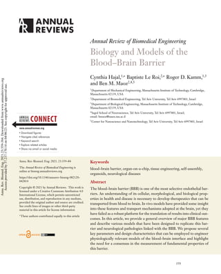
  • 8. TEER TEER Permeability 0 1,000 10–9 10–8 10–7 10–6 10–5 10–4 2,000 3,000 in vivo TEER a b c d RS Rpara Rm Cm RS in vivo permeability, 4–20 kDa Nonbrain/ nonhuman Lines Primary iPSCs Transwell 2D chip models 3D vessel-like structures 3D self- assembled models Model 20 40 60 80 100 120 123 0.80 500 1,000 1,500 2,000 75 2,489 Permeability (×10–7 cm/s) Permeability (cm/s) Cell source Nonbrain/ nonhuman Lines Primary iPSCs Transwell 2D chip models 3D vessel-like structures 3D self- assembled models Model Cell source TEER (Ω/cm 2 ) TEER (Ω/cm2 ) TEER permeability Primary iPSC Animal Cell lines S para m m para para m m mes Figure 3 Comparison of barrier permeability to solutes and TEER in terms of the type of endothelial cells employed (nonbrain, primary, immortalized, and iPSC-derived) and the type of BBB model (cell culture insert, 2D chip, 3D vessel-like, 3D self-assembled). Human iPSC-derived endothelial cells in 3D microfluidic chips exhibit the lowest permeability and highest TEER values. (a) Two methods to assess BBB permeability can be employed (the two equations, and the equivalent circuit, are extensively described in section 4.1 of the Supplemental Material). (b) Graph of permeability and TEER measurements of four cell types used as brain endothelial cells. Each data point represents a different model. In vivo TEER values are from Reference 57, and in vivo permeability values are from Reference 51 (see Supplemental Table 3). (c,d) Heat maps of the highest TEER and lowest permeability values measured in different models (up to 3 days of culture, 4–20 kDa dextrans). Red corresponds to experimental values reaching physiological values (the data, references, and rationale used to plot the graphs are presented in sections 4.2 and 4.3 of the Supplemental Material). Abbreviations: BBB, blood–brain barrier; iPSC, induced pluripotent stem cell; TEER, transendothelial resistance. P, given by P = P0 + Lp(1 − σ )(p − σπ), where Lp is the hydraulic conductivity, p is the pressure differential across the endothelium, σ is the reflection coefficient, π is the osmotic pressure differential, and P0 is the diffusive permeability. TEER, which defines barrier impedance to the passage of small ions through barriers, has also been employed as a quantitative metric to assess BBB permeability to charged solutes. Briefly, one 366 Hajal et al. Annu. Rev. Biomed. Eng. 2021.23:359-384. Downloaded from www.annualreviews.org Access provided by 37.237.170.14 on 04/06/23. See copyright for approved use.
  • 9. Table 1 Solute permeability and TEER: What do they measure? Solute permeability TEER Principle Net exchange of molecules, typically of higher molecular weight, across the endothelial barrier Transport of charged molecules or ions across the endothelial barrier Biological measurement Diffusion and convection of molecules Paracellular transport of ions Typical in vitro values in human BBB models 10−7 cm/s for 4–20 kDa dextrans 1,500 /cm2 Measured in vivo values in animals 4 × 10⁻⁷ cm/s [rat (51)] 1,300 /cm2 [frog (52)] Availability Most in vivo and in vitro models In vivo and in vitro models for which the area of the endothelium is well defined Correlation/translation Although both solute permeability and TEER measure transport of different types of solutes across the barrier, they are negatively correlated. Low permeabilities indicate reduced paracellular transport, which often correlates with restricted ionic transport and high TEER values. Abbreviations: BBB, blood–brain barrier; TEER, transendothelial resistance. can use the voltage and current through a BBB capillary to compute the electrical resistance of the cellular barrier via Ohm’s law. One thus obtains TEER as the product of the cellular layer resis- tance and area; high values indicate restricted ionic transport through the BBB and optimal barrier function (Table 1). Shortly after its first implementation in frog mesentery, the TEER technique was used in rat BBB capillaries, yielding values close to 6,000 /cm2 , significantly higher than those found in in vitro BBB models (48). These discrepancies partly reflect the relatively poor barrier function of in vitro BBB monolayer systems, where ionic transport cannot be fully recapit- ulated. More importantly, voltage levels applied across the BBB endothelia can play a significant role in the resulting TEER values, as shown by Sharabi et al. (49). These authors found that voltages as low as 10 V led to an ∼40% decrease in TEER due to electroporation of the cellular membrane. In addition, probe design, positioning, and shape, as well as anatomical configurations, can result in nonuniform current distributions across the barrier, reducing its effective area and overestimating its TEER (50). Finally, similarly as for solute permeability, TEER values do not provide information about transcellular transport of charged compounds. TEER measurements should be interpreted with caution, since several parameters can significantly affect the results. 3. MODELS OF THE BLOOD–BRAIN BARRIER 3.1. In Vivo Models Much of our basic knowledge regarding the BBB first came from animal models, which elucidated the fundamental mechanisms of regulated transport, barrier permeability, cellular architecture, and associated morphological features (53–55). The clear advantage of these models is their phys- iological relevance, including not only all the structures forming the BBB but also the effects of blood flow or biochemical mediators of function. Thus, animal models have offered tremendous insight into the physiology of brain tissue, particularly in the context of disease modeling. How- ever, challenges remain in the translation of animal findings to human patients. The underlying genetic, molecular, and immunologic differences between human and animal BBBs have limited the use of in vivo models as effective platforms for the development of therapeutics. This is clearly evident in the high failure rates (≥80%) in clinical trials of drugs validated in animals (37). This www.annualreviews.org • Blood–Brain Barrier Models 367 Annu. Rev. Biomed. Eng. 2021.23:359-384. Downloaded from www.annualreviews.org Access provided by 37.237.170.14 on 04/06/23. See copyright for approved use.
  • 10. limitation, in combination with increased ethical concerns and high costs, has spurred the devel- opment and use of in vitro human BBB systems. 3.2. Cell Source The choice of cell source is key to the development of in vitro BBB models. Important con- siderations include availability, human relevance, cost, robustness, reproducibility, batch-to-batch variability, capability for disease modeling, and barrier properties. Decisions regarding the species (e.g., murine, bovine, porcine, simian, or human) and cells used [e.g., cell lines, induced pluripo- tent stem cells (iPSCs), primary cells, or direct/transdifferentiated cells] are critical to the success of a BBB model. Here, we focus on endothelial cells employed to design in vitro BBB models, although other cells (e.g., pericytes, astrocytes, and to a lesser extent neurons) are also important. We note that BBB endothelial cells have critical morphological and functional differences from other endothelial cells, as they possess tighter cell–cell junctions, are highly polarized, and gener- ally exhibit lower values of permeability or higher TEER values compared with capillaries found in other tissues (56, 57). TRs, such as P-glycoprotein and GLUT-1, as well as TJ proteins, such as claudins and occludins, are key elements involved in barrier function of the BBB and need to be taken into account when selecting endothelial cells to engineer an in vitro BBB model. 3.2.1. Primary cells. Primary endothelial cells can be easily isolated from animals or human biopsies and cultured in vitro. These cells are accessible, widely used, and well documented in the literature (58). However, animal primary cells are not fully representative of human cells in either composition or function despite their common characteristics, such as TJ and TR protein expressions. Human primary cells collected from donors and used either in single-cell monolayers or in coculture models have thus been the preferred choice. Although they express high levels of platelet endothelial cell adhesion molecule 1 (PECAM-1), claudin-5, and zonula occludens 1 (ZO-1) (59), human primary cells paradoxically present low TEER values, usually under 100 /cm2 (Figure 3) (60). Finally, two major drawbacks of human primary cells are that they often lose their BBB properties through cell passage (61) and the source heterogeneity significantly reduces the reproducibility of experiments. Human umbilical vein endothelial cells (HUVECs) have been grown in coculture with peri- cytes, astrocytes, and even neurons. Under these conditions, expression of key BBB markers, such as ZO-1 and VE-cadherin, is often upregulated. HUVECs generate stable channels or vascular networks (62). They also exhibit strong barrier function, even for 3 kDa dextran, with permeabil- ity values as low as 2 × 10⁻⁷ cm/s, even though they are not specific to the brain microvasculature (45, 63). These observations suggest that endothelial cells alter their phenotype depending on their coculture conditions and the stromal cells employed in the model. 3.2.2. Immortalized cells. Owing to their low cost and ease of culture, immortalized cell lines are now extensively employed in in vitro BBB models. They retain their properties following cell passage and are genetically identical to one another, thus improving reproducibility in BBB models. Despite these advantages, cell lines generally present poor BBB properties and barrier function and may not be suitable for molecular transport studies (64). In addition, rodent BBB cell lines have been observed to lose their in vivo features, such as the expression of GLUT-1 (65). Human cells lines, in contrast, can present inconsistent protein levels. For example, the TY10 cell line expresses high levels of claudin-5 and VE-cadherin, while the hCMEC/D3 line has the highest expression level of ZO-1 but no claudin-5 (66). These inconsistencies imply that different cell lines might be optimal, depending on the application of the study (67, 68). Because of low TJ 368 Hajal et al. Annu. Rev. Biomed. Eng. 2021.23:359-384. Downloaded from www.annualreviews.org Access provided by 37.237.170.14 on 04/06/23. See copyright for approved use.
  • 11. or TR expression levels, immortalized cell lines are not capable of creating a tight cell layer and may not be relevant for general BBB modeling but only for specific studies such as drug absorption or pathogen transmigration across the BBB. A promising cell source derived from stem cells has garnered increasing attention over the last decade. This includes cells differentiated from embryonic or mesenchymal stem cells (ESCs or MSCs) and iPSCs, which are stem cells generated from a differentiated tissue. Stem cells are usually defined by their capacities to self-renew and to generate multiple different cell types. 3.2.3. Embryonic and mesenchymal stem cells. ESCs are pluripotent stem cells (PSCs) with the ability to differentiate into all cell types. ESCs from the blastocyst can be employed to recapit- ulate the embryonic development of brain microvascular endothelial cells (BMECs), and several embryoid differentiation protocols have been generated for this purpose. Generally, 3D embryoid differentiation protocols enable direct differentiation into the three embryonic layers.Yet the yield with these protocols is low, and there is a need for further isolation and expansion. In contrast, 2D monolayer differentiation protocols offer a high yield but generally result in the generation of a single cell type (69). Various types of human ESC lines can be employed, depending on the desired protein ex- pression of the resulting differentiated cells. For example, H1 and H9 are used for PECAM-1 expression (70), and HES3 and HES4 are employed for VE-cadherin expression (71). ESCs are also used in the context of modeling development in vitro. For example, Weidenfeller et al. (72) demonstrated that undifferentiated neural progenitor cells do not express BBB properties but dif- ferentiated cells do. In addition to ESCs, MSCs, which give rise to embryonic connective tissue, can improve the stabilization of the resulting BBB models. 3.2.4. Induced pluripotent stem cells. Since their development by Yamanaka and colleagues (73), human iPSCs have found application in numerous in vitro models. Yet, there is still no consensus in terms of a single differentiation protocol to produce BMECs from iPSCs (iBMECs). Protocols vary in length, number of steps, and differentiation factors employed, such as retinoic acid and Wnt/β-catenin agonist (74) or hypoxia (75). However, common to all protocols is a succession of steps including iPSC culture, endothelial induction, BMEC specification, and pu- rification (76). iPSCs have the considerable advantage that they can, in principle, produce all the cells com- posing an organ while maintaining genetic consistency among all the differentiated cells, thus enabling the generation of a patient-specific BBB model. In addition, they present excellent bar- rier properties, with strong expression of BBB markers such as ZO-1, claudin-5, and occludin as well as cell activity markers such as GLUT-1 and P-glycoprotein. Finally, their TEER and per- meability values are comparable to those measured in vivo (Figure 3). However, in contrast to cell lines and primary cells, they often have a limited life span in vitro, and processes ranging from dedifferentiation to redifferentiation are complex, time consuming, and very sensitive to experi- mental conditions. As mentioned above, iPSCs show promise for personalized medicine applications, wherein a model based on the patient’s own genotype is constructed (77). For example, Vatine et al. (78) modeled psychomotor retardation with iPSCs from patients deficient in monocarboxylate trans- porter 8. In addition, such models need not focus solely on inducing BMECs but can include all relevant components of the NVU (79). However, one needs to keep in mind several key elements when inducing iBMECs, such as the expression of canonical endothelial cell markers (cadherin-5, PECAM-1, VEGF receptor 2, apelin receptor, endothelial nitric oxide synthase), ability to form www.annualreviews.org • Blood–Brain Barrier Models 369 Annu. Rev. Biomed. Eng. 2021.23:359-384. Downloaded from www.annualreviews.org Access provided by 37.237.170.14 on 04/06/23. See copyright for approved use.
  • 12. tubular networks, low-density lipoprotein uptake, response to angiogenic stimuli, high TEER/low permeability, and expression of critical efflux transporters (80). While human primary cells appear to be the best source of cells for BBB models, they tend to lose their BBB properties over time and present weak barrier properties. In order to overcome these limitations, iPSCs are being employed to build complex BBB models including different cell types with a common genome. Note, however, that iPSCs have a nonnegligible tumorigenic and teratogenic potential. As a result, one must take care to eliminate nonusable or potentially tumorigenic differentiated cells. 3.3. In Vitro Models The development of BBB models with physiological transport properties has been a major chal- lenge since the 1980s. Cells were first cultured in two dimensions by use of a single cell type to mimic the BBB. Gradual increases in the complexity and functionality of in vitro BBB models enabled the generation of sophisticated platforms including multiple interacting cell types that can be dynamically stimulated, as well as the presence of hydrogels to mimic the 3D brain ex- tracellular matrix (ECM). Some of these elements have been included in Transwell® systems and microfluidic platforms. Here, we classify systems as either 2D or 3D; 3D models are defined as those with hydrogels at least 100 μm thick. 3.3.1. 2D static models. In 2D static models, cells are cultured on hard plastic (Figure 2a) or in Transwell inserts without the application of flow (Figure 2b). Historically, the first models of the BBB consisted of monocultures of endothelial cells isolated from brain capillaries. These allowed for the observation of TJ structures via freeze fracture (86). In order to recapitulate the BBB mi- croenvironment and reduce the lack of neighboring cell stimuli, coculture models were developed, notably with a full NVU composition of endothelial cells, astrocytes, pericytes, and neurons (87). Culturing cells on plastic offers several advantages in terms of ease of imaging and environmental control, as well as the presence of several standard protocols. However, hard plastics do not reca- pitulate the interface between vascular and brain parenchymal compartments, the major aspect of the BBB, thus limiting the utility of these models to assess BBB functionality through metabolic, diffusion, or permeability studies. Finally, the stiffness of tissue culture plastics, their flat geome- try, and the lack of ECM in 2D layer systems all limit cell development, alter cell phenotype, and result in poor models with limited potential for clinical translation (88). Transwell systems can overcome these challenges, as they offer the ability to perform per- meability assays, observe cell migration across a membrane, study multicellular interactions, and promote epithelial cell polarity (89). Transwell systems have progressively improved and are now widely used owing to their broad commercial availability. While these systems are static, several parameters can be optimized to improve the resulting models, such as the size of the inserts, their membrane porosity, their thickness, and the type of material (polymer) employed, with or with- out coating (90). Transwell systems allow for the introduction of at least two cell types, usually (a) endothelial cells on top and (b) astrocytes, pericytes, and sometimes neurons either in direct contact with the endothelial cells, when cultured on the opposite side of the membrane, or in indi- rect contact, when cultured at the bottom of the well (81). Transwell models of the BBB have been generated with all types of cells (primary cells, cell lines, and iPSCs), and they enable measurement of barrier properties such as TEER and solute permeability (56). However, their flat geometry limits cellular interactions that could promote barrier properties or the polarization of astrocytes, resulting in subpar BBB models. Hybrid devices containing 3D gels on Transwell inserts have re- cently been developed and are referred to as chip-on-a-Transwell systems (91). Nevertheless, the 370 Hajal et al. Annu. Rev. Biomed. Eng. 2021.23:359-384. Downloaded from www.annualreviews.org Access provided by 37.237.170.14 on 04/06/23. See copyright for approved use.
  • 13. presence of a Transwell insert introduces artifacts and limits the natural interactions between cells and the underlying ECM, resulting in poor recapitulation of the natural morphology of the BBB. 3.3.2. 2D organs-on-a-chip. Standard in vitro models lack cell–cell interactions and do not permit accurate control of hemodynamics (92) or appropriate organization of the microenvi- ronment represented. These limitations have been mitigated by the introduction of organ-on-a- chip (OoC) models in which cells can be compartmentalized in several channels, often separated by a membrane. These systems facilitate cell–cell interactions, the application of fluid flow and controlled shear stresses, and real-time readouts (93). This technology can be used to generate multiorgan systems by linking different OoCs together, producing so-called human-on-a-chip platforms (94–96). Recent developments in microfluidic chip technologies have enabled the in- tegration of sophisticated sensors (e.g., microelectrode arrays, TEER electrodes, oxygen tension probes) and measurements (e.g., mass spectroscopy analyses) (97, 98). Given the complexity of these additions, several OoC systems lack sensors and rely on the use of Transwell systems to per- form parameter measurements such as TEER. The results obtained in Transwell systems might not be representative of those in OoCs, however, and they are sometimes mathematically cor- rected, which can introduce a degree of error (99). TEER values, for example, vary according to the cell type and BBB model employed. Currently, iPSC models (79) present the lowest perme- ability values and the highest resistance, where TEER values reach or exceed physiological values of 1,300 /cm2 (Figure 3c) (52). 2D BBB-on-a-chip (BBBoC) models are defined as linear systems in microfluidic chips that incorporate two or more BBB cells with hydrogels no more than 100 μm thick. Compared with Transwell platforms, BBBoC systems offer greater physiological relevance and complexity and can facilitate the observation of cell migration/protrusion (100) and angiogenesis (85). They have been employed to recapitulate the effects of hypoxia on the BBB by use of both primary cells and iPSC models (Figure 2c) (75, 82). BBBoCs are a powerful tool for studying BBB physiology in terms of membrane protein expression (TJs, AJs, and TRs) or cell–cell interactions such as the formation of astrocyte endfeet (100). Recent studies have incorporated other CNS cells into BBBoC models in order to produce an NVU platform to uncover metabolic pathways between the vasculature and brain parenchyma in health and disease (101, 102). More complex models including five (brain, intestine, liver, kidney, and skeletal muscle) or eight organs (i.e., human-on-a-chip models) have been designed and employed in pharmacological studies (94, 95). Despite these advances in 2D OoC technologies, several challenges remain regarding minia- turizing these systems and increasing their throughput to perform large-scale drug screens (103). Finally, there has been a major push toward 3D models because of their ability to better recapit- ulate organ form and function. 3.3.3. 3D models. Researchers’ attention is shifting to 3D brain models, as they can better mimic the architecture and function of the BBB while offering greater physiological relevance. Several platforms have been proposed to generate human-like brain models, particularly with the use of biocompatible hydrogels, relevant cell types, and techniques drawing from the process of embryogenesis. 3.3.3.1. Monolayer of cells in a gel system. As mentioned above, we define 3D BBB models as those for which the BBB cells are cultured adjacent to a hydrogel with a thickness of at least 100 μm. One example is the model developed by Ahn et al. (104), who created a platform with human brain pericytes and endothelial cells in a microfluidic device with astrocytes on the bottom, seeded in Matrigel (Figure 2f ). The astrocytes extended their AQP-4-expressing endfeet to www.annualreviews.org • Blood–Brain Barrier Models 371 Annu. Rev. Biomed. Eng. 2021.23:359-384. Downloaded from www.annualreviews.org Access provided by 37.237.170.14 on 04/06/23. See copyright for approved use.
  • 14. establish contact with the pericytes and endothelial cells, similar to the behavior observed in vivo (104). The simple design and ease of use of 3D monolayer brain models, coupled with their increased physiological relevance, have been instrumental in the generation of high-throughput in vitro brain models. With the help of multichannel microfluidic chips, investigators have engineered identical BBB platforms for use as functional assay platforms, notably for studies of metastasis or drug transport (105). Importantly, when compared with 2D BBB models, brain en- dothelial cells cultured in 3D platforms exhibit improved TJ expression and increased TEER val- ues, suggesting that the addition of appropriate matrices plays a fundamental role in the integrity and function of the resulting BBB model (106). Additionally, the application of physiological levels of fluid flow and resulting wall shear stresses improve TJ protein expression as well as solute permeability and TEER values (105). In comparison to 2D systems, the ability of 3D brain models to recapitulate physiological BBB hemodynamics is paramount in the function of the barrier. 3.3.3.2. Vessel-like structures in gels. Microfluidic technology has played a significant role in the development of BBB models with increased complexity and realism, giving rise to a new ap- proach to in vitro 3D BBB models: 3D channels embedded in or adjacent to hydrogel compart- ments. These platforms possess several desirable features in terms of morphology, cellular orga- nization, high-throughput capabilities, and improved barrier function. Importantly, they better recapitulate important aspects of BBB morphology consisting of hollow tubular structures lined with endothelial cells and variably cocultured with neural stromal cells, all embedded in a 3D hy- drogel. For example, Marino et al. (83) used two-photon lithography to generate porous tubular structures with an average diameter of 10 μm, which were subsequently coated with mouse BBB endothelial cells (Figure 2d). Similar platforms with increasing levels of complexity have been designed. They incorporate hydrogels with improved physiological relevance (107) and additional BBB cell types such as per- icytes and astrocytes. Both Brown et al. (108) and Adriani et al. (109) employed multichannel microfluidic chips in which one of the channels was lined with primary human brain endothelial cells, with combinations of pericytes, astrocytes, and neurons in an adjacent collagen hydrogel. Al- though collagen type 1 is commonly used in cell culture systems, it is not usually found in the brain parenchyma; the use of more physiological matrices, such as hyaluronic acid– or proteoglycan- based hydrogels, could improve the BBB properties of these models (110). In addition, the mor- phological attributes and cellular organization of these platforms do not fully recapitulate the BBB, either because of their large diameter (typically approximately 500 μm) or because of the lack of stromal cells with appropriate spatial distribution. Herland et al. (84) addressed some of these challenges by generating hollow cylindrical structures in a collagen gel via a pressure-driven viscous fingering method (Figure 2e). The channel lumens (still large, with diameters of 600– 800 μm) were perfused with pericytes or astrocytes followed by human BMECs, which resulted in a decrease of barrier permeability to solutes in comparison to platforms without brain stromal cells (84). To address the remaining challenge of reducing vessel diameter and improving vascular morphology, several groups have generated self-assembled vascular systems that recapitulate in vitro the inherent ability of endothelial cells to self-assemble into vascular networks with small- diameter lumens. 3.3.3.3. Self-assembled vasculatures and the role of emergence. To better recapitulate the mor- phological and functional features of the BBB, researchers have focused on harnessing the natural processes of vascular development in vivo, whereby endothelial precursors coalesce and organize into complex microvascular networks (vasculogenesis) and endothelial cells sprout from preexist- ing blood vessels (angiogenesis) (111). This inherent ability of endothelial cells to form tubular 372 Hajal et al. Annu. Rev. Biomed. Eng. 2021.23:359-384. Downloaded from www.annualreviews.org Access provided by 37.237.170.14 on 04/06/23. See copyright for approved use.
  • 15. structures has been extensively employed to engineer 3D vascular networks with HUVECs and lung stromal cells or placental pericytes; the resulting structures exhibit improved morphology, TJ protein expression, and solute transport machinery in comparison to other 3D tubular structures or 2D models (112). Extrapolating from these models, Bang et al. (113) incorporated astrocytes and neurons into a HUVEC–fibroblast platform to mimic the BBB in three dimensions. While this model includes non-brain-specific cell types, it offers valuable insight into the role astrocytes play in promoting the expression of BBB properties (113). Challenges in isolating tissue-specific human endothelial cells and ensuring that they retain their properties when cultured in vitro have hindered the development of 3D OoC self-assembled models, particularly BBB microvascular platforms. However, certain commercial human BMEC lines isolated from brain biopsy samples do retain their ability to self-lumenize when cultured in appropriate 3D hydrogels. For instance, Lee et al. (85) used human BMECs, primary brain pericytes and astrocytes, and lung fibroblasts to create via angiogenesis in a microfluidic chip a 3D self-assembled BBB model that exhibited improved barrier permeability and increased ex- pression of TJ proteins. In another model (29), self-assembled BBB vasculatures were generated via vasculogenesis using human iPSC-derived endothelial cells, as well as primary brain pericytes and astrocytes embedded simultaneously in a fibrinogen–thrombin gel, without the need for cell types from different organs for vascular support (e.g., lung fibroblasts). The self-assembled BBB vessels exhibited physiological morphologies with diameters as small as 20 μm and decreased so- lute permeability, comparable to levels measured in rat BBB (Figure 3) (Supplemental Tables 2 and 3). These self-assembled vascular systems have revolutionized the field of in vitro BBB models, significantly improving their physiological relevance and allowing for high-resolution spatiotem- poral imaging of key events at the BBB, such as cell diapedesis or solute transport. Despite these advances, challenges regarding the choice of hydrogels in these systems remain. The common use of fibrinogen gels for 3D angiogenic and vasculogenic BBB models does not fully recapitulate the in vivo brain ECM, which comprises mostly hyaluronic acid and other proteoglycans. In addition, fluid flow, which is not included in most in vitro self-assembled BBB models, exhibits specific dy- namic patterns at the brain that promote the upregulation of TJ and transporter proteins (31, 114). Recapitulating BBB hemodynamics, and thus improved transport of gases and nutrients, would be paramount for the maintenance of long-term cultures in vitro, since the current engineered BBB vessels are known to regress after a couple of weeks. 3.3.3.4. Cerebral organoids and the blood–brain barrier. The last ten years have witnessed the development of 3D brain organoids from the differentiation of PSCs, such as iPSCs or ESCs. These systems harness the entire development process to generate structures that recapitulate specific brain regions, such as midbrain organoids or cerebral cortex organoids that house BBB capillaries (115). Cerebral organoids that model neurological diseases such as Alzheimer’s disease (AD) have also been created through the use of patient-derived iPSCs (116). Despite successes in generating brain organoids, these structures still lack appropriate vas- culature. Pham et al. (117) addressed this limitation by engineering discrete vascular sprouts in their patient-derived iPSC cerebral organoids by using differentiated endothelial cells from the same iPSCs. Although a few CD31-positive blood vessel sprouts were observed, they were not connected, and perfusion was not achieved in these structures (117). In another cerebral organoid model, Cakir et al. (118) engineered ESCs to express human ETS variant 2, which reprograms human dermal fibroblasts into endothelial cells, promoting vascularization. However, BBB vessels were observed only at the organoid surface. Homan et al. (119) employed a similar approach, based on the application of fluid flow, to enhance the vascularization of kidney organoids. www.annualreviews.org • Blood–Brain Barrier Models 373 Annu. Rev. Biomed. Eng. 2021.23:359-384. Downloaded from www.annualreviews.org Access provided by 37.237.170.14 on 04/06/23. See copyright for approved use.
  • 16. High-shear-stress flow resulted in the most effective vessel formation in the surrounding ECM in comparison to static conditions and low-shear-stress flow. These results obtained in kidney organoids could be extrapolated to cerebral structures to enhance vascularization, confirming the importance of hemodynamics in BBB modeling and function. These models attest to the signif- icance of design considerations, such as the application of fluid flow, the choice of 3D matrix, and the use of appropriate growth factors in the vascularization of cerebral organoids. Recapitulating organ development at the brain in these structures also offers insight into the development of the brain cortex, particularly in the context of developmental neurological disorders. 3.3.3.5. 3D-printed models. Microfabrication of OoCs can be a long, expensive process that requires clean-room facilities and fabrication engineers and offers little flexibility in modifying the design of the chip. Moreover, OoCs are not truly 3D organ models. 3D printing is now a well- established technique that overcomes these issues.3D printing,or additive manufacturing,consists of layer-by-layer production of a computer-aided design file. In order to print organ models, hy- drogels are often used as bioinks, which are networks of polymeric and hydrophilic molecules such as gelatin (collagen) derivatives like gelatin methacryloyl (120), hyaluronic acid, or alginate (121). These bioinks integrate cells such as neurons, astrocytes, HUVECs, or glioblastoma cells (122). In order to print different types of cells, either multinozzle printers can be used simultaneously (123) or a single-nozzle printer can be employed to sequentially print multilayered bioinks in which each layer contains a different cell type (124). Several groups have generated BBBoC models of glioblastoma using 3D printing of com- partmentalized concentric tumor/vascular compartment structures in order to study the effect of drugs and radial oxygen gradients on tumor progression (123). The cells were surrounded by polydimethylsiloxane to ensure chip integrity (123). Other researchers have built tubular printed vessels-on-a-chip with excellent properties (e.g., TEER of 1,500 /cm2 ) (125). Finally, bioinspired synthetic scaffolds have been designed for the coculture of endothelial cells and neurons via two-photon lithography (83). 3D-printed models differ from microfluidic ones in terms of tissue maturation and self-assembly. Two techniques are generally used to print vessels: indirect printing, which involves the use of a sacrificial material employed to generate the vessel lumens (126), and direct printing, where lumens are assembled without sacrificial structures. Vascular lumens can be formed by seeding cells either inside the preformed, multilayer tubes (127) or within porous materials (128). 4. BLOOD–BRAIN BARRIER–RELATED DISEASES: CURRENT MODELS AND IMPROVEMENTS Multiple neurological or brain diseases are known to affect the BBB through the secretion of inflammatory cytokines that degrade TJs (129). Because of the limited spatiotemporal resolution of in vivo models and the challenge of translating results from animal to human studies, several groups have shifted their efforts to the development of in vitro human BBB disease systems. These platforms also enable the generation of high-throughput identical models that can be used for studies pertaining to drug delivery across the BBB. 4.1. Alzheimer’s Disease Models According to the two-hit vascular hypothesis of AD, BBB damage is thought to be the initial insult that results in diminished brain perfusion and neuronal injury (the first hit), followed by amyloid-β (Aβ) accumulation due to faulty clearance and increased antibody production (the sec- ond hit) (130). In an early 2D Transwell model of AD with patient-derived BBB endothelial cells, 374 Hajal et al. Annu. Rev. Biomed. Eng. 2021.23:359-384. Downloaded from www.annualreviews.org Access provided by 37.237.170.14 on 04/06/23. See copyright for approved use.
  • 17. Mackic et al. (131) uncovered the role of the endothelial cell receptor for advanced glycation end products and scavenger receptor in the transcytosis of Aβ-40 into the brain parenchyma. More recently, Robert et al. (132) used a 3D in vitro vessel-like structure of brain endothelial cells, lined with smooth muscle cells and astrocytes, to show that apolipoprotein E2 (APOE2) can reduce the accumulation of amyloid plaques. While these models provide useful insight into the mechanisms of accumulation and clearance of Aβ, they do not fully recapitulate AD pathology in the context of neuron–BBB interaction and BBB disruption. Microfluidic technologies have addressed some of these challenges. Recently, Shin et al. (133) developed a 3D microfluidic model with a vessel-like structure consisting of primary brain en- dothelial cells adjacent to neural progenitor cells. In this model, mutations in the amyloid precur- sor protein and presenilin-1 genes in neurons resulted in their robust secretion of Aβ as well as an observable deposition at the endothelial surface, corresponding to an increase in barrier perme- ability. In another 3D AD BBB model of endothelial cells, pericytes, and astrocytes differentiated from the same iPSCs in Matrigel, APOE4 pericytes exhibited upregulated calcineurin signaling, which is associated with cerebral amyloid angiopathy, a common precursor to AD (134). Despite their improved throughput and physiological relevance, 3D AD BBB systems, which are cultured for only a few weeks, remain limited in their ability to recapitulate barrier dysfunction and plaque deposition, which occur over the course of multiple years in humans. The emergence of cerebral organoid models that can be cultured for several months might provide a way to model and study long-term progressing neurological disorders such as AD. 4.2. Stroke Models Strokes, primarily ischemic in nature, are characterized by disruptions of the BBB wherein hy- poxia and ROS production during the ischemic insult lead to TJ breakdown and subsequent ionic shifts in the brain parenchyma (135). Hundreds of drugs have been reported to effectively improve outcomes in experimental animal models. However, almost all of them have failed to translate into successful therapies for humans (136). Several groups have thus developed in vitro human BBB models of stroke to better recapitulate this pathology. For instance, Chen et al. (137) created a BBB stroke Transwell model with primary brain endothelial cells and astrocytes subjected to oxygen and glucose deprivation, showing that the increase of Sirtuin-1 following stroke and ischemia was cor- related with ROS production and subsequent inflammation and cell damage. Similarly, Chaitanya et al. (138) subjected a Transwell BBB model with primary brain endothelial cells, astrocytes, and neurons to various metabolic stressors, such as hypoxia, glucose deprivation, and reoxygenation. These stressors induced brain capillary tube formation and angiogenesis in vitro and promoted lymphocyte adhesion via increased expression of adhesion markers on the BBB endothelial cells (138). Although these models could be employed as high-throughput platforms for drug screen- ing, they remain severely limited in their ability to mimic the architecture and physiology of the BBB. More importantly, current human in vitro stroke models primarily mimic the lack of oxygen and glucose at the brain, disregarding other key features of strokes such as inflammatory cytokine production and microglial activation (135). 4.3. Brain Cancer Models Given the poor prognoses of primary brain cancers, the prevalence of metastatic tumors in the brain, and the lack of in vitro brain tumor platforms that incorporate BBB vessels, there is a significant need for models that can aid in the understanding of cancer progression and drug delivery across the BBB. In one recent example, Plummer et al. (139) designed a high-throughput glioblastoma–neuron–astrocyte tumor platform to test drug transport and found that therapeutics www.annualreviews.org • Blood–Brain Barrier Models 375 Annu. Rev. Biomed. Eng. 2021.23:359-384. Downloaded from www.annualreviews.org Access provided by 37.237.170.14 on 04/06/23. See copyright for approved use.
  • 18. selectively target the glioblastoma cells. More recently, in vitro primary brain cancer models that integrate vasculature and tumor have been designed. Ngo Harley (140) developed a coculture model of glioma cells with HUVECs and lung fibroblasts in gelatin and hyaluronic acid and observed that the addition of cellular and matrix elements led to a decrease in the response of glioma cells to therapeutics. Similarly, Ozturk et al. (141) designed a long-term 3D in vitro model of glioblastoma tumor spheroids surrounded by bioprinted HUVEC gelatin channels to study drug delivery over time. Although these models are useful to screen for therapeutic toxicity, the use of HUVECs or lung fibroblasts does not fully recapitulate cell types found in the brain, and the 3D architecture of these systems could be improved to replicate the in vivo BBB vasculature in contact with tumors. Given the prevalence of metastatic brain tumors originating from various primary sites, mod- els have been designed to elucidate the mechanisms employed by cancer cells to cross the BBB. For example, Bos et al. (142) observed that increased expression of the sialyltransferase gene ST6GALNAC5 promotes extravasation in a HUVEC–astrocyte Transwell system. In another study using a high-throughput microfluidic platform with brain endothelial cells and astrocytes in collagen, Xu et al. (105) found that lung and breast tumor cells exhibit greater transmigration potential in comparison to melanoma and liver cancer cells. While these high-throughput models allow for high-resolution spatiotemporal imaging of tumor extravasation, their physiological rel- evance remains limited, particularly in terms of the cell types employed and the 3D architecture of the BBB capillaries. 4.4. Infectious Disease Models Although the BBB is a highly restrictive endothelial barrier, bacteria and viruses are still able to enter the parenchyma. Two general mechanisms are thought to be responsible for their trans- port: increased barrier permeability following infection and active transport via receptor-mediated transport (143). Infectious diseases known to target the BBB include Zika virus disease, malaria, and, more recently, coronavirus disease 2019 (COVID-19), yet in vitro models of these pathologies are limited. The use of patient data revealed that SARS-CoV-2, the virus that causes COVID-19, employs the S1 unit of its spike protein to attach to angiotensin-converting enzyme 2 receptor on cells and infect the patient (144). Buzhdygan et al. (145) used primary brain endothelial cells to engineer 2D and 3D BBB models showing that the spike protein increases inflammation, barrier permeability to dextran, and expression of adhesion molecules on brain endothelial cells, sug- gesting cellular activation and BBB breakdown. Despite its simple architecture and lack of brain stromal cells, the COVID-19 in vitro platform offers useful and important information about this globally significant disease. 4.5. Other Diseases: In Vitro and Computational Models Several other neurological diseases are known to predominantly affect the BBB (129, 146, 147). Al- though not discussed extensively in this review, these include neurodegenerative disorders such as Parkinson’s and Huntington’s diseases, as well as epilepsy and mental disorders such as schizophre- nia, clinical depression, and anxiety disorder. In vivo animal models, although useful to understand the genetic and phenotypic profiles of these diseases, often fail in their translation to meaningful clinical results. Yet, the use of in vitro platforms to recapitulate these pathologies remains limited. Lim et al. (148) designed a 2D in vitro model of Huntington’s disease by using patient iPSC- derived endothelial cells. They uncovered BBB deficits in terms of angiogenic signaling, transcy- tosis protein expression, and overall barrier permeability. Similar models that recapitulate other neurological diseases are necessary to bridge the gap between in vivo models and clinical outcomes. 376 Hajal et al. Annu. Rev. Biomed. Eng. 2021.23:359-384. Downloaded from www.annualreviews.org Access provided by 37.237.170.14 on 04/06/23. See copyright for approved use.
  • 19. Despite significant advances in in vitro technologies, the complexity of the BBB and its trans- port characteristics cannot be entirely captured by these systems. Several scientists have employed computational models to address these limitations and investigate various BBB pathologies such as brain tumors, AD, and traumatic brain injury (149). These computational approaches, ranging from molecular dynamics to partial differential equation–based models, finite-element methods, and pharmacokinetics, are used to investigate drug design and delivery at the brain, as well as BBB pathologies. For instance, Adhikari et al. (150) designed an in silico model of traumatic brain injury and its effect on lipid bilayer damage at the BBB by using a coarse-grained molecular dy- namics simulation. Their simulation showed that low-intensity shock waves in the presence of nanobubbles can break claudin–TJ interactions between adjacent endothelial cells of the BBB, thus damaging the barrier. While current simulations are relatively straightforward to modulate in comparison to complex cellular systems, they do not fully recapitulate all the relevant properties of the BBB, particularly in the context of neurological diseases. Large data sets of readily available 3D images of the BBB and information about its properties have certainly promoted the genera- tion of improved computational models of the brain in health and disease, notably through the use of machine learning and artificial intelligence technologies. These novel techniques will foster the development of more complex BBB simulations that can incorporate patient-specific pathologies, thus allowing for the evaluation of disease progression or drug delivery with greater ease. 5. CHALLENGES AND FUTURE DIRECTIONS As the gatekeeper of the brain, the BBB has attracted tremendous interest for its role in both health and disease. This requires the design of relevant in vitro human models that recapitulate its key morphological and functional properties. Despite significant recent advances in the ability to engineer novel in vitro systems, major challenges remain, such as incorporating all the relevant cell types of the BBB, employing an appropriate 3D hydrogel to recapitulate the brain ECM, and achieving vascularization in vitro to emulate transport across this barrier, notably for therapeu- tics in the context of neurological diseases. Future efforts and advances in the vascularization of cerebral organoids that incorporate all the relevant BBB cells in a brainlike matrix will certainly provide platforms that can accurately recapitulate the human BBB. More importantly, there is a need to further improve permeability measurement techniques in order to capture changes in barrier integrity in health and disease. Next-generation in vitro BBB models with tunable trans- port properties will certainly provide new avenues to modulate barrier function and recapitulate neurological pathologies with greater accuracy. In addition, connecting in vitro brain models with other OoCs and perfusing the interconnected systems with patient plasma or blood will reca- pitulate upstream effects from other organs on the BBB or systemic effects from perfused blood on the multi-OoCs. Yet, prior to engineering these complex physiological BBB models, it will be crucial to characterize the cells of the brain and their specific markers and genotypes. Current techniques rely mostly on in vitro characterization tools following isolation, sorting, and culture of cells obtained from fresh brain tissue, where loss of function and markers is inevitable. The development of novel mapping techniques for gene expression in vivo will allow for the genera- tion of accurate baseline characteristics of the different cell types of the BBB. These can then be employed as benchmarks for in vitro BBB models, notably for the generation of stem cell–derived brain-specific cells that can be engineered to overexpress relevant BBB-specific genes. DISCLOSURE STATEMENT R.D.K. is a cofounder of AIM Biotech, a company that markets microfluidic systems, and receives research support from Biogen, Amgen, and Gore. The other authors are not aware of any www.annualreviews.org • Blood–Brain Barrier Models 377 Annu. Rev. Biomed. Eng. 2021.23:359-384. Downloaded from www.annualreviews.org Access provided by 37.237.170.14 on 04/06/23. See copyright for approved use.
  • 20. affiliations, memberships, funding, or financial holdings that might be perceived as affecting the objectivity of this review. ACKNOWLEDGMENTS We apologize to our colleagues whose research we could not cite or discuss owing to space lim- itations. C.H. is supported by a Ludwig Center for Molecular Oncology Graduate Fellowship. C.H. and R.D.K. acknowledge funding from the National Cancer Institute (U01 CA202177) and the National Science Foundation (CBET-0939511). B.L. is supported by a PhD fellowship from the Biomedical Engineering Department of Tel Aviv University. B.L. and B.M.M. acknowledge funding from the Azrieli Foundation, the Israel Science Foundation (2248/19), and the European Research Council (SweetBrain 851765). LITERATURE CITED 1. Stern L, Gautier R. 1921. Récherches sur le liquide céphalo-rachidien. I. Les rapports entre le liquide céphalo-rachidien et la circulation sanguine. Arch. Int. Physiol. 17:138–92 2. Ehrlich P.1885.Das Sauerstoff-Bedürfniss des Organismus: Eine farbenanalytische Studie.Berlin: Hirschwald 3. Spatz H. 1934. Die Bedeutung der vitalen Färbung für die Lehre vom Stoffaustausch zwischen dem Zentralnervensystem und dem übrigen Körper. Arch. Psychiatrie Nervenkrankh. 101:267–358 4. Reese TS, Karnovsky MJ. 1967. Fine structural localization of a blood-brain barrier to exogenous per- oxidase. J. Cell Biol. 34:207–17 5. Saunders NR, Dreifuss JJ, Dziegielewska KM, Johansson PA, Habgood MD, et al. 2014. The rights and wrongs of blood-brain barrier permeability studies: a walk through 100 years of history. Front. Neurosci. 8:404 6. Risau W. 1997. Mechanisms of angiogenesis. Nature 386:671–74 7. Daneman R, Agalliu D, Zhou L, Kuhnert F, Kuo CJ, Barres BA. 2009. Wnt/β-catenin signaling is re- quired for CNS, but not non-CNS, angiogenesis. PNAS 106:641–46 8. Lindahl P, Johansson BR, Levéen P, Betsholtz C. 1997. Pericyte loss and microaneurysm formation in PDGF-B-deficient mice. Science 277:242–45 9. Mizee MR,Wooldrik D,Lakeman KAM,van het Hof B,Drexhage JAR,et al.2013.Retinoic acid induces blood-brain barrier development. J. Neurosci. 33:1660–71 10. Hupe M, Li MX, Kneitz S, Davydova D, Yokota C, et al. 2017. Gene expression profiles of brain en- dothelial cells during embryonic development at bulk and single-cell levels. Sci. Signal. 10:eaag2476 11. Levison SW, de Vellis J, Goldman JE. 2005. Astrocyte development. In Developmental Neurobiology, ed. MS Rao, M Jacobson, pp. 197–222. New York: Springer. 4th ed. 12. Ormestad M, Astorga J, Carlsson P. 2004. Differences in the embryonic expression patterns of mouse Foxf1 and -2 match their distinct mutant phenotypes. Dev. Dyn. 229:328–33 13. Yamamoto S, Muramatsu M, Azuma E, Ikutani M, Nagai Y, et al. 2017. A subset of cerebrovascular pericytes originates from mature macrophages in the very early phase of vascular development in CNS. Sci. Rep. 7:3855 14. Dore-Duffy P, Cleary K. 2011. Morphology and properties of pericytes. In The Blood–Brain and Other Neural Barriers, ed. S Nag, pp. 49–68. New York: Humana 15. Nag S. 2011. Morphology and properties of astrocytes. Methods Mol. Biol. 686:69–100 16. Armulik A, Genové G, Mäe M, Nisancioglu MH, Wallgard E, et al. 2010. Pericytes regulate the blood- brain barrier. Nature 468:557–61 17. Hall CN, Reynell C, Gesslein B, Hamilton NB, Mishra A, et al. 2014. Capillary pericytes regulate cere- bral blood flow in health and disease. Nature 508:55–60 18. Hill RA, Tong L, Yuan P, Murikinati S, Gupta S, Grutzendler J. 2015. Regional blood flow in the nor- mal and ischemic brain is controlled by arteriolar smooth muscle cell contractility and not by capillary pericytes. Neuron 87:95–110 378 Hajal et al. Annu. Rev. Biomed. Eng. 2021.23:359-384. Downloaded from www.annualreviews.org Access provided by 37.237.170.14 on 04/06/23. See copyright for approved use.
  • 21. 19. Mishra A, Reynolds JP, Chen Y, Gourine AV, Rusakov DA, Attwell D. 2016. Astrocytes mediate neu- rovascular signaling to capillary pericytes but not to arterioles. Nat. Neurosci. 19:1619–27 20. Chow BW, Nuñez V, Kaplan L, Granger AJ, Bistrong K, et al. 2020. Caveolae in CNS arterioles mediate neurovascular coupling. Nature 579:106–10 21. Alarcon-Martinez L, Villafranca-Baughman D, Quintero H, Kacerovsky JB, Dotigny F, et al. 2020. In- terpericyte tunnelling nanotubes regulate neurovascular coupling. Nature 585:91–95 22. Haruwaka K, Ikegami A, Tachibana Y, Ohno N, Konishi H, et al. 2019. Dual microglia effects on blood brain barrier permeability induced by systemic inflammation. Nat. Commun. 10:5816 23. Hanisch UK, Kettenmann H. 2007. Microglia: active sensor and versatile effector cells in the normal and pathologic brain. Nat. Neurosci. 10:1387–94 24. Zonta M, Angulo MC, Gobbo S, Rosengarten B, Hossmann KA, et al. 2003. Neuron-to-astrocyte sig- naling is central to the dynamic control of brain microcirculation. Nat. Neurosci. 6:43–50 25. Li YN, Pan R, Qin XJ, Yang WL, Qi Z, et al. 2014. Ischemic neurons activate astrocytes to disrupt endothelial barrier via increasing VEGF expression. J. Neurochem. 129:120–29 26. Charabati M, Rabanel JM, Ramassamy C, Prat A. 2020. Overcoming the brain barriers: from immune cells to nanoparticles. Trends Pharmacol. Sci. 41:42–54 27. Weiss N, Miller F, Cazaubon S, Couraud PO. 2009. The blood-brain barrier in brain homeostasis and neurological diseases. Biochem. Biophys. Acta Biomembr. 1788:842–57 28. Rodriguez-Baeza A, Reina-De La Torre F, Ortega-Sanchez M, Sahuquillo-Barris J. 1998. Perivascular structures in corrosion casts of the human central nervous system: a confocal laser and scanning electron microscope study. Anat. Rec. 252:176–84 29. Campisi M, Shin Y, Osaki T, Hajal C, Chiono V, Kamm RD. 2018. 3D self-organized microvascular model of the human blood-brain barrier with endothelial cells, pericytes and astrocytes. Biomaterials 180:117–29 30. Cipolla MJ. 2016. The Cerebral Circulation. San Rafael, CA: Morgan Claypool. 2nd ed. Colloq. Ser. Integr. Syst. Physiol. Mol. Funct. 31. Follain G, Osmani N, Azevedo AS, Allio G, Mercier L, et al. 2018. Hemodynamic forces tune the arrest, adhesion, and extravasation of circulating tumor cells. Dev. Cell 45:33–52 32. Destefano JG, Jamieson JJ, Linville RM, Searson PC. 2018. Benchmarking in vitro tissue-engineered blood–brain barrier models. Fluids Barriers CNS 15:32 33. Zarrinkoob L, Ambarki K, Wahlin A, Birgander R, Carlberg B, et al. 2016. Aging alters the dampening of pulsatile blood flow in cerebral arteries. J. Cereb. Blood Flow Metab. 36:1519–27 34. Webb AJ, Simoni M, Mazzucco S, Kuker W, Schulz U, Rothwell PM. 2012. Increased cerebral arterial pulsatility in patients with leukoaraiosis: Arterial stiffness enhances transmission of aortic pulsatility. Stroke 43:2631–36 35. Jessen NA, Munk ASF, Lundgaard I, Nedergaard M. 2015. The glymphatic system: a beginner’s guide. Neurochem. Res. 40:2583–99 36. Andreone BJ, Chow BW, Tata A, Lacoste B, Ben-Zvi A, et al. 2017. Blood-brain barrier permeability is regulated by lipid transport–dependent suppression of caveolae-mediated transcytosis. Neuron 94:581– 94 37. Hajal C, Campisi M, Mattu C, Chiono V, Kamm RD. 2018. In vitro models of molecular and nano- particle transport across the blood-brain barrier. Biomicrofluidics 12:042213 38. Barar J, Rafi MA, Pourseif MM, Omidi Y. 2016. Blood-brain barrier transport machineries and targeted therapy of brain diseases. BioImpacts 6:225–48 39. Rapoport SI. 2000. Osmotic opening of the blood-brain barrier. Cell. Mol. Neurobiol. 20:217–30 40. Yuan W, Lv Y, Zeng M, Fu BM. 2009. Non-invasive measurement of solute permeability in cerebral microvessels of the rat. Microvasc. Res. 77:166–73 41. Shi L, Zeng M, Sun Y, Fu BM. 2014. Quantification of blood-brain barrier solute permeability and brain transport by multiphoton microscopy. J. Biomech. Eng. 136:031005 42. Schaaf MB, Houbaert D, Meçe O, Agostinis P. 2019. Autophagy in endothelial cells and tumor angio- genesis. Cell Death Differ. 26:665–79 43. Pieper C,Pieloch P,Galla HJ.2013.Pericytes support neutrophil transmigration via interleukin-8 across a porcine co-culture model of the blood-brain barrier. Brain Res. 1524:1–11 www.annualreviews.org • Blood–Brain Barrier Models 379 Annu. Rev. Biomed. Eng. 2021.23:359-384. Downloaded from www.annualreviews.org Access provided by 37.237.170.14 on 04/06/23. See copyright for approved use.
  • 22. 44. Fazakas C, Wilhelm I, Nagyoszi P, Farkas AE, Haskó J, et al. 2011. Transmigration of melanoma cells through the blood-brain barrier: role of endothelial tight junctions and melanoma-released serine pro- teases. PLOS ONE 6:e20758 45. Offeddu GS, Possenti L, Loessberg-Zahl JT, Zunino P, Roberts J, et al. 2019. Application of trans- mural flow across in vitro microvasculature enables direct sampling of interstitial therapeutic molecule distribution. Small 15:1902393 46. Abbott NJ, Patabendige AA, Dolman DE, Yusof SR, Begley DJ. 2010. Structure and function of the blood-brain barrier. Neurobiol. Dis. 37:13–25 47. Banks WA. 2009. Characteristics of compounds that cross the blood-brain barrier. BMC Neurol. 9:S3 48. Butt AM, Jones HC, Abbott NJ. 1990. Electrical resistance across the blood-brain barrier in anaes- thetized rats: a developmental study. J. Physiol. 429:47–62 49. Sharabi S, Bresler Y, Ravid O, Shemesh C, Atrakchi D, et al. 2019. Transient blood–brain barrier disrup- tion is induced by low pulsed electrical fields in vitro: an analysis of permeability and trans-endothelial electric resistivity. Drug Deliv. 26:459–69 50. Elbrecht DH, Long CJ, Hickman JJ. 2016. Transepithelial/endothelial electrical resistance (TEER) the- ory and applications for microfluidic body-on-a-chip devices. J. Rare Dis. Res. Treat. Open 1:46–52 51. Deosarkar SP, Prabhakarpandian B, Wang B, Sheffield JB, Krynska B, Kiani MF. 2015. A novel dynamic neonatal blood-brain barrier on a chip. PLOS ONE 10:e0142725 52. Crone C, Olesen SP. 1982. Electrical resistance of brain microvascular endothelium. Brain Res. 241:49– 55 53. Wiranowska M, Wilson TC, Beneze KS, Prockop LD. 1988. A mouse model for the study of blood-brain barrier permeability. J. Neurosci. Methods 26:105–9 54. Rapoport SI, Bachman DS, Thompson HK. 1972. Chronic effects of osmotic opening of the blood-brain barrier in the monkey. Science 176:1243–44 55. Sohet F, Daneman R. 2013. Genetic mouse models to study blood-brain barrier development and func- tion. Fluids Barriers CNS 10:1 56. Srinivasan B, Kolli AR, Esch MB, Abaci HE, Shuler ML, Hickman JJ. 2015. TEER measurement tech- niques for in vitro barrier model systems. J. Lab. Autom. 20:107–26 57. Crone C, Christensen O. 1981. Electrical resistance of a capillary endothelium. J. Gen. Physiol. 77:349–71 58. Helms HC, Abbott NJ, Burek M, Cecchelli R, Couraud PO, et al. 2015. In vitro models of the blood- brain barrier: an overview of commonly used brain endothelial cell culture models and guidelines for their use. J. Cereb. Blood Flow Metab. 36:862–90 59. Lacombe O, Videau O, Chevillon D, Guyot AC, Contreras C, et al. 2011. In vitro primary human and animal cell-based blood–brain barrier models as a screening tool in drug discovery.Mol.Pharm.8:651–63 60. Gaston JD, Bischel LL, Fitzgerald LA, Cusick KD, Ringeisen BR, Pirlo RK. 2017. Gene expression changes in long-term in vitro human blood-brain barrier models and their dependence on a Transwell scaffold material. J. Healthc. Eng. 2017:5740975 61. Cecchelli R, Berezowski V, Lundquist S, Culot M, Renftel M, et al. 2007. Modelling of the blood–brain barrier in drug discovery and development. Nat. Rev. Drug Discov. 6:650–61 62. Bertassoni LE, Cecconi M, Manoharan V, Nikkhah M, Hjortnaes J, et al. 2014. Hydrogel bioprinted microchannel networks for vascularization of tissue engineering constructs. Lab Chip 14:2202–11 63. Offeddu GS, Haase K, Gillrie MR, Li R, Morozova O, et al. 2019. An on-chip model of protein para- cellular and transcellular permeability in the microcirculation. Biomaterials 212:115–25 64. Watanabe T, Dohgu S, Takata F, Nishioku T, Nakashima A, et al. 2013. Paracellular barrier and tight junction protein expression in the immortalized brain endothelial cell lines bEND.3,bEND.5 and mouse brain endothelial cell 4. Biol. Pharm. Bull. 36:492–95 65. Rist RJ,Romero IA,Chan MW,Couraud PO,Roux F,Abbott NJ.1997.F-actin cytoskeleton and sucrose permeability of immortalised rat brain microvascular endothelial cell monolayers: effects of cyclic AMP and astrocytic factors. Brain Res. 768:10–18 66. Eigenmann DE, Xue G, Kim KS, Moses AV, Hamburger M, Oufir M. 2013. Comparative study of four immortalized human brain capillary endothelial cell lines, hCMEC/D3, hBMEC, TY10, and BB19, and optimization of culture conditions, for an in vitro blood-brain barrier model for drug permeability studies. Fluids Barriers CNS 10:33 380 Hajal et al. Annu. Rev. Biomed. Eng. 2021.23:359-384. Downloaded from www.annualreviews.org Access provided by 37.237.170.14 on 04/06/23. See copyright for approved use.
  • 23. 67. Wevers NR, Kasi DG, Gray T, Wilschut KJ, Smith B, et al. 2018. A perfused human blood–brain barrier on-a-chip for high-throughput assessment of barrier function and antibody transport. Fluids Barriers CNS 15:23 68. Poller B, Gutmann H, Krähenbühl S, Weksler B, Romero I, et al. 2008. The human brain endothelial cell line hCMEC/D3 as a human blood-brain barrier model for drug transport studies. J. Neurochem. 107:1358–68 69. Xu M, He J, Zhang C, Xu J, Wang Y. 2019. Strategies for derivation of endothelial lineages from human stem cells. Stem Cell Res. Ther. 10:200 70. Lippmann ES, Azarin SM, Kay JE, Nessler RA, Wilson HK, et al. 2012. Derivation of blood-brain barrier endothelial cells from human pluripotent stem cells. Nat. Biotechnol. 30:783–91 71. Orlova VV, Van Den Hil FE, Petrus-Reurer S, Drabsch Y, Ten Dijke P, Mummery CL. 2014. Genera- tion, expansion and functional analysis of endothelial cells and pericytes derived from human pluripotent stem cells. Nat. Protoc. 9:1514–31 72. Weidenfeller C, Svendsen CN, Shusta EV. 2007. Differentiating embryonic neural progenitor cells in- duce blood-brain barrier properties. J. Neurochem. 101:555–65 73. Takahashi K, Tanabe K, Ohnuki M, Narita M, Ichisaka T, et al. 2007. Induction of pluripotent stem cells from adult human fibroblasts by defined factors. Cell 131:861–72 74. Qian T, Hernday SE, Bao X, Olson WR, Panzer SE, et al. 2019. Directed differentiation of human pluripotent stem cells to podocytes under defined conditions. Sci. Rep. 9:48–50 75. Park TE, Mustafaoglu N, Herland A, Hasselkus R, Mannix R, et al. 2019. Hypoxia-enhanced Blood- Brain Barrier Chip recapitulates human barrier function and shuttling of drugs and antibodies. Nat. Commun. 10:2621 76. Workman MJ, Svendsen CN. 2020. Recent advances in human iPSC-derived models of the blood–brain barrier. Fluids Barriers CNS 17:30 77. Linville RM, DeStefano JG, Sklar MB, Xu Z, Farrell AM, et al. 2019. Human iPSC-derived blood- brain barrier microvessels: validation of barrier function and endothelial cell behavior. Biomaterials 190/191:24–37 78. Vatine GD, Al-Ahmad A, Barriga BK, Svendsen S, Salim A, et al. 2017. Modeling psychomotor retarda- tion using iPSCs from MCT8-deficient patients indicates a prominent role for the blood-brain barrier. Cell Stem Cell 20:831–43 79. Delsing L, Herland A, Falk A, Hicks R, Synnergren J, Zetterberg H. 2020. Models of the blood-brain barrier using iPSC-derived cells. Mol. Cell. Neurosci. 705:103533 80. Lu TM, Redmond D, Magdeldin T, Nguyen DHT, Snead A, et al. 2019. Human induced pluripotent stem cell–derived neuroectodermal epithelial cells mistaken for blood-brain barrier–forming endothelial cells. bioRxiv 699173. https://doi.org/10.1101/699173 81. Stone NL, England TJ, O’Sullivan SE. 2019. A novel Transwell blood brain barrier model using primary human cells. Front. Cell. Neurosci. 13:230 82. Vatine GD,Barrile R,Workman MJ,Sances S,Barriga BK,et al.2019.Human iPSC-derived blood-brain barrier chips enable disease modeling and personalized medicine applications. Cell Stem Cell 24:995– 1005 83. Marino A, Tricinci O, Battaglini M, Filippeschi C, Mattoli V, et al. 2018. A 3D real-scale, biomimetic, and biohybrid model of the blood-brain barrier fabricated through two-photon lithography. Small 14:1702959 84. Herland A, Van Der Meer AD, FitzGerald EA, Park TE, Sleeboom JJ, Ingber DE. 2016. Distinct contri- butions of astrocytes and pericytes to neuroinflammation identified in a 3D human blood-brain barrier on a chip. PLOS ONE 11:e0150360 85. Lee S, Chung M, Lee S, Jeon NL. 2020. 3D brain angiogenesis model to reconstitute functional human blood–brain barrier in vitro. Biotechnol. Bioeng. 117:748–62 86. Bowman PD, Ennis SR, Rarey KE, Lorris Betz A, Goldstein GW. 1983. Brain microvessel endothelial cells in tissue culture: a model for study of blood–brain barrier permeability. Ann. Neurol. 14:396–402 87. Rauh J, Meyer J, Beuckmann C, Galla HJ. 1992. Development of an in vitro cell culture system to mimic the blood-brain barrier. Prog. Brain Res. 91:117–21 www.annualreviews.org • Blood–Brain Barrier Models 381 Annu. Rev. Biomed. Eng. 2021.23:359-384. Downloaded from www.annualreviews.org Access provided by 37.237.170.14 on 04/06/23. See copyright for approved use.
  • 24. 88. Shafaie S, Hutter V, Brown MB, Cook MT, Chau DY. 2017. Influence of surface geometry on the culture of human cell lines: a comparative study using flat, round-bottom and v-shaped 96 well plates. PLOS ONE 12:e0186799 89. Dehouck M, Meresse S, Delorme P, Fruchart J, Cecchelli R. 1990. An easier, reproducible, and mass- production method to study the blood–brain barrier in vitro. J. Neurochem. 54:1798–801 90. Bischel LL, Coneski PN, Lundin JG, Wu PK, Giller CB, et al. 2016. Electrospun gelatin biopapers as substrate for in vitro bilayer models of blood-brain barrier tissue. J. Biomed. Mater. Res. A 104:901–9 91. Sip CG, Bhattacharjee N, Folch A. 2014. Microfluidic Transwell inserts for generation of tissue culture– friendly gradients in well plates. Lab Chip 14:302–14 92. Wang X, Xu B, Xiang M, Yang X, Liu Y, et al. 2020. Advances on fluid shear stress regulating blood-brain barrier. Microvasc. Res. 128:103930 93. Zhang B,Korolj A,Lai BFL,Radisic M.2018.Advances in organ-on-a-chip engineering.Nat.Rev.Mater. 3:257–78 94. Vernetti L, Gough A, Baetz N, Blutt S, Broughman JR, et al. 2017. Functional coupling of human mi- crophysiology systems: intestine, liver, kidney proximal tubule, blood-brain barrier and skeletal muscle. Sci. Rep. 7:42296 95. Novak R, Ingram M, Marquez S, Das D, Delahanty A, et al. 2020. Robotic fluidic coupling and interro- gation of multiple vascularized organ chips. Nat. Biomed. Eng. 4:407–20 96. Herland A, Maoz BM, Das D, Somayaji MR, Prantil-Baun R, et al. 2020. Quantitative prediction of human pharmacokinetic responses to drugs via fluidically coupled vascularized organ chips. Nat. Biomed. Eng. 4:421–36 97. Junaid A, Mashaghi A, Hankemeier T, Vulto P. 2017. An end-user perspective on organ-on-a-chip: assays and usability aspects. Curr. Opin. Biomed. Eng. 1:15–22 98. Esch EW, Bahinski A, Huh D. 2015. Organs-on-chips at the frontiers of drug discovery. Nat. Rev. Drug Discov. 14:248–60 99. Odijk M, Van Der Meer AD, Levner D, Kim HJ, Van Der Helm MW, et al. 2015. Measuring direct current trans-epithelial electrical resistance in organ-on-a-chip microsystems. Lab Chip 15:745–52 100. Brown TD, Nowak M, Bayles AV, Prabhakarpandian B, Karande P, et al. 2019. A microfluidic model of human brain (μHuB) for assessment of blood brain barrier. Bioeng. Transl. Med. 4:e10126 101. Maoz BM, Herland A, FitzGerald EA, Grevesse T, Vidoudez C, et al. 2018. A linked organ-on-chip model of the human neurovascular unit reveals the metabolic coupling of endothelial and neuronal cells. Nat. Biotechnol. 36:865–74 102. Herland A, Maoz BM, FitzGerald EA, Grevesse T, Vidoudez C, et al. 2020. Proteomic and metabolomic characterization of human neurovascular unit cells in response to methamphetamine. Adv. Biosyst. 4:1900230 103. Probst C, Schneider S, Loskill P. 2018. High-throughput organ-on-a-chip systems: current status and remaining challenges. Curr. Opin. Biomed. Eng. 6:33–41 104. Ahn SI, Sei YJ, Park HJ, Kim J, Ryu Y, et al. 2020. Microengineered human blood–brain barrier platform for understanding nanoparticle transport mechanisms. Nat. Commun. 11:175 105. Xu H, Li Z, Yu Y, Sizdahkhani S, Ho WS, et al. 2016. A dynamic in vivo–like organotypic blood-brain barrier model to probe metastatic brain tumors. Sci. Rep. 6:36670 106. Katt ME,Linville RM,Mayo LN,Xu ZS,Searson PC.2018.Functional brain-specific microvessels from iPSC-derived human brain microvascular endothelial cells: the role of matrix composition on monolayer formation. Fluids Barriers CNS 15:7 107. Rauti R, Renous N, Maoz BM. 2019. Mimicking the brain extracellular matrix in vitro: a review of current methodologies and challenges. Isr. J. Chem. 60:1141–51 108. Brown JA, Pensabene V, Markov DA, Allwardt V, Neely MD, et al. 2015. Recreating blood-brain barrier physiology and structure on chip: a novel neurovascular microfluidic bioreactor.Biomicrofluidics 9:054124 109. Adriani G, Ma D, Pavesi A, Kamm RD, Goh EL. 2017. A 3D neurovascular microfluidic model consist- ing of neurons, astrocytes and cerebral endothelial cells as a blood-brain barrier. Lab Chip 17:448–59 110. Ruoslahti E. 1996. Brain extracellular matrix. Glycobiology 6:489–92 382 Hajal et al. Annu. Rev. Biomed. Eng. 2021.23:359-384. Downloaded from www.annualreviews.org Access provided by 37.237.170.14 on 04/06/23. See copyright for approved use.
  • 25. 111. Peak CW, Cross L, Singh A, Gaharwar AK. 2015. Microscale technologies for engineering complex tissue structures. In Microscale Technologies for Cell Engineering, ed. A Singh, AK Gaharwar, pp. 3–25. New York: Springer 112. Chen MB, Whisler JA, Fröse J, Yu C, Shin Y, Kamm RD. 2017. On-chip human microvasculature assay for visualization and quantification of tumor cell extravasation dynamics. Nat. Protoc. 12:865–80 113. Bang S, Lee SR, Ko J, Son K, Tahk D, et al. 2017. A low permeability microfluidic blood-brain barrier platform with direct contact between perfusable vascular network and astrocytes. Sci. Rep. 7:8083 114. Wang YI, Abaci HE, Shuler ML. 2017. Microfluidic blood–brain barrier model provides in vivo–like barrier properties for drug permeability screening. Biotechnol. Bioeng. 114:184–94 115. Qian X, Song H, Ming GL. 2019. Brain organoids: advances, applications and challenges. Development 146:dev166074 116. Raja WK, Mungenast AE, Lin YT, Ko T, Abdurrob F, et al. 2016. Self-organizing 3D human neural tissue derived from induced pluripotent stem cells recapitulate Alzheimer’s disease phenotypes. PLOS ONE 11:e0161969 117. Pham MT,Pollock KM,Rose MD,Cary WA,Stewart HR,et al.2018.Generation of human vascularized brain organoids. NeuroReport 29:588–93 118. Cakir B, Xiang Y, Tanaka Y, Kural MH, Parent M, et al. 2019. Engineering of human brain organoids with a functional vascular-like system. Nat. Methods 16:1169–75 119. Homan KA, Gupta N, Kroll KT, Kolesky DB, Skylar-Scott M, et al. 2019. Flow-enhanced vasculariza- tion and maturation of kidney organoids in vitro. Nat. Methods 16:255–62 120. Yue K, Trujillo-de Santiago G, Alvarez MM, Tamayol A, Annabi N, Khademhosseini A. 2015. Synthesis, properties,and biomedical applications of gelatin methacryloyl (GelMA) hydrogels. Biomaterials 73:254– 71 121. Zhang Z, Jin Y, Yin J, Xu C, Xiong R, et al. 2018. Evaluation of bioink printability for bioprinting applications. Appl. Phys. Rev. 5:041304 122. Bajaj P, Schweller RM, Khademhosseini A, West JL, Bashir R. 2014. 3D biofabrication strategies for tissue engineering and regenerative medicine. Annu. Rev. Biomed. Eng. 16:247–76 123. Yi HG, Jeong YH, Kim Y, Choi YJ, Moon HE, et al. 2019. A bioprinted human-glioblastoma-on-a-chip for the identification of patient-specific responses to chemoradiotherapy. Nat. Biomed. Eng. 3:509–19 124. Chen N, Zhu K, Zhang YS, Yan S, Pan T, et al. 2019. Hydrogel bioink with multilayered interfaces im- proves dispersibility of encapsulated cells in extrusion bioprinting. ACS Appl. Mater. Interfaces 11:30585– 95 125. Grifno GN, Farrell AM, Linville RM, Arevalo D, Kim JH, et al. 2019. Tissue-engineered blood-brain barrier models via directed differentiation of human induced pluripotent stem cells. Sci. Rep. 9:13957 126. Yang L, Shridhar SV, Gerwitz M, Soman P. 2016. An in vitro vascular chip using 3D printing-enabled hydrogel casting. Biofabrication 8:035015 127. Pi Q, Maharjan S, Yan X, Liu X, Singh B, et al. 2018. Digitally tunable microfluidic bioprinting of multilayered cannular tissues. Adv. Mater. 30:1706913 128. Lee JB, Kim DH, Yoon JK, Park DB, Kim HS, et al. 2020. Microchannel network hydrogel induced ischemic blood perfusion connection. Nat. Commun. 11:615 129. Rosenberg GA. 2012. Neurological diseases in relation to the blood–brain barrier. J. Cereb. Blood Flow Metab. 32:1139–51 130. Sweeney MD, Sagare AP, Zlokovic BV. 2018. Blood-brain barrier breakdown in Alzheimer disease and other neurodegenerative disorders. Nat. Rev. Neurol. 14:133–50 131. Mackic JB, Stins M, McComb JG, Calero M, Ghiso J, et al. 1998. Human blood-brain barrier receptors for Alzheimer’s amyloid-β 1-40 asymmetrical binding, endocytosis, and transcytosis at the apical side of brain microvascular endothelial cell monolayer. J. Clin. Investig. 102:734–43 132. Robert J, Button EB, Yuen B, Gilmour M, Kang K, et al. 2017. Clearance of β-amyloid is facilitated by apolipoprotein E and circulating high-density lipoproteins in bioengineered human vessels. eLife 6:e29595 133. Shin Y, Choi SH, Kim E, Bylykbashi E, Kim JA, et al. 2019. Blood–brain barrier dysfunction in a 3D in vitro model of Alzheimer’s disease. Adv. Sci. 6:1900962 www.annualreviews.org • Blood–Brain Barrier Models 383 Annu. Rev. Biomed. Eng. 2021.23:359-384. Downloaded from www.annualreviews.org Access provided by 37.237.170.14 on 04/06/23. See copyright for approved use.