Call Girls Service Jaipur {9521753030 } ❤️VVIP BHAWNA Call Girl in Jaipur Raj...
5. The Blood-Brain Barrier. David S. Younger. Neurol Clinics 2019_37_235-248.pdf
1. The Blood-Brain Barrier
Implications for Vasculitis
David S. Younger, MD, MPH, MS
a,b,
*
INTRODUCTION
The past decade has witnessed an expansion of knowledge in the properties
possessed by the blood-brain barrier (BBB) in health and disease summarized in
several excellent recent reviews.1–5
In essence, the neurovascular unit of the BBB is
composed of capillary vascular and neural cells, extracellular matrix components,
and a variety of immune cells that mediate local immunity contained in the neurovas-
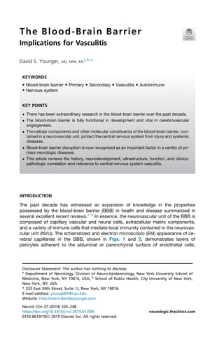
cular unit (NVU). The schematized and electron microscopic (EM) appearance of ce-
rebral capillaries in the BBB, shown in Figs. 1 and 2, demonstrates layers of
pericytes adherent to the abluminal or parenchymal surface of endothelial cells,
Disclosure Statement: The author has nothing to disclose.
a
Department of Neurology, Division of Neuro-Epidemiology, New York University School of
Medicine, New York, NY 10016, USA; b
School of Public Health, City University of New York,
New York, NY, USA
* 333 East 34th Street, Suite 1J, New York, NY 10016.
E-mail address: youngd01@nyu.edu
Website: http://www.davidsyounger.com
KEYWORDS
Blood-brain barrier Primary Secondary Vasculitis Autoimmune
Nervous system
KEY POINTS
There has been extraordinary research in the blood-brain barrier over the past decade.
The blood-brain barrier is fully functional in development and vital in cerebrovascular
angiogenesis.
The cellular components and other molecular constituents of the blood-brain barrier, con-
tained in a neurovascular unit, protect the central nervous system from injury and systemic
diseases.
Blood-brain barrier disruption is now recognized as an important factor in a variety of pri-
mary neurologic diseases.
This article reviews the history, neurodevelopment, ultrastructure, function, and clinico-
pathologic correlation and relevance to central nervous system vasculitis.
Neurol Clin 37 (2019) 235–248
https://doi.org/10.1016/j.ncl.2019.01.009 neurologic.theclinics.com
0733-8619/19/ª 2019 Elsevier Inc. All rights reserved.
2. together surrounded by a layer of basal lamina composed of extracellular matrix pro-
tein molecules. The end-feet of neighboring astrocyte processes ensheathes the
blood vessels. Monolayers of adjacent endothelial cells that form tight junction (TJ)
strands, shown in Fig. 1, connect adjacent endothelial cells by adhesions of trans-
membrane (occludin, claudin, and junctional associated molecules [JAM]) across
the intercellular space, whereas cytoplasmic scaffolding and regulatory proteins,
such as zona occludens type 1 and 2 (ZO-1, ZO-2), provide linkage to the actin cyto-
skeleton and initiate several signaling mechanisms via protein-protein interactions.
Endothelia BBB cells are also linked by adherens junctions composed of vascular
Fig. 1. (A) A capillary in the human BBB over an endothelial TJ. (B) The insert shows the mo-
lecular composition of tight and adherens junctions. See text for details. (From Daneman R.
The blood-brain barrier in health and disease. Ann Neurol 2012;72:649; with permission.)
Fig. 2. EM of a capillary in the adult murine BBB. Endothelial cells are held together by TJs
(red arrow). L, lumen. (From Daneman R. The blood-brain barrier in health and disease. Ann
Neurol 2012;72:648–72; with permission.)
Younger
236
3. endothelial (VE)-cadherin, which mediates cell-cell adhesion interactions, linking
adherens junctions to the actin cytoskeleton via catenins.2,3
Perivascular macro-
phages that reside between astrocyte end-feet and the vessel wall, mast cells asso-
ciated with specific regions of the central nervous system (CNS), resident microglia
that act as antigen-presenting cells, circulating leukocytes that can penetrate the
intact BBB via interactions with endothelial cell adhesion molecules (CAM) to mediate
bidirectional cross-talk between immune cells and endothelium for normal surveil-
lance, constitute the extended neurovascular unit.2
Breakdown or disruption of the BBB that accompanies a variety of inflammatory and
autoimmune, neoplastic, infectious, and neurodegenerative CNS disorders, notably
stroke, multiple sclerosis, brain trauma, human immune virus, infection, and Alzheimer
disease. These disorders are associated with the abnormal entry of plasma compo-
nents, immune molecules, and cellular elements that lead to further neural dysfunction
and varying degrees of irreversible neural degeneration. Although there is little known
about the role of BBB breakdown in primary and secondary CNS vasculitis, future prog-
ress could lead to improved understanding of primary and secondary forms of CNS
vasculitis6
with the prospect of even improving the outcome of 2 potentially devastating
disorders, childhood and adult primary angiitis of the CNS.7–9
According to Weiss and
colleagues,10
further understanding of the BBB could envision the use of new therapeu-
tic strategies that bypass it, taking advantage of the selective expression of membrane-
bound proteins expressed by brain endothelia cells or circulating leukocytes to target
new drugs as well as improve the effectiveness of conventional systemic immunosup-
pression. This article reviews the history, neurodevelopment, ultrastructure, function,
and clinicopathologic correlation and relevance to CNS vasculitis.
HISTORICAL BACKGROUND
In 1885, Ehrlich11
provided the first suggestion of the presence of a barrier when a
parenteral injection of vital dye into the bloodstream of mice penetrated practically
every systemic organ except the brain, turning them dark purplish-blue, leaving the
brain and spinal cord pale white-yellow. Ehrlich himself thought that this difference
was due to a low binding affinity. The existence of a barrier at the level of the cerebral
vessels was postulated by Bield and Kraus12
and later by Goldman13
and Lewandow-
sky14
at the turn of the twentieth century, who jointly interpreted their experience in
favor of a true BBB, later termed Blut-Hirn-Schranke. In 1967, Reese and Karnovsky15
demonstrated a structural barrier to an intravenous injection of horseradish peroxi-
dase (HRP) demonstrating exogenous peroxidase to the lumina of blood vessels
and in some micropinocytotic vesicles within endothelial cells, but none beyond the
vascular endothelium. The relatively scarce number of vesicles was a morphologic
feature of a functioning BBB. Their findings localized at a fine structural level a barrier
composed of the plasma membrane and the cell body of endothelial cells and TJ be-
tween adjacent cells of the cerebral cortex. In 1969, EM studies by Brightman and
Reese16
in the mouse conclusively demonstrated endothelial and epithelia TJ that
occluded the interspaces between blood and parenchyma or cerebral ventricles,
constituting the ultrastructural basis for the blood-brain and blood-cerebrospinal fluid
barriers. Feder17
noted active exclusion of the small electron-dense tracer microper-
oxidase by intact TJ after parenteral injection supplementing the findings of Reese and
Karnovsky.15
Nagy and colleagues18
examined fracture faces of cerebral endothelium
in normal and hyperosmolar mannitol-treated rat brains to elucidate the organization
of TJ in various segments of the cerebral vascular bed and the structural basis of
BBB opening in hyperosmotic conditions. Their findings provided no direct evidence
Blood-Brain Barrier—Implications for Vasculitis 237
4. for the structural basis of BBB opening in hyperosmolar mannitol-treated rat brains,
noting extended TJ regions in capillaries and postcapillary venues. Shivers and co-
workers19
studied isolated rat brain capillaries using freeze-fracture images of interen-
dothelial ZO revealing complex arrays of intramembrane ridges and grooves
characteristic of TJ. The ZO of these capillary endothelial cells were considered
very tight.
NEURODEVELOPMENT
In contrast to neuronal development, the vascular system undergoes blood vessel
formation through the 2 distinct processes: vasculogenesis and angiogenesis. The
former commences with endothelial differentiation from angioblasts to vascular
plexuses, whereas the latter is associated with sprouting of new vessels from exist-
ing ones. Both vasculogenesis and angiogenesis are influenced by vascular endo-
thelial growth factor (VEGF), a major attractive molecule for extending blood
vessels, especially endothelial tip cells,20
as well as by other molecules, blood
flow, and contact with surrounding tissues. Found at the growing end of extending
vessels, the shaft of extending endothelial tip cells is composed of an endothelial
cell chain made of stalk cells similar to the axon growth cone and its associated
axonal shaft. In vivo imaging using green fluorescent protein (GFP) depicts the
advancing endothelial tip cell navigating the environment and sprouting from exist-
ing vasculature.21
Several experimental observations have suggested the importance of the neurode-
velopment of the BBB.5
First, there are shared molecular and cellular mechanisms in both neurogenesis and
angiogenesis.22,23
Various axon guidance pathways from members of the 4 major
families of axon guidance ligand-receptor pairs, including Slit/Robo, semaphoring/
plexin/neuropilin, Netrin/Unc5/DCC, and Ephrin/Eph, that mediate complex cellular
navigational programs within axons as both chemoattractants and repellents, also
direct angiogenic tip cells toward their final destinations. Neuropilin-1 is necessary
for endothelial tip cell guidance in the developing CNS.24
Moreover, axonal terminal
arborization parallels vessel sprouting. Similar to hypoxic tissue that secretes VEGF
via hypoxia-inducible factor, a transcription factor that promotes cell survival through
the downstream activation of numerous genes including VEGF,25
axonal terminals
devoid of synaptic input secrete nerve growth factor, the expression of which is down-
regulated when innervation occurs.
Second, there is coregulation of these 2 systems in developing embryonic and adult
brains.22,26,27
Stubbs and colleagues26
found that blood vessels provided a support-
ing niche in regions of adult neurogenesis. The investigators26
used Tbr2-GFP trans-
genic mice that served as a correlate for the expression of the intermediate progenitor
cell (IPC) T-box transcription factor Tbr2, to examine the proximity of dividing cells in
the subventricular zone (SVZ) and ventricular zone of the shaking rat Kawasaki and
reeler mutant mouse in relation to blood vessels throughout neurogenesis. Their find-
ings, which included the extension of neuritis toward and along labeled blood vessels,
supported the notion of vascular-neuronal interactions in development. Javaherian
and Kriegstein,27
who likewise studied IPC in the SVZ of embryonic Swiss Webster
mouse cortices, used confocal microscopy to image the vast network of capillaries
in the SV and SVZ. The investigators27
noted that Tbr2 cells divided near vascular
branch points, suggesting endothelial tip cells contributed to the neurogenic niche
for IPC, with ectopic overexpression of VEGF-A in a pattern that followed that of blood
vessel development. These findings indicated that the developing cortical vasculature
Younger
238
5. provided a microenvironment within the SVZ in which IPC accumulated and divided
during neurogenesis.
Third, a structural and functional BBB complete with TJ appears as soon as cerebral
vessels penetrate the CNS parenchyma.28,29
Johansson and colleagues28
explained
that the widely held view that the BBB was immature during development stemmed
from teleologic interpretations and experimental observations of high cerebrospinal
fluid protein levels in fetal cerebrospinal fluid and the apparent passive passage of bio-
markers during development. Instead, the blood–cerebrospinal fluid barrier, like the
BBB, is functionally and morphologically mature from very early in development.
The investigators maintain that inconsistent terminology used in the literature, such
as leaky, immature, and developing, used to describe the barrier gives a connotation
of TJ that is more permeable than their adult counterparts without evidence to support
this concept. Mølgȧrd and Saunders30
noted well-formed complex TJ across cerebral
endothelial cells in human embryos and fetuses by freeze fracture and thin-section EM
by 8 weeks of age, commensurate with the differentiation of brain capillaries. Efflux
transporters are likewise expressed in cerebral endothelial and choroid plexus epithe-
lial cells early in the fetal and postnatal rats.31
Ballabh and colleagues32
studied the
expression and quantification of endothelial TJ molecules, including claudin-5,
occluding, and JAM, by immunohistochemistry and Western blot analysis in blood
vessels of germinal matrix, cortex, and white matter of fetuses and premature infants
gestational age 16 to 40 weeks. The investigators32
noted no significant decrease in
the expression of the endothelial TJ molecules claudin-5, occludin, and JAM-1 as a
function of gestational age in germinal matrix compared with cortex and white matter,
suggesting that they were unlikely to be responsible for germinal matrix fragility and
vulnerability to hemorrhage in premature infants. These findings are consistent with
the concept that TJ molecules develop and perhaps mature early during human gesta-
tion. Ballabh and colleagues33
observed that a paucity of TJ or pericytes coupled with
incomplete coverage of blood vessels by astrocyte end-feet, could instead account
for the observed fragility of blood vessels in the germinal matrix of premature infants.
Braun and colleagues34
found that pericytes coverage and density were less in the
germinal matrix vasculature than in the cortex or white matter in human fetuses, pre-
mature infants, and premature rabbit pups. Although VEGF suppression significantly
enhanced pericyte coverage in germinal matrix, it remained less than in other brain
regions.
NEUROBIOLOGY AND CELLULAR INTERACTIONS
Endothelial Cell Interactions
The existence of the endothelial cell was first surmised by William Harvey, first
observed by Marcello Malpighi in blood capillaries using compound microscopy in
the nineteenth century, and later by EM in the mid-twentieth century, revealing the
presence of plasmalemmal vesicles or caveolae. The ability to culture EC later
permitted even more detailed investigation of their activation and function in vivo.
Derived from mesoderm via the differentiation of hemangioblasts and angioblasts,
there are a few protein/messenger RNA marker candidates, including platelet/
endothelial cell adhesion molecule (PECAM)-1 in monocytes and VE-cadherin in fetal
stem cells. Endothelial cells of the BBB not only provide a physical barrier between the
systemic circulation and the brain but also assure the selective inward passage of
ions, nutrients, and neuropeptides via specialized transport mechanisms. Sodium,
potassium, chloride, hydrogen, bicarbonate, and calcium ions are transported across
the BBB via transporters located mainly along the luminal surface of endothelial
Blood-Brain Barrier—Implications for Vasculitis 239
6. cells, including the sodium and potassium adenosine triphosphate (ATP) -dependent
transport pump, the sodium-potassium-chloride cotransporter, sodium-proton, chlo-
ride-bicarbonate, and sodium calcium exchanges that assure optimal levels of brain
electrolyte levels and intracellular pH. The transport of essential nutrients is assured
by members of the solute-linked carriers (SLC) superfamily, located variably along
the luminal and abluminal membrane, including glucose transporter-1, monocarbox-
ylic acid-1, excitatory, organic acid, cation, amine, and choline transporters, respec-
tively, to transport lactate and ketone bodies as alternative energy neuronal sources,
and sodium-independent or -dependent removal of glutamate, aspartate, glutamine,
histidine, and asparagine from the interstitial compartment of the brain. Other specific
carrier-mediated transporters mediate the passage of transferrin, low-density lipopro-
teins, leptin, immunoglobulin G, insulin, and growth factors via receptor-mediated
transcytosis via binding of the protein to specific receptors on the endothelial cell sur-
face followed by endocytosis of the ligand-receptor complex with passage across the
cytoplasm and exocytosis at the opposite side of the cell35
and via the formation of
caveolae or vesicle formation for the transport of macromolecules.36
Transmigration
of cellular elements across endothelial cells of the BBB during inflammation, including
leukocytes, neoplastic cells, and pathogenic viruses, bacteria and yeasts, investi-
gated in experimental animal models using HRP, highlighted the role of caveolae as
minitransporters of the CNS.36
Unique systems of modified caveolae that fuse
together forming transendothelial cell channels and later vesiculocanalicular or
vesiculotubular structures or vesiculovacuolar organelles appear to be an important
gateway to the CNS in damaged endothelial cell populations.36
Transportation of
potentially toxic endogenous or xenobiotic lipid-soluble nonpolar molecules from
the brain to the blood is accomplished by transporters located along the luminal mem-
brane, such as the ATP binding cassette transporter P-glycoprotein 1 (multidrug resis-
tance protein 1 or ATP-binding cassette subfamily B member 1), respectively,
important in the distribution of CNS tumor drug treatment37
and the active efflux of
the anti–human immunodeficiency virus type 1 (HIV1) nucleoside drug abacavir at
the BBB,38–40
and breast-cancer resistance protein and multidrug resistance related
protein (MRP) 1, 2, 4, and 5 efflux transporter pumps that serve as defense mecha-
nisms and determinate bioavailability and concentration of many CNS drugs important
in the treatment of CNS cancers,41
such as the novel tyrosine kinase inhibitor dasati-
nib,42
and the efflux transportation of the protease inhibitor lopinavir that contributes
to its poor oral bioavailability in the treatment of HIV1.43
The neuroinflammation and
progression of damage associated with focal cerebral ischemia appear to be modu-
lated by upregulation of other MRP protein molecules that activate Toll-like receptor
signaling contributing to neuroinflammation and progression of ischemic cerebral
damage.44
Transendothelial migration of circulating leukocytes involves a multistep process.
Leukocyte adhesion molecules expressed on the surface of EC initiate binding of leu-
kocytes as a beginning step in the in their entry in brain tissue, which later includes roll-
ing adhesion to EC, firm adhesion, and transmigration. Although less well understood,
the molecular mechanism is thought to involve endothelial CAM, including CD99,
PECAM-1/CD31, vascular CAM-1 (VCAM-1) (important in firm adhesion); junctional
adhesion molecule-1; and expression of leukocyte adhesion molecules E- and
P-selectin (rolling adhesion); cytokine responsiveness so noted in situ and in cell cul-
ture,45,46
and expression of the integrins alpha-4 and beta-2. Inflamed capillary endo-
thelia support transmigration of different subsets of leukocytes. There are 2 routes for
leukocytes to pass through endothelial cells: the so-called paracellular route, or
through the endothelial cell itself or transcellular route. The BBB with its abundance
Younger
240
7. of TJ complexes relies primarily on the transcellular route as it does for solute and fluid
transport. *Neutrophil recruitment is partially dependent on ICAM-1, and express
L-selectin and lymphocyte function-associated antigen-1 but not chemokine C motif
receptor 7 may explain why granulocytes roll but do not arrest for transmigration in
high endothelial venules.
Pericyte Interactions
Brain endothelial cells are exposed to a myriad of pericyte interactions47
in the regu-
lation of brain angiogenesis, endothelial cell TJ formation, as well as the differentiation,
microvascular vasodynamic capacity, structural stability, and neuroimmunologic
network operations of the intact BBB.48
Rouget49
first ascribed capillary contractility
to pericytes, but Zimmerman50
named the cell and described its morphologic aspects.
The presence of smooth muscle cells in association with pericytes and the absence of
a smooth muscle layer from capillaries and postcapillary venues influenced early
views ascribing contractile properties to narrow capillaries, hence regulating micro-
vascular flow even though several subsequent experimental studies failed to substan-
tiate it.51,52
Smooth muscle actin was conclusively demonstrated in pericytes by
immunocytochemistry using smooth muscle a-actin isoform-specific antibodies and
immunogold labeling in conjunction with EM, noting that smooth muscle a-actin
expression in capillaries was limited exclusively to pericytes and not present in endo-
thelial cells.53
Because the histochemical localization of smooth muscle a-actin is
demonstrated in precapillaries and not in midcapillaries, it has been suggested that
smooth muscle a-actin–containing capillaries are involved in contractility and the con-
trol of capillary blood flow in the BBB.54
Unlike other perivascular cells, they lie within the microvessel basal lamina and
contribute to its formation, and typical CNS pericytes are flattened or elongated,
stellate-shaped solitary cells with multiple cytoplasmic processes encircling the capil-
lary endothelium and contacting a large abluminal vessel area. Brain pericytes are
characterized by granular deposits present in lysosomes that strongly react with
acid phosphatase, a finding that led to consideration of a phagocytic role.55
They
rapidly phagocytose an intravenous injection of HRP, which can be used as a pericyte
histochemical stain. The number of granular lysosomes in brain pericytes increases
with disruption of the BBB. Several other markers have been used in the identification
of pericytes, including smooth muscle a-actin, desmin, polydendrocytes (NG2 cells),
platelet-derived growth factor receptor-b, aminopeptidase A and N, regulator of
G-protein signaling 5, and the promoter trap transgene XlacZ4.56
An active role of pericytes in the BBB was inferred from the localization of g-glutamyl
transpeptidase (GGTP) in brain capillary endothelial cells and pericytes, both in vivo
and in vitro.57
This heterodimeric glycoprotein distributed on the external surface of
the cell catalyzes the transfer of g-glutamyl from glutathione to accept peptides and
functionally appears to be concerned with transport of large neutral amino acids
across the BBB. Detectable amounts of GGTP are found in other regions of the brain
with an intact BBB but not in those that lack one, such as the median eminence.
Abnormal platelet-derived growth factor (PDGF)-B and PDGF-b signaling play a crit-
ical role in the recruitment of pericytes to newly formed vessels, and when deficient,
as in knockout of pdgfb and pdbfrb, lead to perinatal death due to vascular dysfunc-
tion with associate vascular leakage and hemorrhage.
Pericyte-endothelia cell signaling factors have been identified. Sphingosine-
1-phosphate signaling triggers cytoskeletal, adhesive, and junctional changes,
affecting cell migration, proliferation, and survival.58
Angiopoietin-Tie2 signaling in
the vascular wall is involved in reciprocal communication between endothelial cell
Blood-Brain Barrier—Implications for Vasculitis 241
8. and pericytes, such as may be seen in ang1-or tie2-null mice deficient in Ang1, which
leads to defective angiogenesis and poorly organized BM and reduced coverage and
detachment of pericytes. Conversely, overexpression of Ang1 leads to expanded and
stabilized leakage-resistant microvasculature.59,60
The importance of CNS pericytes has been underscored by their proposed role in
neuroimmunologic networks associated with BBB function. First, CNS pericytes
may be actively involved in the regulation of leukocyte transmigration, antigen presen-
tation, and T-cell activation. They constitutively express low levels of VCAM-1 and
ICAM-1, which have costimulatory activity in main histocompatibility cell class II
dependent antigen presentation, and leukocytes cluster on pericytes in culture,48
suggesting a role in inflammation. Smooth muscle pericytes present antigen in vivo
and differentially activate Th1 and Th2 CD4 T cells. Moreover, CNS pericytes pro-
duce several immunoregulatory cytokines, including interleukin (IL) 1b and
granulocyte-macrophage colony stimulatory factor.61
Transforming growth factor
(TGF)-b produced in an active form in pericytes/endothelial cocultures may function
as an endogenous immunoregulator at the BBB.62
It is therefore of interest that
TFG-b1 inhibits cytokine-induced CNS endothelial cell activation in isolated rat CNS
microvessels.63
To further emphasize the importance of pericyte interactions in asso-
ciation with endothelial cells, there are no known genetic human diseases due to peri-
cytes deficiency.
Astrocyte Interactions
The intimate relationship of astrocytes and blood vessels was appreciated by Ramon y
Cajal64
and Golgi65
in the late nineteenth century. Since then, ultrastructural studies
have shown that astrocytic end-feet in the perivascular astroglial sheath leads to a com-
plete covering of brain microvessels.66
Signaling at the gliovascular interface is facilitated
by astrocyte-specific proteins and channels in astrocyte end-feet, including acquaporin-
4, connexin 43, purinergic receptors, and potassium channels.67
Moreover, ultrastruc-
tural studies have demonstrated that processes of vasoactive neurons for the regulation
of cerebrovascular tone, in particular, those expressing noradrenaline, synapse onto as-
trocytes rather than directly onto blood vessels.68
Altogether, these findings support the
observationthatastrocytes, oneof the morenumerouscells in theCNS, areimportantde-
terminants of the intact BBB and crucial as well for ionic homeostasis, neurotransmitter
uptake, synapse formation, and neurodevelopment. Zhang and Barres69
have reviewed
the differences in astrocyte morphology, developmental origin, gene expression profile,
physiologic properties, function, and response to injury and disease. Two essential roles
of astrocytes, in neurovascular coupling and the regulation of lymphocyte trafficking
across the BBB, have been extensively studied.
All signaling molecules targeted to the cerebral vasculature must first act on or pass
through astrocytes in order to reach smooth muscle cells in the vessel wall. It is now
recognized that neurotransmitter-mediated signaling has a key role in regulating cere-
bral blood flow, and that much of this control is mediated by astrocytes70
; moreover,
cerebral blood flow may be controlled by capillaries as well as by arterioles. The glial
and neuronal control of cerebral blood flow has been studied in brain slices.71
Koehler
and colleagues72
demonstrated that electrical field stimulations in brain slices led to an
increase in intracellular calcium in astrocyte cell bodies, which, when transmitted to
perivascular end-feet, was followed by a decrease in vascular smooth muscle calcium
oscillations and arteriolar dilation. The increase in astrocyte calcium after neuronal
activation was in part mediated by activation of metabotropic glutamate receptors.
Calcium signaling in vitro was influenced by adenosine acting on A2B receptors and
by epoxyeicosatrienoic acids (EET) shown to be synthesized in astrocytes. Moreover,
Younger
242
9. prostaglandins, EET, arachidonic acid, and potassium ions are candidate mediators of
communication between astrocyte end-feet and vascular smooth muscle. Astrocytes
appear to be capable of transmitting signals to pial arterioles on the brain surface to
ensure adequate blood flow to feeding arterioles; therefore, these cells play an impor-
tant role in the coupling of dynamic changes in cerebral blood flow in association with
neuronal activity.
Koehler and colleagues72
have provided insight into the morphologic aspects of
neurovascular coupling at the capillary level of the BBB. At least one astrocyte end-
foot process contacts a blood vessel, and those abutting capillaries and larger vessels
express connexin-43 and purinergic P2Y receptors, which together permit Ca21
in-
creases to be transmitted 60 mm or more along the abluminal side of the vessel
wall. Astrocytic cells are therefore in a unique position for sensing neuronal activity,
integrating that information, and communicating with blood vessels in brain paren-
chyma. Although neurons do not directly innervate intraparenchymal vascular smooth
muscle, subpopulations of GABAergic interneurons come into close contact with
astrocyte foot processes and elicit vasodilation. Such neurons might modulate
vascular function through stimulation of nitric oxide synthase activity, release of vaso-
active peptides, or an astrocyte signaling mechanism.
Hudson and coworkers73
studied trafficking of peripheral blood mononuclear cells
(PBMC) across feline brain endothelial cells (FBEC) in cell culture system after the
addition of combinations of different configurations of astrocytes and microglia in a
model of feline immunodeficiency virus. The addition of astrocytes to FBEC signifi-
cantly increased the adherence of PBMC, which was suppressed by the addition of
microglia, whereas the latter alone had no effect on PBMC adherence. Whereas all
PBMC showed some level of trafficking across FBEC, monocytes and B cells were
significantly increased if astrocytes were present. The exposure of astrocytes notably
increased the percentage of trafficking CD8 T cells from 24% to 64%, whereas micro-
glia led to a significant reversal in the preferential trafficking of CD8 cells in the pres-
ence of astrocytes. Astrocytes are capable of secreting various cytokines and
chemokines in the upregulation of adhesion molecules and T-cell ligands in intact
endothelial cells, such as ICAM, VCAM, E-selectin, and PECAM. Human cocultured
human endothelial cells and astrocytes increase the expression of ICAM-1 due to in-
flammatory activation by hypoxia in vitro.74
Other studies have demonstrated that as-
trocytes are a source of IL-6, tumor necrosis factor-a, and monocyte chemoattractant
protein-1, which contribute to the CNS inflammatory response.75
Trafficking of PBMC
along the endothelial cell of the BBB is a complex mechanism that involves major sub-
sets of immune cells and relies heavily on astrocyte, microglia, and endothelial cell in-
teractions; moreover, astrocytes appear to be an active factor in the recruitment of
immune cells, whereas microglia appear to curtail this activity.
IMPLICATIONS FOR CEREBRAL VASCULITIS
There is an extensive literature of BBB biology in health and in widely differing neuro-
logic disorders, including stroke,76
epilepsy,77
multiple sclerosis,78
Alzheimer dis-
ease,79
motor neuron disease,80
Parkinson disease,81
trauma,82
glioblastoma,83
HIV
encephalitis,84
and neuropsychiatric systemic lupus erythematosus (NPSLE).85
Be-
tween 40% and 70% of patients with SLE have involvement of the CNS,86
yet unlike
systemic and primary CNS vasculitides, the role of the BBB in CNS lupus has been the
subject of intense study using animal models and human clinical data.
The BBB is crucial because it maintains the brain internal milieu constant, allowing
optimal neuronal function. Loss of BBB integrity leads to influx of inflammatory cells,
Blood-Brain Barrier—Implications for Vasculitis 243
10. and molecules, such as autoantibodies, causing brain injury. Earlier studies using the
well-established MRL/lpr mouse model that reflects the events that occur in human
CNS lupus revealed loss of BBB integrity with worsening disease87,88
due to chronic
activation of the complement cascade with the generation of the anaphylatoxins, C3a
and C5a, and aggravated C5a/C5aR signaling. In cell culture, C5a causes neuronal
cells to become apoptotic and binds to 2 receptors, the G-coupled, C5aR1, and the
alternate receptor, C5aR2.89
C5a/C5aR1 signaling mediates several biological pro-
cesses, including chemotaxis and degranulation of mast cells, basophils, neutrophils,
and eosinophils. It increases vascular permeability, increases generation of reactive
oxygen species, and enhances production of cytokines from monocytes and macro-
phages. Whether C5a/C5aR signaling is protective or neurotoxic depends on the
setting, with increases in circulating C5a leading to poor outcomes in CNS lupus.89
Hopkins and colleagues90
measured levels of complement anaphylatoxin split prod-
ucts, C3a and C5a, in the circulation of patients with SLE who were followed serially.
Means complement levels were significantly higher during periods of lupus flare
compared with those during a stable period, with the highest levels seen in patients
with CNS involvement. Pathologic specimens from 2 cases who died during an acute
lupus flare revealed neutrophils occluding the cerebral and systemic intestinal vessels.
C5a also contributes to cellular apoptosis in lupus,91
wherein treatment with the
C5aRant, inhibiting C5a/C5aR signaling, results in significant and substantial de-
creases in brain pathologic condition in MRL/lpr mice, leaving upstream potentially
protective complement activation events intact. There was evidence for a relationship
between complement activation, inflammation, and neuronal viability in lupus brain tis-
sue. C5aR1 is present predominantly on blood myeloid cells, but is also constitutively
expressed on several brain cell types, including endothelial cells, which contribute to
BBB integrity. Such studies show the possible neuroprotective role for C5aR antago-
nists in MRL/lpr mice and indicate potential future avenues of research for systemic
and primary CNS vasculitides.
SUMMARY
There has been extraordinary research in the BBB over the past decade. Once consid-
ered a static anatomic barrier to the traffic of molecules in and out of the CNS, the BBB
of children and adults is now recognized to be fully functional and vital to both cere-
brovascular angiogenesis and normal homeostatic maintenance. The cellular compo-
nents and other molecular constituents of the BBB, contained in the NVU, protect the
CNS from injury and disease by limiting the passage of toxins, pathogens, and inflam-
matory effectors of the immune system. The implications of BBB disruption in cerebral
vasculitis have yet to be appreciated; however, comparisons to CNS lupus may lead to
a better understanding of the neural mechanisms involved in disease pathogenesis
and BBB integrity, and efficacious neuroprotective treatment strategies.
REFERENCES
1. Daneman R. The blood-brain barrier in health and disease. Ann Neurol 2012;72:
648–72.
2. Benarroch EE. Blood-brain barrier. Neurology 2012;78:1268–76.
3. Hawkins BR, Davis TP. The blood-brain barrier/Neurovascular unit in health and
disease. Pharmacol Rev 2005;57:173–85.
4. Weiss N, Miller F, Cazaubon S, et al. The blood-brain barrier in brain homeostasis
and neurological diseases. Biochim Biophys Acta 2009;1788:842–57.
Younger
244
11. 5. Neuwelt EA, Bauer B, Fahlke C, et al. Engaging neuroscience to advance trans-
lational research in brain barrier biology. Nat Rev Neurosci 2011;12:169–82.
6. Younger DS. Adult and childhood vasculitis of the nervous system. In:
Younger DS, editor. Chapter 14. Motor disorders. 3rd edition. New York: Rothstein
Publishing; 2013. p. 235–80.
7. Twilt M, Benseler SM. The spectrum of CNS vasculitis in pediatrics and adults.
Nat Rev Rheumatol 2011;8:97–107.
8. Hajj-Ali RA, Singhal AB, Benseler S, et al. Primary angiitis of the CNS. Lancet
Neurol 2011;10:561–72.
9. Salvarani C, Brown RD Jr, Calamia KT, et al. Primary central nervous system
vasculitis: analysis of 101 patients. Ann Neurol 2007;62:442–51.
10. Weiss N, Miller F, Cazaubon S, et al. Implications of the blood-brain barrier in
neurological diseases: part II. Rev Neurol (Paris) 2009;165:1010–22 [in French].
11. Ehrlich P. The requirement of the organism for oxygen. An analytical study with
the aid of dyes. In: Himmelweit F, Marquardt M, Dale H, editors. The collected pa-
pers of Paul Ehrlich. London: Pergamon Press; 1957. p. 433–96 [English
translation].
12. Bield A, Kraus B. Über eine bisher unbekannte toxische Wirkung der Gallensau-
ren auf das Zentralnervensystem. Zhl Inn Med 1898;19:1185–200.
13. Goldmann EE. Vitalfarbung am Zentral-nervensystem. Abh Preuss Akad Wis-
sensch. Physkol Mathem Klasse 1913;1:1–60.
14. Lewandowsky M. Zür lehre der cerebrospinal flüssigkeit. Z Klin Med 1900;40:
480–94.
15. Reese TS, Karnovsky MJ. Fine structural localization of a blood-brain barrier to
exogenous peroxidase. J Cell Biol 1967;34:207–17.
16. Brightman MW, Reese TS. Junctions between intimately apposed cell mem-
branes in the vertebrate brain. J Cell Biol 1969;40:648–77.
17. Feder N. Microperoxidase: an ultrastructural tracer of low molecular weight. J Cell
Biol 1971;51:339–43.
18. Nagy Z, Peters H, Huttner I. Fracture faces of cell junctions in cerebral endothe-
lium during normal and hyperosmotic conditions. Lab Invest 1984;50:313–22.
19. Shivers RR, Betz AL, Goldstein GW. Isolated rat brain capillaries possess intact,
structurally complex, interendothelial tight junctions: freeze-fracture verification of
tight junction integrity. Brain Res 1984;324:313–22.
20. Gerhardt H, Golding M, Fruttiger M, et al. VEGF guides angiogenic sprouting uti-
lizing endothelial tip cell filopodia. J Cell Biol 2003;161:1163–77.
21. Lawson ND, Weinstein BM. In vivo imaging of embryonic vascular development
using transgenic zebrafish. Dev Biol 2002;248:307–18.
22. Tam SJ, Watts RJ. Connecting vascular and nervous system development: angio-
genesis and the blood-brain barrier. Annu Rev Neurosci 2010;33:379–408.
23. Carmeliet P, Tessier-Lavigne M. Common mechanisms of nerve and blood vessel
wiring. Nature 2005;436:193–200.
24. Gerhardt H, Ruhrberg C, Abramsson A, et al. Neuropilin-1 is required for endo-
thelial tip cell guidance in the developing central nervous system. Dev Dyn
2004;231:503–9.
25. Pugh CW, Ratcliffe PJ. Regulation of angiogenesis by hypoxia: role of the HIF sys-
tem. Nat Med 2003;9:677–84.
26. Stubbs D, DeProto J, Nie K, et al. Neurovascular congruence during cerebral
cortical development. Cereb Cortex 2009;19:i32–41.
27. Javaherian A, Kriegstein A. A stem cell niche for intermediate progenitor cells of
the embryonic cortex. Cereb Cortex 2009;19:i70–7.
Blood-Brain Barrier—Implications for Vasculitis 245
12. 28. Johansson PA, Dziegielewska KM, Liddelow SA, et al. The blood-CSF barrier ex-
plained: when development is not immaturity. Bioassays 2008;30:237–48.
29. Ek CJ, Dziegielewska KM, Stolp H, et al. Functional effectiveness of the blood-
brain barrier to small water soluble molecules in developing and adult opossum
(Monodelphis domestica). J Comp Neurol 2006;496:13–26.
30. Møllgȧrd K, Saunders NR. The development of human blood-brain and blood-
CSF barriers. Neuropathol Appl Neurobiol 1986;12:337–58.
31. Ek CJ, Wong A, Liddelow SA, et al. Efflux mechanisms at the developing brain
barriers: ABC-transporters in the fetal and postnatal rat. Toxicol Lett 2010;197:
51–9.
32. Ballabh P, Hu F, Kumarasiri M, et al. Development of tight junction molecules in
blood vessels of germinal matrix, cerebral cortex, and white matter. Pediatr
Res 2005;58:791–8.
33. Ballabh P, Braun A, Nedergaard M. The blood-brain barrier: an overview. Struc-
ture, regulation, and clinical implications. Neurobiol Dis 2004;16:1–13.
34. Braun A, Xu H, Hu F, et al. Paucity of pericytes in germinal matrix vasculature of
premature infants. J Neurosci 2007;27:12012–24.
35. Kreuter J. Drug delivery to the central nervous system by polymeric nanopar-
ticles: What do we know? Adv Drug Deliv Rev 2014;71:2–14.
36. Lossinsky AS, Shivers RR. Structural pathways for macromolecular and cellular
transport across the blood-brain barrier during inflammatory conditions. Histol
Histopathol 2004;19:535–64.
37. Dai H, Marbach P, Lemaire M, et al. Distribution of ST1-571 to the brain is limited
by P-glycoprotein-mediated efflux. J Pharmacol Exp Ther 2003;304:1085–92.
38. Shaik N, Giri N, Pan G, et al. P-glycoprotein-mediated active efflux of the anti-
HIV1 nucleoside abacavir limits cellular accumulation and brain distribution.
Drug Metab Dispos 2007;35:2076–85.
39. Zhang C, Kwan P, Zuo Z, et al. The transport of antiepileptic drugs by P-glycopro-
tein. Adv Drug Deliv Rev 2012;64:930–42.
40. Aronica E, Sisodiya SM, Gorter JA. Cerebral expression of drug transporters in
epilepsy. Adv Drug Deliv Rev 2012;64:919–29.
41. Kerb R, Hoffmeyer S, Brinkmann U. ABC drug transporters: hereditary polymor-
phisms and pharmacological impact in MDR1, MRP1 and MRP2. Pharmacoge-
nomics 2001;2:51–64.
42. Chen Y, Agarwal S, Shaik NM, et al. P-glycoprotein and breast cancer resistance
protein influence brain distribution of dasatinib. J Pharmacol Exp Ther 2009;330:
956–63.
43. Agarwal S, Pai D, Mitra AK. Both P-gp and MRP2 mediate transport of lopinavir, a
protease inhibitor. Int J Pharm 2007;339:139–47.
44. Ziegler G, Prinz V, Albrecht MW, et al. Mrp-8 and -14 mediate CNS injury in focal
cerebral ischemia. Biochim Biophys Acta 2009;1792:1198–204.
45. Petzelbauer P, Bender JR, Wilson J, et al. Heterogeneity of dermal microvascular
endothelial cell antigen expression and cytokine responsiveness in situ and in
cell culture. J Immunol 1993;151:5062–72.
46. Milstone DS, O’Donnell PE, Stavrakis G, et al. E-selectin expression and stimula-
tion by inflammatory mediators are developmentally regulated during embryo-
genesis. Lab Invest 2000;80:943–54.
47. Armulik A, Abramssson A, Betsholtz C. Endothelial/pericytes interactions. Circ
Res 2005;97:512–23.
48. Balabanov R, Dore-Duffy P. Mini-review. Role of the CNS microvascular pericytes
in the blood-brain barrier. J Neurosci Res 1998;53:637–44.
Younger
246
13. 49. Rouget C. Mémoire sur le dévelopement, la structure et les propriéties physiolo-
giques des capillaires sanguins et lymphatiques. Arch Physiol Normale Pathol
1873;5:603–61.
50. Zimmerman K. Die feinere Bau der Blutcapillaren. A Anat Entwickl 1923;68:
29–109.
51. Diaz-Flores LR, Gutierrez H, Varela N, et al. Microvascular pericytes: a review of
their morphological and functional characteristics. Histol Histopathol 1991;6:
269–86.
52. Tilton RG. Capillary pericytes: perspectives and future trends. J Electon Microsc
Tech 1991;19:327–44.
53. Bandopadhyay R, Orte C, Lawrenson JG, et al. Contractile proteins in pericytes
at the blood-brain and blood-retinal barriers. J Neurocytol 2001;30:35–44.
54. Boado RJ, Pardridge WM. Differential expression of a-actin mRNA and immuno-
reactive protein in brain microvascular pericytes and smooth muscle cells.
J Neurosci Res 1994;39:430–5.
55. Farrell CR, Steward PA, Farrell CL, et al. Pericytes in human cerebral microvascu-
lature. Anat Rec 1987;218:466–9.
56. Gerhardt H, Betsholtz C. Endothelial-pericyte interactions in angiogenesis. Cell
Tissue Res 2003;314:15–23.
57. Frey AB, Meckelein H, Weiler-Guttler B, et al. Pericytes of the brain microvascu-
lature express g-glutamyl transpeptidase. Eur J Biochem 1991;202:421–9.
58. Allende ML, Proia RL. Sphingosine-1-phosphate receptors and the development
of the vascular system. Biochim Biophys Acta 2002;1582:222–7.
59. Suri C, McClain J, Thurston G, et al. Increased vascularization in mice overex-
pressing angiopoietin-1. Science 1998;282:468–71.
60. Thurston G, Suri C, Smith K, et al. Leakage-resistant blood vessels in mice tran-
genically overexpressing angiopoietin-1. Science 1999;286:2511–4.
61. Fabry Z, Fitzsimmons K, Herlein J, et al. Production of cytokines interleukin 1 and
6 by murine brain microvessel endothelium and smooth muscle pericytes.
J Neuroimmunol 1993;47:23–34.
62. Dore-Duffy P, Balabanov R, Rafols J, et al. The recovery phase of acute experi-
mental autoimmune encephalomyelitis in rats corresponds to development of
endothelial cell unresponsiveness to interferon gamma activation. J Neurosci
Res 1996;44:223–34.
63. Dore-Duffy P, Balabanov R, Washington R, et al. Transforming growth factor-b1
inhibits cytokine-induced CNS endothelial cell activation. Mol Chem Neuropathol
1994;22:161–75.
64. Ramon y Cajal S. Algunas conjeturas sobre el mecanismo anatomico de la idea-
cion, asociacion y atencion. Rev Med Cir Pract 1895;36:497–508.
65. Golgi C. Sulla fina anatomia degli organi central del sistema nervosa. Milan
(Italy): Hoepli; 1986.
66. Mathiisen TM, Lehre KP, Danbolt NC, et al. The perivascular astroglial sheath pro-
vides a complete covering of the brain microvessels: an electron microscopic 3D
reconstruction. Glia 2010;58:1094–103.
67. Price DL, Ludwig JW, Mi H, et al. Distribution of rSlo Ca21-activated K1 channels
in rat astrocyte perivascular endfeet. Brain Res 2002;956:183–93.
68. Hamel E. Perivascular nerves and the regulation of cerebrovascular tone. J Appl
Physiol (1985) 2006;100:1059–64.
69. Zhang Y, Barres BA. Astrocyte heterogeneity: an underappreciated topic in
neurobiology. Curr Opin Neurobiol 2010;20:588–94.
Blood-Brain Barrier—Implications for Vasculitis 247
14. 70. Petzold GC, Murthy VN. Role of astrocytes in neurovascular coupling. Neuron
2011;71:782–97.
71. Attwell D, Buchan AM, Charpak S, et al. Glial and neuronal control of brain blood
flow. Nature 2010;468:232–43.
72. Koehler RC, Gebremedhin D, Harder DR. Role of astrocytes in cerebrovascular
regulation. J Appl Physiol (1985) 2006;100:307–17.
73. Hudson LC, Bragg DC, Tompkins MB, et al. Astrocytes and microglia differentially
regulate trafficking of lymphocyte subsets across brain endothelial cells. Brain
Res 2005;1058:148–60.
74. Zhang W, Smith C, Howlett D, et al. Inflammatory activation of human brain endo-
thelial cells by hypoxic astrocytes in vitro is mediated by IL-1beta. J Cereb Blood
Flow Metab 2000;20:967–78.
75. Dong Y, Benveniste EN. Immune function of astrocytes. Glia 2001;36:180–90.
76. Jiao H, Wang Z, Liu Y, et al. Specific role of tight junction proteins claudin-5,
occluding, and ZO-1 of the blood brain barrier in a focal cerebrovascular
ischemic insult. J Mol Neurosci 2011;44:130–9.
77. Oby E, Janigro D. the blood-brain barrier and epilepsy. Epilepsia 2006;47:
1761–74.
78. Alvarez JI, Cayrol R, Prat A. Disruption of central nervous system barriers in mul-
tiple sclerosis. Biochim Biophys Acta 2011;1812:252–64.
79. Bowman GL, Kaye JA, Moore M, et al. Blood-brain barrier impairment in Alzheimer
disease: stability and functional significance. Neurology 2007;68:1809–14.
80. Garbuzova-Davis S, Haller E, Saporta S, et al. Ultrastructure of blood-brain barrier
and blood-spinal barrier in SOD1 mice modeling ALS. Brain Res 2007;1157:126–37.
81. Faucheux BA, Bonnet AM, Agid Y, et al. Blood vessels change in the mesenceph-
alon of patients with Parkinson’s disease. Lancet 1999;353:981–2.
82. Tompkins O, Shelef I, Kaizerman I, et al. Blood-brain barrier disruption in post-
traumatic epilepsy. J Neurol Neurosurg Psychiatry 2008;79:774–7.
83. Ishihara H, Kubota H, Lindberg RL, et al. Endothelial cell barrier impairment
induced by glioblastoma and transforming growth factor beta2 involves matrix
metalloproteinases and tight junction proteins. J Neuropathol Exp Neurol 2008;
67:435–48.
84. Roberts TK, Buckner CM, Berman JW. Leukocyte transmigration across the
blood-brain barrier: perspectives on neuroAIDS. Front Biosci 2010;15:478–536.
85. Huizinga TW, Diamond B. Lupus and the central nervous system. Lupus 2008;17:
376–9.
86. Mahajan SD, Tutino VM, Redae Y, et al. C5a induces caspase-dependent
apoptosis in brain vascular endothelial cells in experimental lupus. Immunology
2016;148:407–19.
87. Jacob A, Hack B, Chen P, et al. C5a/CD88 signaling alters blood–brain barrier
integrity in lupus through nuclear factor-kB. J Neurochem 2011;119:1041–51.
88. Jacob A, Hack B, Chiang E, et al. C5a alters blood–brain barrier integrity in
experimental lupus. FASEB J 2010;24:1682–8.
89. Mahajan SD, Tutino VM, Redae Y, et al. C5a induces caspase-dependent
apoptosis in brain vascular endothelial cells in experimental lupus. Immunology
2016;148:407–19.
90. Hopkins P, Belmont HM, Buyon, et al. Increased levels of plasma anaphylatoxins
in systemic lupus erythematosus predict flares of the disease and may elicit
vascular injury in lupus cerebritis. Arthritis Rheum 1988;31:632–41.
91. Jacob A, Hack B, Bai T, et al. Inhibition of C5a receptor alleviates experimental
CNS lupus. J Neuroimmunol 2010;221:46–52.
Younger
248
![together surrounded by a layer of basal lamina composed of extracellular matrix pro-
tein molecules. The end-feet of neighboring astrocyte processes ensheathes the
blood vessels. Monolayers of adjacent endothelial cells that form tight junction (TJ)
strands, shown in Fig. 1, connect adjacent endothelial cells by adhesions of trans-
membrane (occludin, claudin, and junctional associated molecules [JAM]) across
the intercellular space, whereas cytoplasmic scaffolding and regulatory proteins,
such as zona occludens type 1 and 2 (ZO-1, ZO-2), provide linkage to the actin cyto-
skeleton and initiate several signaling mechanisms via protein-protein interactions.
Endothelia BBB cells are also linked by adherens junctions composed of vascular
Fig. 1. (A) A capillary in the human BBB over an endothelial TJ. (B) The insert shows the mo-
lecular composition of tight and adherens junctions. See text for details. (From Daneman R.
The blood-brain barrier in health and disease. Ann Neurol 2012;72:649; with permission.)
Fig. 2. EM of a capillary in the adult murine BBB. Endothelial cells are held together by TJs
(red arrow). L, lumen. (From Daneman R. The blood-brain barrier in health and disease. Ann
Neurol 2012;72:648–72; with permission.)
Younger
236](data:image/gif;base64,R0lGODlhAQABAIAAAAAAAP///yH5BAEAAAAALAAAAAABAAEAAAIBRAA7)